
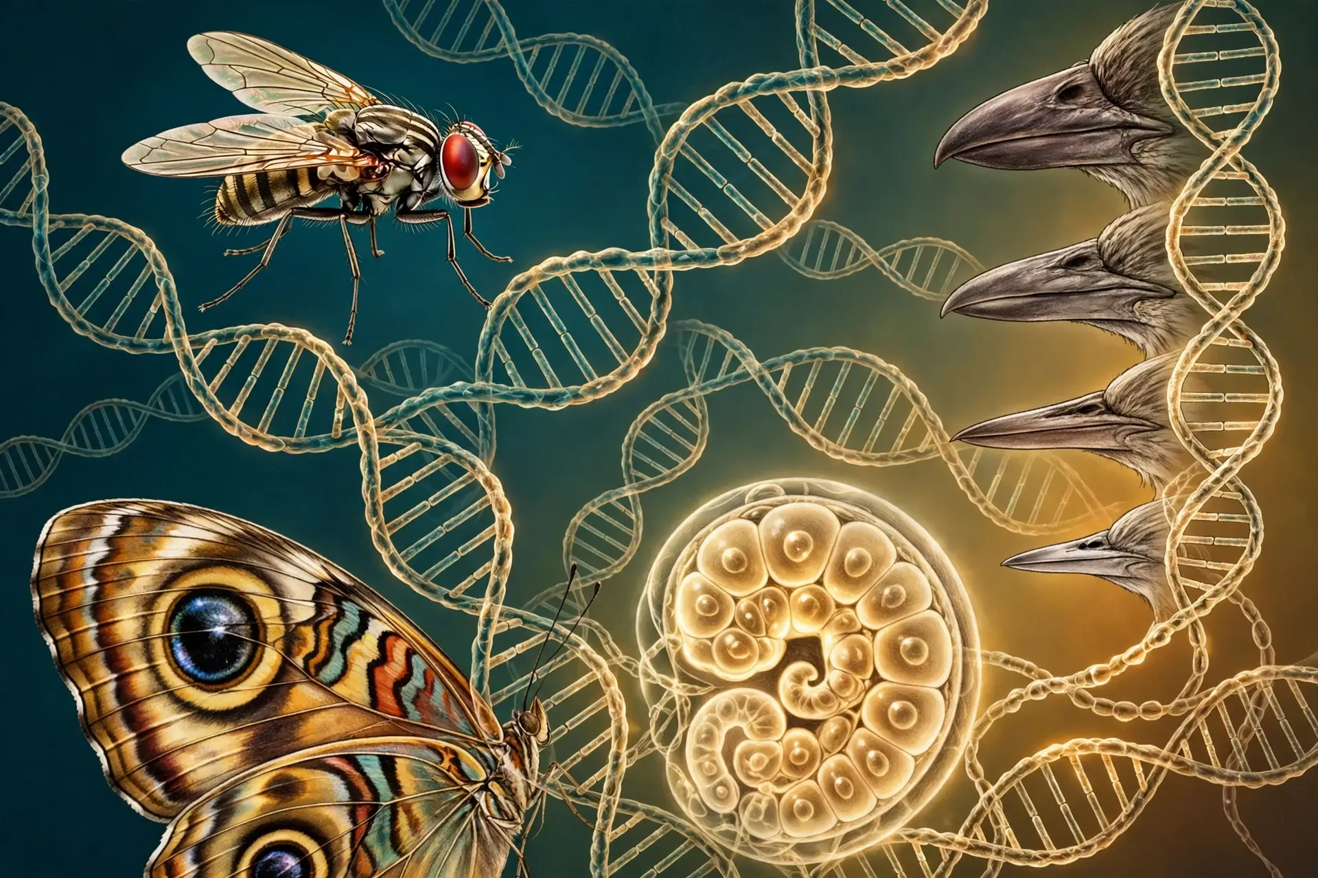
1. The Fly With Legs on Its Head
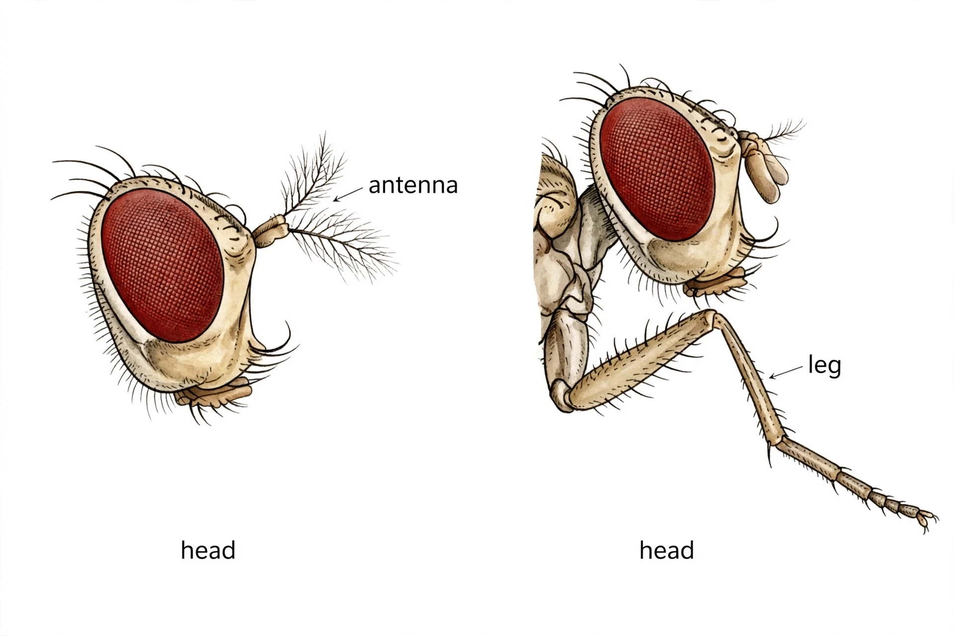
The fly in the vial looked wrong in a way that took a second to register. It had the right size, the right wings, the right bristly body. Then I looked at its head, and the head had legs on it. Not stumps, not nubs — proper legs, with joints and bristles and little claws, growing out of the front of the head where the antennae should have been. The fly was alive, walking around on six ordinary legs, plus two more that belonged nowhere.
That fly was not a monster from a horror film. It was a laboratory fruit fly, Drosophila melanogaster, carrying a mutation called Antennapedia — the homeotic mutant at the heart of evo-devo. And the strangest thing about it was not that it had legs on its head. The strangest thing was how good those legs were. They were perfectly formed legs. Every joint was in the right place, every bristle pointed the right way. The leg knew exactly how to be a leg. It simply did not know it was supposed to be an antenna.
That single observation cracks open the deepest question in evo-devo. If a leg can grow in the wrong place and still be a perfect leg, then the instructions for "leg" and the instructions for "here" must be separate things. The embryo must carry two kinds of information: a set of building programmes for parts, and a set of switches that decide where each programme runs. Change the switch, and the same programme runs somewhere else. The body plan — the whole arrangement of head, thorax, abdomen, limbs, and organs — is not drawn directly by the genes. It is drawn by a small set of regulatory genes that tell other genes when and where to work.
Evo-devo is the science of those switches. Its full name is evolutionary developmental biology, and it asks a question that Darwin's theory left open for more than a century: how do changes in development produce evolutionary changes in body form? Natural selection explains why a trait spreads through a population. Evo-devo explains where the trait comes from in the first place — how a beak gets deeper, how a pelvis vanishes, how a leg ends up on a head, and how the same genes can build a fly, a fish, and a human.
Here is the answer in two sentences. Animals share a small, ancient toolkit of regulatory genes — Hox genes above all — that assign positional identity to the embryo, telling each segment what it is supposed to become. Evolution mostly works not by inventing new toolkit genes, but by rewiring when, where, and how strongly those genes are switched on, and that rewiring is enough to sculpt every body plan on Earth, including yours.
The road runs through 19 sections. It starts with that fly and the definition of evo-devo, then meets the Hox genes themselves: what they are, how many there are, and the eerie map they draw on the chromosome. Then comes the hierarchy that switches Hox genes on, the homeotic mutants that proved segment identity is a decision, and the experiment in which a mouse gene built a fly eye. The middle of the article is the mechanism — gene networks, enhancers, and the three dials of timing, place, and proportion — followed by four case studies: finch beaks, stickleback pelvises, legless snakes and whales, and butterfly eyespots. The final stretch follows the toolkit from fins to fingers, asks what it did and did not do in the Cambrian explosion, clears up the common mistakes and myths, and closes with why this science matters for medicine and for the reader's own body.
1.1 The scene, slowed down
Picture the vial again. A fruit fly is about three millimetres long, small enough that you need a microscope to see the details. Under the lens, the normal fly's antenna is a short, feathery structure, built for smelling. The mutant's replacement is unmistakably a leg: a coxa, a femur, a tibia, a tarsus with five segments, and a claw at the end. It even has the bristle pattern of a second thoracic leg — the middle leg of the fly's three pairs.
Nothing else about the fly is wrong. Its wings are normal. Its other six legs are normal. It eats, walks, and mates. The mutation changed one decision in one place: in the head segment, the cells were told "you are a leg segment" instead of "you are an antenna segment". Everything downstream of that decision — the whole leg-building programme — then ran perfectly, because the programme itself was untouched.
That is the first lesson of evo-devo, and it is worth sitting with. Development is modular. The programme for building a leg is a self-contained unit that can be switched on wherever the embryo tells it to run. The embryo does not build a leg by following a blueprint of the whole fly. It builds a leg by running a local programme, and the address of that programme is set by a handful of regulatory genes.
1.2 Why a misplaced leg matters
A misplaced leg matters because it shows that evolution has a shortcut. If body plans were built by thousands of genes each doing one tiny job, changing a body plan would need thousands of coordinated mutations — an unlikely event. But if body plans are built by a small number of switches that control whole programmes, then a single mutation in a switch can move, resize, or duplicate an entire structure in one step. That is how a beak can deepen, a pelvis can vanish, and a limb can be lost — not by rewriting the whole animal, but by changing one instruction about where or when a programme runs.
This is also why the same genes keep showing up in wildly different animals. The leg-building programme in a fly and the limb-building programme in a mouse share deep similarities, because both are run by descendants of the same ancient regulatory genes. The toolkit is old. The deployments are new. That is the sentence to carry through the rest of this article: new bodies come mostly from old genes used in new ways.
1.3 The question this article answers
The question is simple to state and took a century to answer: how does an embryo know what goes where? The answer, built up over the next eighteen sections, has four parts. First, Hox genes assign positional identity along the head-to-tail axis, in the same order as they sit on the chromosome. Second, a hierarchy of earlier genes switches the Hox genes on in the right segments. Third, the Hox genes and their partners control whole networks of building genes through enhancers — small DNA switches that say where and when. Fourth, evolution tweaks those enhancers, and the timing and place of expression, to produce new forms from old parts. Small changes in the instructions, large changes in the animal — that is the story of evo-devo, and it connects a fly's head to your own hands. For how small changes accumulate into new species, see the companion article on speciation.
2. What Is Evo-Devo?
Evo-devo is short for evolutionary developmental biology: the study of how changes in development produce evolutionary changes in body form. It is a young field with old roots, and its central claim is disarmingly simple. The diversity of animal bodies — the wings, beaks, fins, legs, eyes, and antennae — does not come mainly from new genes. It comes from old genes used in new ways: switched on in a different place, at a different time, or at a different strength. Where a gene is expressed matters as much as which gene it is.
2.1 The plain definition
Put plainly, evo-devo asks how an embryo builds a body, and how small changes in that building process produce the differences between species. Every animal starts as a single cell. That cell divides, its descendants specialise, and a body plan emerges: head at one end, tail at the other, segments in between, limbs and organs in their places. The instructions for that process are written in DNA, but they are not a blueprint. They are more like a recipe with conditional steps — "if you are in the second thoracic segment, build a leg; if you are in the head, build an antenna" — and the conditions are set by regulatory genes.
The field's name gives the game away. It joins two sciences that spent most of the twentieth century working apart: evolutionary biology, which explains how populations change over generations, and developmental biology, which explains how a single cell becomes an organism. Evo-devo is the bridge between them. It says that to understand evolution, you must understand development, because development is where new forms are actually made.
2.2 The old puzzle
Darwin knew embryos held clues. In On the Origin of Species he noted that the embryos of different animals often look more alike than the adults do, and he took that as evidence of shared ancestry. But the Modern Synthesis — the version of evolutionary theory that dominated the twentieth century — had no real room for development. It treated genes as units that change in frequency in populations, and it treated the embryo as a black box: genes went in, adult traits came out, and what happened inside the box was somebody else's problem.
Embryologists, for their part, worked on the box itself. They mapped how embryos develop, discovered the signals that pattern tissues, and described the process in exquisite detail — but they rarely connected it to evolution. The two fields that evo-devo would later join used different languages, attended different meetings, and published in different journals. A geneticist studying natural selection and an embryologist studying a frog embryo could spend whole careers without meeting.
That separation left a hole in the theory. Natural selection explains why a trait spreads once it exists, but it does not explain where new traits come from. A beak that is a millimetre deeper is easy to explain as the result of selection on small variations. But where does the variation itself come from? How does a developmental process produce a deeper beak, a longer limb, or an extra pair of wings? For most of the twentieth century, the honest answer was: nobody knew.
2.3 The reunion
The reunion began in the early 1980s, and it began with a surprise. When researchers cloned the homeotic genes of the fruit fly — the genes behind the Antennapedia and bithorax mutants — they found that these genes all shared a short DNA sequence, about 180 letters long, that coded for a DNA-binding protein domain. They called it the homeobox. Then they used it as a probe, fishing in the DNA of other animals for the same sequence. What they found overturned everyone's expectations: the same sequence turned up in worms, in frogs, in mice, and in humans. The genes that build a fly's body plan have counterparts in every animal with a head and a tail.
That discovery, made in 1984 in two laboratories working independently — Walter Gehring's group in Basel and Thomas Kaufman's group in Indiana — was the first hard evidence that development runs on shared machinery. The genes were not just similar. They were the same genes, inherited from a common ancestor that lived more than half a billion years ago, and still doing the same job: telling the embryo where its head ends and its tail begins.
The 1990s completed the reunion. The 1995 Nobel Prize went to three developmental geneticists — Edward Lewis, Christiane Nüsslein-Volhard, and Eric Wieschaus — for working out how genes control early embryonic development in the fly. And in the same decade, the Pax6 experiment showed that a mouse gene could build a fly eye. By the turn of the century, the message was clear: the toolkit is ancient, the deployments are new, and the history of animal form is largely the history of regulatory change.
2.4 The core claim in one sentence
Here is the core claim of evo-devo, in a single sentence: evolution rewrites the instructions for using genes far more often than it invents new genes. A new body plan rarely needs a new protein. It needs an old protein switched on in a new place, at a new time, or in a new amount. That is why a fly and a human can share the genes that build their bodies and still look nothing alike — the genes are the same, but the wiring is different.
That claim also explains why evo-devo matters beyond the laboratory. Birth differences in humans — a missing iris, a malformed limb, a misplaced organ — often trace back to the same regulatory genes and the same switches. When a Hox gene is broken or a Pax6 switch fails, the result is not a subtle biochemical defect. It is a body built wrong in a visible, structural way. Understanding how the switches work is the first step toward understanding what goes wrong when they do not — and, one day, toward fixing them. For the wider story of how populations change, see the companion article on natural selection, and for the very beginning of the story, the article on the origin of life.
3. Hox Genes: The Architects of the Body Plan
If evo-devo has a central character, it is the Hox gene. Hox genes are the architects of the body plan: the regulatory genes that give each part of the embryo its positional identity, telling a segment whether it should become head, thorax, or abdomen, and which structures it should grow. They do not build the structures themselves. They issue the orders, and batteries of downstream genes do the building.
3.1 What a Hox gene is
A Hox gene codes for a transcription factor — a protein that binds to DNA and switches other genes on or off. Think of it as a foreman on a building site. The foreman does not lay bricks or pour concrete. The foreman reads the plan, points at a crew, and says "you, build a wall here". A Hox protein does the same thing at the molecular level: it binds to the control regions of dozens or hundreds of target genes and tells them to run, or to stay quiet, in a particular segment of the embryo.
That is why a single Hox gene can have such a dramatic effect. Switching one Hox gene on in the wrong segment does not change one protein. It changes the entire programme that the segment runs. The antenna becomes a leg not because one gene changed, but because the Hox protein Antennapedia switched on the whole leg-building network in the head. The power of Hox genes is the power of hierarchy: one switch at the top, hundreds of genes below it.
3.2 The homeobox: a 180-letter signature
Every Hox gene carries a distinctive piece of DNA called the homeobox: a sequence about 180 letters long that codes for a 60-amino-acid DNA-binding region called the homeodomain. The homeodomain folds into a helix-turn-helix shape that grips DNA at specific sites. It is the part of the protein that does the reading — and it is so similar across animals that a fly Hox protein can recognise the same DNA sites as a human one.
Hox genes are a subset of a much larger family. Homeobox genes — genes that carry a homeobox — number more than 200 in humans, and they do many jobs: patterning the brain, the limbs, the gut, the blood. Only 39 of them are Hox genes in the strict sense, the ones that pattern the head-to-tail axis. The rest are cousins, related by descent, doing related jobs in other parts of the body. That family resemblance is itself evidence of deep history: the homeobox is an ancient invention, duplicated and diversified over hundreds of millions of years into a whole toolkit of patterning genes.
The homeobox was discovered in 1984, when two laboratories independently found the same 180-letter sequence hiding inside the fly's homeotic genes. The Basel group, led by Walter Gehring, and the Indiana group, led by Thomas Kaufman, both realised at once that they had found something bigger than a fly gene. In June 1983, even before the homeobox papers appeared in 1984, McGinnis and Levine's "zoo blot" experiment — a single probe washed over DNA from many animals — showed homeobox sequences in worms, grasshoppers, and mammals. The same developmental genes exist across the animal kingdom. That was the moment evo-devo stopped being a speculation and became a science.
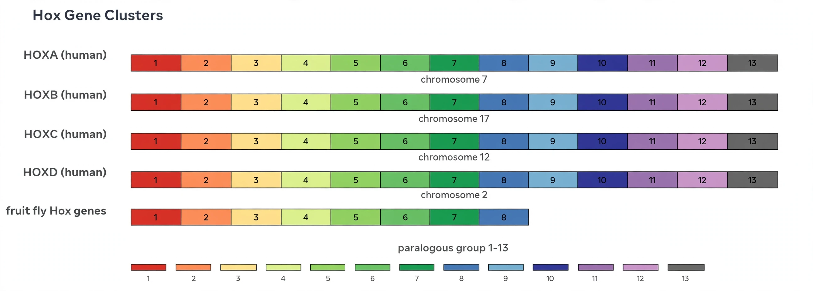
3.3 The counts: 8 in the fly, 39 in us
The numbers are worth holding onto, because they tell the story of the toolkit's history. A fruit fly has 8 Hox genes, arranged in two clusters on the same chromosome: the Antennapedia complex, with five genes (labial, proboscipedia, Deformed, Sex combs reduced, Antennapedia), and the bithorax complex, with three (Ultrabithorax, abdominal-A, Abdominal-B). Eight genes, and they pattern the entire fly — head, thorax, abdomen, and all the segments in between.
A mouse or a human has 39 Hox genes, arranged in four clusters — HOXA, HOXB, HOXC, and HOXD — sitting on four different chromosomes (7, 17, 12, and 2 in humans). The 39 genes fall into 13 paralogous groups, numbered HOX1 to HOX13, so that each cluster carries a subset of the same thirteen positions: HOXA has eleven genes, HOXB ten, HOXC nine, and HOXD nine, with some positions lost in some clusters. The fly's eight genes correspond to a subset of the same thirteen positions — the fly and the human are running versions of the same system, one compact, one expanded.
That expansion is the first clue evo-devo offers to how vertebrate bodies got their complexity. More clusters mean more copies of each positional gene, and more copies mean more room for each copy to specialise. A single ancestral Hox gene that patterned a whole region could, after duplication, split its job between two daughters — one handling the front of the region, one the back — and each daughter could then be rewired independently. Duplication does not create new functions by itself. It creates the freedom for new functions to evolve.
3.4 How four clusters arose from one
The four vertebrate clusters arose by two rounds of whole-genome duplication from a single ancestral cluster — the same cluster the fly still carries in its compact form. Somewhere in early vertebrate history, the entire genome was copied, then copied again, so that one Hox cluster became two, then four. Most of the duplicated genes were later lost, but the Hox clusters kept most of theirs: 39 surviving genes from an ancestral set that probably numbered around thirteen.
Teleost fish went one step further. Their lineage suffered an extra whole-genome duplication, so a zebrafish carries seven Hox clusters instead of four — and 48 Hox genes, only nine more than a human, because many duplicated copies were later lost.
Species | Hox genes | Clusters | Notes |
|---|---|---|---|
Fruit fly (Drosophila melanogaster) | 8 | 2 complexes | Antennapedia complex (5 genes) + bithorax complex (3 genes), both on chromosome 3 |
Mouse and human | 39 | 4 | HOXA (chromosome 7), HOXB (17), HOXC (12), HOXD (2); 13 paralogous groups |
Zebrafish | 48 | 7 | Extra whole-genome duplication in the teleost lineage; more clusters, not more complexity |
Sea anemone (a cnidarian) | 7 Hox genes | 1 Hox cluster (+ 1 ParaHox cluster) | Hox genes exist outside the Bilateria — the toolkit predates heads and tails |
Table 1: Hox clusters across species. The fly's single compact set and the human's four clusters are versions of the same ancestral system, expanded by genome duplication. Sources: Hox gene overview and the vertebrate Hox literature cited in the Sources section.
For the wider story of the genome that carries these clusters, see the companion article on the human genome.
4. Collinearity: The Chromosome Is a Map of the Body
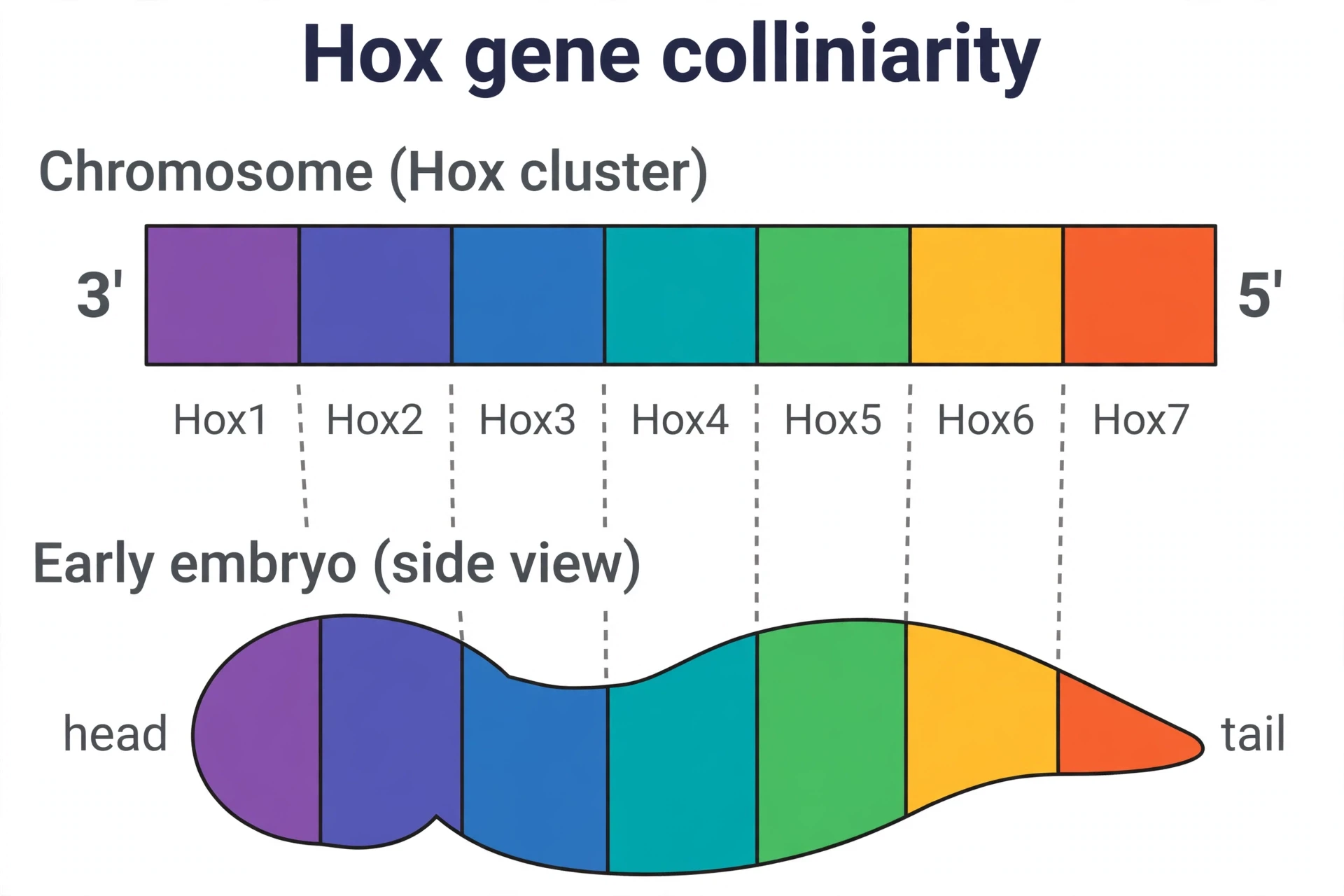
Here is the strangest fact about Hox genes, and the one that first told scientists they were looking at something fundamental. The order of the Hox genes on the chromosome matches the order of the body parts they control. The gene at one end of the cluster acts in the head. The gene at the other end acts in the tail. Genes in the middle act in the middle. The chromosome is a map of the body, laid out in the same order as the animal itself.
4.1 Spatial collinearity: street numbers on a road
Spatial collinearity is the rule that the 3′-to-5′ order of Hox genes along the chromosome corresponds to the head-to-tail order of their expression in the embryo. The 3′ genes — the ones at the "start" of the cluster — switch on in the front of the embryo. The 5′ genes switch on at the back. A gene's address on the chromosome predicts its address in the body.
An analogy helps. Imagine a road with house numbers running from 1 to 13. House 1 is at the north end, house 13 at the south end, and every house in between sits in order. The Hox cluster is that road, and the embryo is the town it maps. To find out which part of the body a Hox gene controls, you can look at where it sits on the chromosome — and, remarkably, that works. The map is not a metaphor evo-devo invented. It is a physical fact about how the genes are arranged.
Edward Lewis discovered this rule before anyone had sequenced a single Hox gene. Working only with mutants, he mapped which parts of the fly's body each gene in the bithorax complex controlled, and he noticed that the genetic order and the body order matched. He called it the colinearity rule. When the genes were finally cloned and sequenced in the 1980s, the rule turned out to be literally true at the level of DNA: the genes sit on the chromosome in the same order as the segments they pattern.
4.2 Temporal collinearity: the same order in time
The map runs in time as well as in space. In vertebrate embryos, Hox genes switch on in the same 3′-to-5′ order over developmental time: the 3′ genes first, the 5′ genes later. The embryo reads the cluster from one end to the other, like a tape playing in sequence, and each gene's moment of activation matches its position on the chromosome.
There is even a third form, quantitative collinearity: where several Hox genes are switched on in the same cells, the most posterior one — the 5′-most — is expressed most strongly, and it dominates the cell's identity. The embryo does not just read the genes in order. It reads them with a volume control that rises toward the tail, so that the last gene to speak is also the loudest.
4.3 How Lewis read the map from mutants
Lewis's achievement deserves a pause, because it shows what genetics could do before DNA sequencing existed. He spent decades — from the 1940s to the 1970s — collecting fly mutants with misplaced body parts, crossing them, and mapping the mutations to positions on the chromosome. From the pattern of the mutants alone, he worked out that the bithorax complex contained a series of genes arranged in the same order as the body segments they controlled, and that they interacted in complex, overlapping ways to specify each segment.
In 1978 he published the synthesis in Nature: "A gene complex controlling segmentation in Drosophila". The paper proposed that the bithorax complex held at least eight genes coding for substances that control levels of thoracic and abdominal development, and that their order on the chromosome matched their order of action in the body. It was a theory built entirely from mutant phenotypes — from flies with wrong body parts — and it turned out to be right in its essentials. The Nobel Prize, seventeen years later, was in large part a recognition of that work.
4.4 The honest limit
Collinearity is a strong pattern, not an unbreakable law, and the honest version of the story includes its limits. In mouse mutants, researchers have shown that the timing mechanism and the spatial patterning phase can be uncoupled: the clock that switches Hox genes on in order can be separated from the later process that sets their spatial boundaries. The two usually run together, but they are distinct mechanisms, and breaking one does not necessarily break the other.
That matters for evo-devo for two reasons. First, it keeps the science honest — collinearity is a rule with exceptions, and the exceptions are interesting in their own right. Second, it shows how evolution can tinker: if timing and space are partly independent, then evolution can change one without changing the other, and that independence is itself a source of new forms.
5. From One Egg to Segments: The Genes Upstream of Hox
Hox genes do not switch themselves on. They are the middle of a hierarchy, not the top of it. Above them sits a cascade of earlier genes that divides the embryo into regions, then into segments, then into segments with polarity — and only then hands each segment its identity card. The story of that cascade is the story of the 1995 Nobel Prize, and it begins with two young scientists, a microscope built for two, and a decision that looked reckless at the time.
5.1 The Heidelberg screen
In the late 1970s, Christiane Nüsslein-Volhard and Eric Wieschaus took up independent research positions at the European Molecular Biology Laboratory in Heidelberg. Both wanted to answer the same question: how does a fertilised egg become a segmented embryo? Nobody had ever tried to answer it systematically. The number of genes involved might have been enormous. The chances of success were, in the Nobel committee's own words, very uncertain.
Their strategy was saturation mutagenesis: treat flies with chemicals that mutate genes at random, damaging roughly half of the fly's genes, and then look at the embryos. Two people sat at a microscope built so both could examine the same embryo at once, and for more than a year they examined embryo after embryo, classifying every malformation they found. The work was slow, meticulous, and — by the standards of the time — audacious. They were trying to find every gene that matters for building a body, by breaking genes one at a time and watching what went wrong.
The 1980 paper that came out of it reported 15 loci whose mutation altered the segmental pattern of the larva. The full screen, published in three papers in 1984, eventually found only about 120 zygotic genes required to specify the larval body plan — out of roughly 14,000 protein-coding genes in the fly. The body plan, in other words, is built by a small fraction of the genome. The rest of the genes do other jobs.
5.2 Gap, pair-rule, segment polarity: three tiers
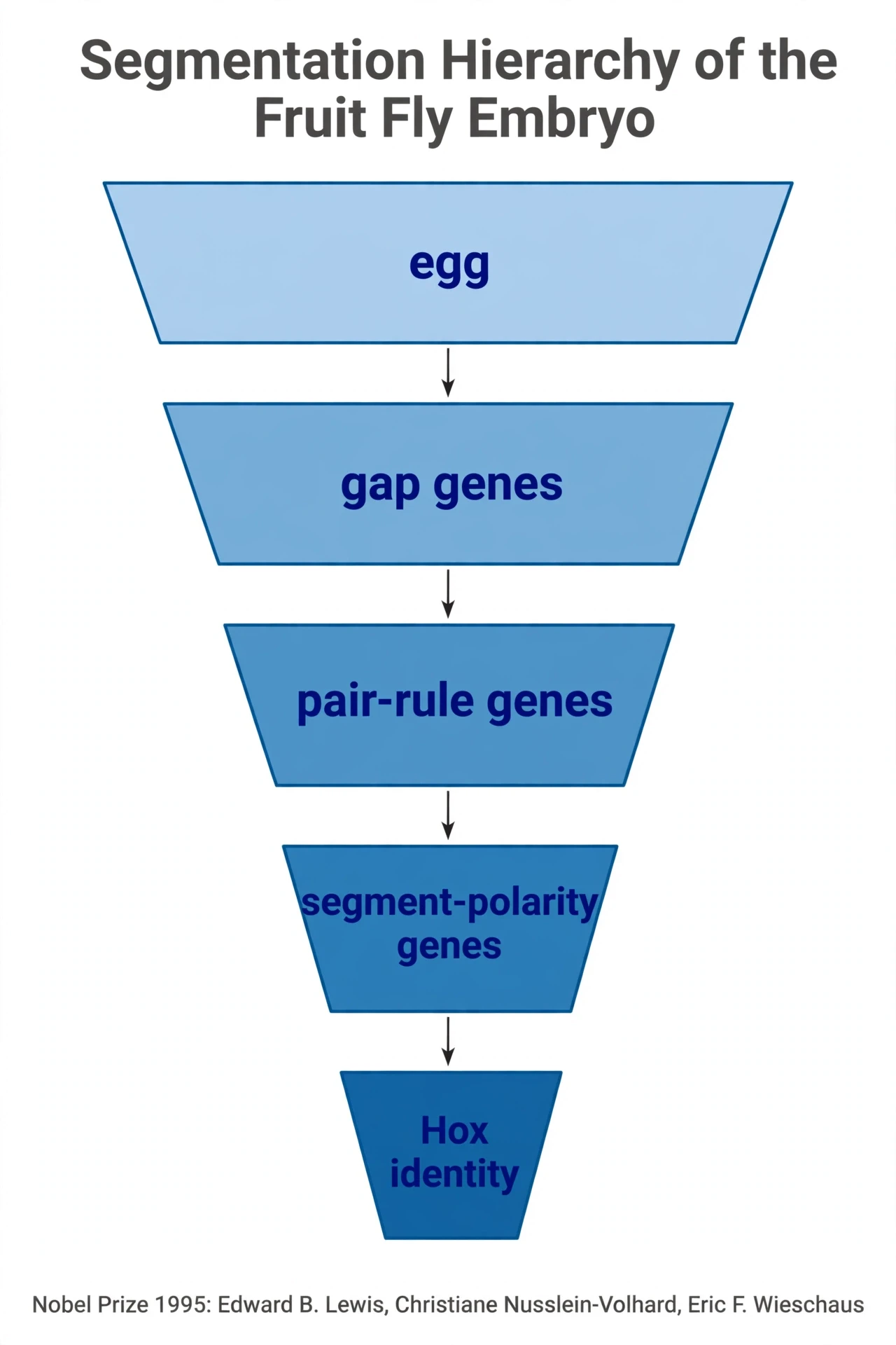
The 15 genes fell into three tiers, each narrowing the map. Gap genes act first and divide the embryo into broad regions; losing a gap gene deletes a whole block of segments. Pair-rule genes act next, and each one affects every second segment — the classic example is even-skipped, whose loss leaves an embryo with only odd-numbered segments, as if every other stripe had been erased. Segment-polarity genes act last, setting the head-to-tail polarity within each individual segment; losing one scrambles the front-to-back organisation of every segment at once.
The hierarchy is a funnel. The egg starts as a uniform ball. Maternal signals set up gradients. Gap genes read the gradients and carve out regions. Pair-rule genes read the regions and draw stripes. Segment-polarity genes read the stripes and give each segment a front and a back. At each step the map gets finer, and the number of cells that share the same instructions gets smaller. By the end of the funnel, every segment has a unique combination of active genes — a molecular postcode — and that postcode is what the Hox genes read.
5.3 Where Hox fits
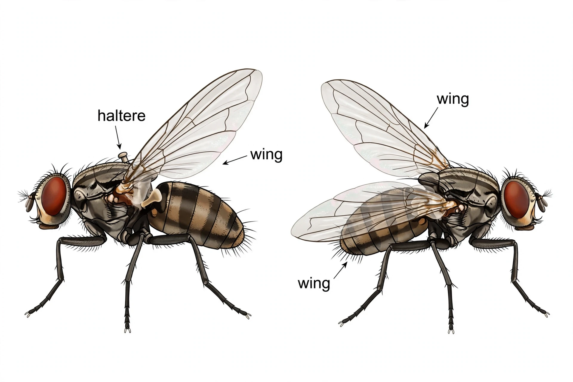
Hox genes sit at the end of that funnel. The segment-polarity tier hands each segment an identity card, and the Hox genes are the readers of the card: they switch on in the segments whose postcode matches them, and they switch on the batteries of downstream genes that build the structures proper to that segment. A segment that expresses Antennapedia builds a leg. A segment that expresses Ultrabithorax builds a haltere instead of a wing. The Hox gene does not decide the segment's position — the earlier tiers do that. It decides what the segment becomes.
That division of labour is the key to the whole system evo-devo describes. Position and identity are separate decisions, made by separate genes, at separate times. And because they are separate, evolution can change one without changing the other: move the identity decision, and a leg appears on a head; move the position decision, and the whole map shifts. The modularity of development is what makes evolutionary change possible in large, coordinated steps.
5.4 The 1995 Nobel Prize
On 9 October 1995, the Nobel Assembly at the Karolinska Institute awarded the Nobel Prize in Physiology or Medicine jointly to Edward B. Lewis, Christiane Nüsslein-Volhard, and Eric F. Wieschaus. The division of labour was clear: Nüsslein-Volhard and Wieschaus had identified and classified the small number of genes that determine the body plan and segment formation; Lewis had worked out how genes control the further development of individual segments into specialised organs.
Lewis was 77 when the prize was announced. He had been working on the bithorax complex since the 1940s — more than half a century of fly genetics, much of it done before anyone knew what a Hox gene was at the molecular level. Nüsslein-Volhard was 52 and Wieschaus 48, and their Heidelberg screen had been done when they were in their thirties, at the very start of their independent careers. The prize recognised a collaboration across generations: one scientist who mapped the system from mutants, and two who found the genes that run it.
The contrast with the companion article on genetic drift is instructive. Drift is evolution by chance, acting on many genes with small effects. The Heidelberg screen found the opposite: a small number of genes with large, visible, decisive effects on the body plan. Both forces operate. But the body plan is built by the second kind — and that is why a single mutation can move a leg onto a head.
6. Homeotic Mutants: When the Identity Card Is Wrong
The word "homeotic" comes from the Greek homeosis, meaning "becoming like" — and it describes exactly what these mutants do. A homeotic mutation replaces one body part with another, perfectly formed, in the wrong place. The transformed structure is not a deformed mess. It is a normal structure, built to the correct specification, that has simply been assigned the wrong address. The mutants are evo-devo's proof that segment identity is a decision — and that decisions can be changed.
6.1 What "homeotic" means
A homeotic transformation is a swap, not a breakage. When a gene that says "build a wing here" is switched off, the segment does not fail to build anything. It builds what the next gene in line tells it to build. The result is a coherent, functional structure — a wing, a leg, an antenna — in a place where it does not belong. The embryo's building programmes are intact. Only the addressing has changed.
That distinction matters. A mutation that breaks a gene usually produces something damaged or missing. A homeotic mutation produces something complete and well-made, in the wrong place. The difference tells you that the mutated gene was not a building gene at all. It was a regulatory gene — a switch — and the thing it regulated was identity itself.
6.2 Antennapedia: the leg on the head
The Antennapedia mutation is a gain of function: the Antp gene, which normally switches on in the second thoracic segment (the middle leg segment) and is absent from the head, gets switched on in the head instead. The head segment's cells read the Antp signal, conclude they are a leg segment, and run the leg-building programme. The result is the fly from section 1: a leg, complete with joints and bristles, growing where the antenna should be.
The reverse experiment is just as telling. Lose Antp function in the leg segment, and the leg transforms into an antenna. The switch works in both directions, because identity is a state the cells are in, not a property of the cells themselves. Change the state, and the same cells build a different structure. The cells are not committed to being leg cells or antenna cells. They are committed to following whatever instruction is active, and Antp is the instruction that says "leg".
6.3 Bithorax and Ultrabithorax: the four-winged fly
The bithorax story is the mirror image, and it is the one that captured the Nobel committee's imagination. A normal fly has two wings, on the second thoracic segment, and a pair of small knobbed balancing organs called halteres on the third thoracic segment. The halteres are the evolutionary remnants of a second pair of wings: flies are descended from four-winged insects, and the hindwings shrank into gyroscopic sensors.
Lose the function of Ultrabithorax (Ubx), the Hox gene that marks the third thoracic segment, and that segment forgets it is the third segment. It behaves like a copy of the second segment — and it grows a second pair of wings. The result is a four-winged fly, a living throwback to the fly's four-winged ancestors. Force Ubx on in the second segment instead, and the reverse happens: the wing becomes a haltere.
The Nobel press release described it in a sentence worth quoting exactly, because it captures the whole idea in plain language:
"In this type of bizarre disturbance of the body plan, cells in one region behave as though they were located in another." — Nobel Prize press release, 9 October 1995
Cells behaving as though they were located in another place. That is what a homeotic mutation is. The cells are healthy, the building programmes are healthy, and only the address is wrong.
6.4 The 1915 origin: credit where it is due
The first bithorax mutant was isolated in 1915 by Calvin Bridges, working in Thomas Hunt Morgan's fly laboratory at Columbia University — decades before Lewis began his work on the complex. Bridges found a fly whose halteres had partly transformed toward wings, and he named the mutation bithorax because the fly seemed to have two thoraxes. Lewis characterised the complex, mapped its genes, and worked out the colinearity rule. But he did not discover the mutant, and the distinction is worth keeping straight. Science is a relay, not a solo sprint, and the baton passed from Bridges to Lewis across more than thirty years.
6.5 What the mutants prove
The mutants prove three things, and each one matters for the rest of this article. First, segment identity is a decision made by regulatory genes, and it can be re-decided — by mutation in the laboratory, and by evolution in the wild. Second, the building programmes are modular: a leg programme can run anywhere, because it is a self-contained unit switched on by a Hox signal. Third, large evolutionary changes do not require large numbers of mutations. A single change in a regulatory switch can move, duplicate, or delete an entire structure in one step.
That third point is the one that changes how you think about evolution — and it is the central insight of evo-devo. The old picture was gradual: thousands of tiny changes, each one barely visible, accumulating over millions of years. The evo-devo picture adds a second mode: a small number of regulatory changes, each one producing a large, coordinated, visible difference. Both modes operate, and evo-devo studies the second as carefully as the first. But the large jumps are the ones that build new body plans — and they are the reason a fly can grow legs on its head.
Today, scientists can make such switches deliberately. The companion article on CRISPR explains how gene editing lets researchers turn regulatory genes on and off in chosen tissues — the same kind of switch, now under human control.
7. Deep Homology: Why a Mouse Gene Can Build a Fly Eye
If the Antennapedia fly is the most startling image in evo-devo, the experiment in this section is the most startling result. In 1995, researchers took a gene from a mouse, put it into a fruit fly, and the fly grew eyes — fly eyes, in the wrong places, built by a mouse switch. The experiment showed that the genetic machinery for making eyes is so deeply conserved that it works across hundreds of millions of years of separate evolution. It also gave evo-devo one of its most important concepts: deep homology.
7.1 The experiment
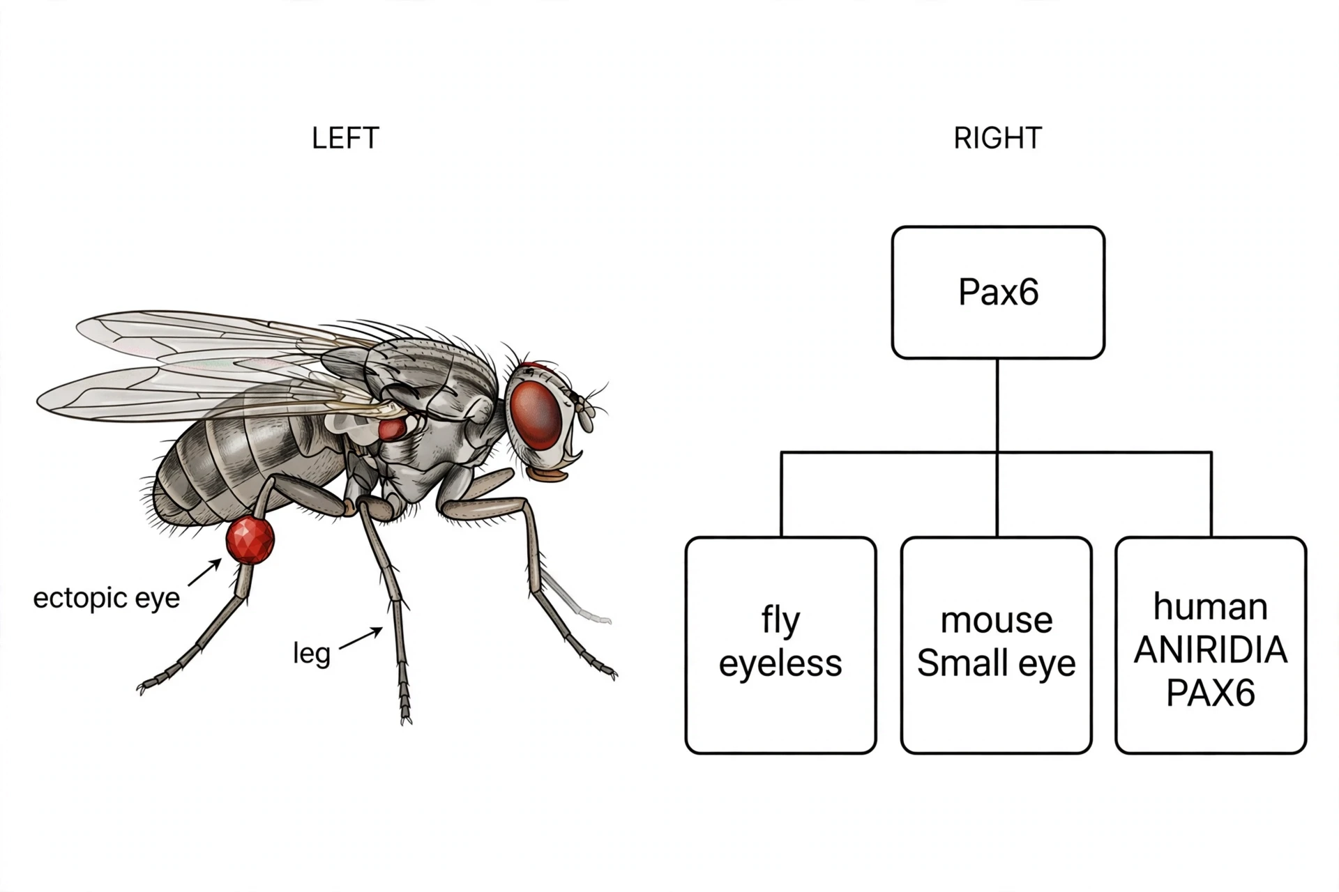
The experiment was done by Georg Halder, Patrick Callaerts, and Walter Gehring, and published in Science in 1995. They took the fly gene eyeless — so named because flies that lack it have no eyes — and switched it on in tissues where it does not normally act: the imaginal discs that become the wing, the leg, and the antenna. The result was eyes growing on wings, on legs, and on antennae. Not vague eye-like blobs, but fully formed compound eyes, with ommatidia and photoreceptors, the same structures a normal fly eye has.
The eyes were in the wrong place, but they were the right eyes. The wing disc that received the eyeless signal built a fly eye, not a wing with eye-like features. The programme for building an eye, like the programme for building a leg, is a self-contained unit. Switch it on anywhere competent to respond, and it runs to completion.
I remember the first time I saw the photograph of that fly, an eye staring out from the middle of a leg, and thinking that it looked like a Photoshop trick. It was not a trick. It was a demonstration that the instructions for building an eye are portable — and that the gene that carries them is ancient.
7.2 Pax6: the same gene in fly, mouse, and human
The fly gene eyeless turned out to be the same gene as the mouse gene Small eye and the human gene PAX6. Mutations in each one block eye development in that animal: flies without eyeless have no eyes; mice without Pax6 have small or missing eyes; humans with a broken copy of PAX6 are born with aniridia — the absence of the iris — and often with other eye defects. One gene, three animals, the same job: switching on eye development.
That is already remarkable. But the cross-species experiment made it more so. When the researchers expressed the mouse Pax6 gene in a fly, the fly grew ectopic eyes — fly eyes, not mouse eyes. When fly eyeless was expressed in a frog embryo, it induced vertebrate eye structures. Only the switch travelled between species. The rest of the programme — the hundreds of genes that actually build the eye, and the details of what kind of eye gets built — stayed local. The mouse gene said "build an eye here". The fly's own machinery decided what a fly eye looks like.
7.3 The cross-species swap, and what it means
The swap works because Pax6 sits at the top of a network, not because it builds eyes by itself. Pax6 switches on a cascade of downstream genes — sine oculis, eyes absent, dachshund, and others in the fly; a related set in vertebrates — and those genes do the building. The network is what is conserved, in outline, across animals. The details differ, because each lineage has rewired the network in its own way over hundreds of millions of years.
That is the key insight of evo-devo. The gene is the same, but the programme it controls has diverged. A fly eye and a human eye are different organs, built by different downstream networks, with different optics and different cell types. What they share is the switch at the top, inherited from a common ancestor that had some kind of light-sensitive structure. The switch is homologous. The organs are not.
7.4 Deep homology, defined
That combination — homologous genetic machinery running non-homologous structures — is what Shubin, Tabin, and Carroll called deep homology in 1997, in a paper on fossils, genes, and the evolution of animal limbs (Bolker and Raff had used the term independently in 1996). Insect eyes and vertebrate eyes are not the same organ. They evolved independently, from different starting points, and they work differently. But they share a deeply conserved light-sensing programme, built around Pax6, that predates their split. The homology is not in the organ. It is in the machinery underneath it.
Deep homology explains a puzzle that used to trouble evolutionary biologists: how can two structures that evolved independently share the same genes? The answer is that the genes were there first. The common ancestor of flies and humans already had a Pax6-based light-sensing system. When the two lineages independently evolved complex eyes, they both recruited the same ancient machinery to the job — not because they inherited eyes from each other, but because they inherited the toolkit from the same ancestor, and the toolkit was the natural starting point for building an eye.
7.5 The "master control gene" caveat
Pax6 is often called a "master control gene", and the phrase is useful shorthand — but it needs a caveat. Pax6 does not build an eye by itself, and it cannot make an eye just anywhere. It needs competent tissue: cells that are already able to respond to its signal, with the right downstream genes available and the right cellular context in place. Switch Pax6 on in the wrong tissue, and nothing happens. Switch it on in a wing disc, and you get an eye — because the wing disc has the competence to respond.
That caveat is important, because it is the difference between a switch and a blueprint. A blueprint contains the whole design. A switch contains one instruction: start here. The rest of the design lives in the network below the switch, and in the tissue that receives it. Evo-devo is the science of switches, networks, and tissues together — never of a single gene acting alone.
The eye's own evolutionary story, including the long debate over how many times eyes evolved, is told in the companion article on the evolution of the eye. For the broader pattern of unrelated species arriving at similar solutions, see convergent evolution.
8. The Toolkit and Its Operating Instructions: Gene Networks and Enhancers
By now the pattern at the heart of evo-devo is clear: a small set of ancient genes, used over and over, in different places and times, builds every animal body. This section names the toolkit, explains how it is wired, and introduces the part of the genome where most evolutionary change actually happens — not in the genes themselves, but in the switches that control them.
8.1 The toolkit
The genetic toolkit is a small set of deeply conserved regulatory genes, reused in many animals and many body parts. Its members include the Hox genes, Pax6, Distal-less (Dll), Sonic hedgehog (Shh), Bmp, Wnt, Hedgehog, and Notch — a roster of a few dozen gene families that pattern the body axis, the limbs, the eyes, the gut, and the nervous system. The same genes turn up in a fly's wing, a fish's fin, and a human hand, doing recognisably similar jobs.
The toolkit is small, and that is the point. An animal does not need a separate gene for every structure. It needs a few versatile switches, combined in different ways, at different times, in different places. The combinatorial power of a small toolkit is enormous: a dozen switches, each of which can be on or off in each tissue at each stage, generates far more combinations than any animal will ever use. Evolution does not need to invent new switches. It needs to find new combinations of the ones it already has.
8.2 Gene regulatory networks: switches controlling switches
Development is controlled by gene regulatory networks: hierarchies and circuits of transcription factors switching each other on and off. A Hox protein or a Pax6 protein sits near the top of such a network. It switches on a set of intermediate regulators, which switch on further regulators, which finally switch on the "realisator" genes — the ones that actually build tissue: the collagens, the muscle proteins, the pigments, the enzymes.
The network structure is what makes development both robust and evolvable — and it is why evo-devo treats the network, not the single gene, as the unit of evolutionary change. Robust, because a network with many steps can absorb small disturbances — if one regulator is slightly late, another can compensate. Evolvable, because a change at the top of the network propagates downward and changes the whole output, while a change at the bottom changes only a small detail. Evolution can tune the system at any level: a big change at the top, a fine adjustment at the bottom, or a rewiring in the middle.
8.3 Enhancers: the switches that say where and when
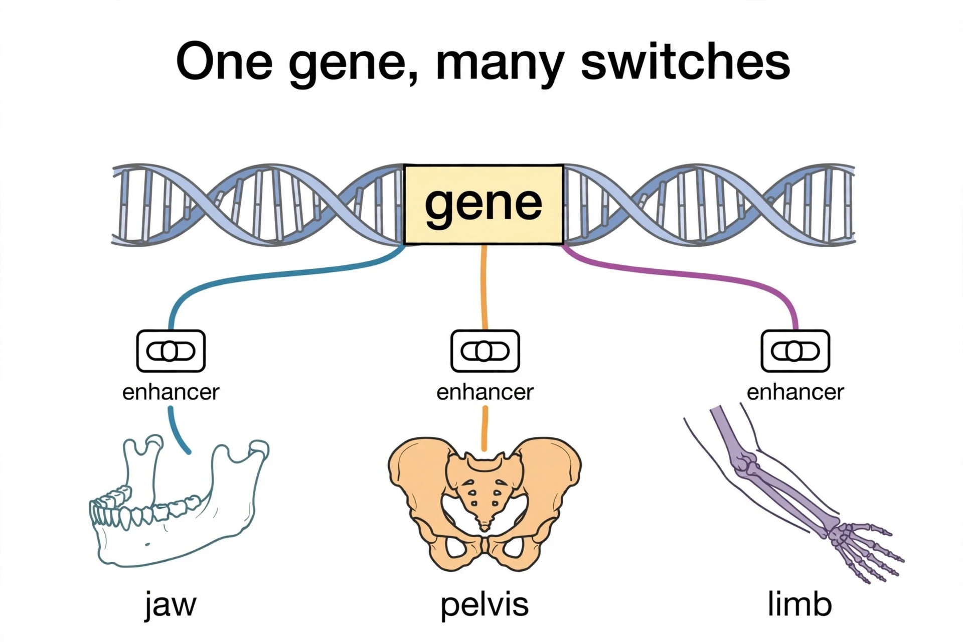
Most of the evolutionary change in the toolkit happens not in the protein-coding genes, but in their enhancers — short stretches of non-coding DNA near a gene that control when and where that gene is switched on. An enhancer is a switch with an address: it says "turn this gene on in the pelvis, and only in the pelvis", or "turn this gene on in the jaw, and only in the jaw". A single gene can have many enhancers, each one responsible for one deployment.
The fly's bithorax complex is the classic illustration. The complex spans more than 300 kilobases of DNA, but it contains only three protein-coding Hox genes — Ubx, abd-A, and Abd-B. Almost all of the rest is regulatory DNA: enhancers, silencers, and insulators that control those three genes in different segments, at different times, in different tissues. The genes are a small part of the complex. The instructions for using them are most of it.
8.4 Modularity: one enhancer per job
Modularity is the property that makes enhancers so powerful for evolution. Because each enhancer controls one deployment of a gene, changing one enhancer changes one deployment without breaking the gene's other jobs. The gene Pitx1, which the next sections will return to, works in the pelvis, the jaw, and the pituitary gland. Breaking the Pitx1 protein would damage all three — a disaster. Deleting the enhancer that switches Pitx1 on in the pelvis removes the pelvis and nothing else. The jaw and the pituitary keep working, because their enhancers are untouched.
That is how evolution avoids pleiotropy — the problem that one gene does many jobs, so changing it breaks many things at once. Enhancers solve the problem by splitting the gene's jobs into separate, independently changeable switches. Evolution can tinker with one switch at a time, testing each change in isolation, without risking the whole animal. Modularity is what makes large evolutionary changes survivable — and it is the reason evo-devo puts enhancers at the centre of the story.
8.5 Co-option: old genes hired for new jobs
The same modularity enables co-option: the recruitment of an old gene, or an old network, for a new job. A gene that patterns legs can be switched on in a wing, and — because the wing tissue is competent to respond — it starts building an eyespot there instead. A gene that patterns the gut can be recruited to pattern a new organ. The gene does not change. Its address changes, and the new address gives it a new job.
Co-option is the theme of the butterfly eyespot case study in section 14, and a central theme of evo-devo that runs through the whole article. Every new structure in animal evolution is, at some level, an old programme running in a new place. The toolkit does not grow by invention. It grows by redeployment — and the record of those redeployments is written in the enhancers, one switch at a time.
That regulatory logic reaches beyond the DNA code itself. The companion article on epigenetics explores another layer of control over when genes are used — and how the environment can influence it.
9. Changing the Timing: Heterochrony and Neoteny
Evolution has three dials for reshaping a body plan: when a programme runs, where it runs, and how much it runs. This section covers the first dial — timing. A genetically controlled change in the timing, rate, or duration of a developmental process is called heterochrony, and it is one of the most powerful ways to produce a new form from an old programme. Change when something happens, and everything downstream shifts with it — the first of evo-devo's three dials.
9.1 Heterochrony, defined
Heterochrony means a change in developmental timing relative to an ancestor. The term was introduced by Ernst Haeckel in 1875, and given its modern sense by Gavin de Beer in 1930. It covers several distinct patterns: a process can start earlier or later, run faster or slower, or stop sooner or later than it did in the ancestor. Each pattern has its own name, and each produces a different kind of change in the adult.
The power of heterochrony is that it is cheap. It does not require new genes or new structures. It requires a change in the schedule — a signal that arrives a day late, a growth phase that lasts a week longer — and the whole body plan shifts in response. A small change in timing, amplified through development, can produce a large change in the adult form.
9.2 Neoteny and paedomorphosis: the axolotl
The most famous example of heterochrony is the axolotl, Ambystoma mexicanum, a salamander from the lakes of Mexico that never grows up — at least, not in the usual sense. An axolotl reaches sexual maturity while still looking like a larva: it keeps its external gills, its finned tail, and its aquatic lifestyle for its whole life. It is an adult that has kept its juvenile traits, a condition called neoteny, a form of paedomorphosis — the retention of juvenile features in the sexually mature adult.
The axolotl is not stuck. Give it thyroid hormone — thyroxine — in the laboratory, and it metamorphoses into a land-going salamander, losing its gills and growing lungs that work on land. The adult programme is still there, in the genome, waiting. What changed in evolution was the timing: the signal that triggers metamorphosis was delayed, or the tissue's response to it was muted, and the larval form became the adult form. One hormonal shift, and the animal's whole way of life changed with it.
Neoteny is not a curiosity confined to salamanders. It is a recurring theme in evolution, and the human case is the one that matters most to us.
9.3 The human case: prolonged brain development
Human development is, in several respects, a story of extended timing. Compared with other primates, our brain development runs for much longer: the human brain keeps growing at foetal rates well after birth, while a chimpanzee's brain growth slows much earlier. Our childhood is extended for years beyond that of any other ape. We stay dependent, and keep learning, for far longer than our relatives do.
The trade is real. A long childhood is expensive — years of feeding and protecting an offspring that cannot feed or protect itself. But it buys something no other ape has: an extended period in which the brain is still plastic, still forming connections, still learning from the world. Our large brains and our long childhoods are two sides of the same heterochronic coin. We did not evolve a new kind of brain. We evolved a new schedule for growing the brain we already had.
That is a general claim, and it should be stated as one. The precise week-by-week comparisons between human and chimpanzee brain growth are still being refined, and the numbers vary between studies. What is solid is the pattern: human brain development is prolonged relative to other primates, and that prolongation is central to what makes us human. The details belong to the specialists. The pattern belongs to everyone.
9.4 Peramorphosis: the opposite direction
Heterochrony runs in both directions. Neoteny slows development down or stops it early. Peramorphosis does the opposite: development is extended or accelerated, so the adult goes further along the ancestral path than the ancestor did. A structure that was modest in the ancestor becomes exaggerated in the descendant, because its growth phase lasts longer.
Both directions use the same machinery. The difference is only in which way the dial is turned. And the dial is turned by the same kind of change — a tweak in the timing of a regulatory signal — that drives the rest of evo-devo. Timing is a dial, not a destiny.
Neoteny — slower somatic development; the adult keeps juvenile traits. Example: the axolotl, sexually mature with larval gills.
Progenesis — earlier reproduction; development stops early and the animal matures at a smaller, more juvenile form. Example: some salamander populations that breed as larvae.
Hypermorphosis — extended development; growth continues longer than in the ancestor. Example: the extended growth of the human brain and the prolonged human childhood.
For the human side of this story — the fossils, the migrations, and the long childhood in context — see the companion article on human evolution. For why a long life and a slow life history matter at all, see why do we age.
10. Changing the Place and the Proportions: Heterotopy and Allometry
Timing is one of evo-devo's three dials. Place is the second, and proportion is the third. Together they complete the mechanism trio — when, where, and how much — and they set up the finch case study that follows. If heterochrony changes the schedule, heterotopy changes the address, and allometry changes the scale.
10.1 Heterotopy: the same gene, a new place
Heterotopy is a change in the place of gene expression: the same gene switched on in a new location, or a structure developing somewhere it did not develop before. The Antennapedia fly is the textbook case: Antp expressed in the head, where it does not belong, produces a leg in the wrong place. The butterfly eyespot is the evolutionary case: Distal-less, a leg-patterning gene, switched on in a new spot on the wing, produces an eyespot where no eyespot existed before.
Heterotopy is powerful for the same reason heterochrony is: it reuses an existing programme. The gene does not need to learn a new job. It runs its old job in a new place, and the new place gives the old job a new meaning. A leg programme in the head makes a leg on the head. A leg programme in a wing spot makes an eyespot. The programme is the same. The address is different.
10.2 Allometry: shape as a matter of scaling
Allometry is differential growth: body parts growing at different rates, so that shape changes as size changes. A beak that grows faster in depth than in length becomes deep and crushing. A beak that grows faster in length than in depth becomes long and probing. The parts are the same. The proportions are different.
Allometry is the quietest of the three dials, and the most pervasive. Almost every difference between related species involves some allometry — a limb slightly longer, a skull slightly broader, a beak slightly deeper. The differences look small, but they add up: a few percent here, a few percent there, and the result is a different animal, eating different food, in a different niche.
10.3 How the three combine
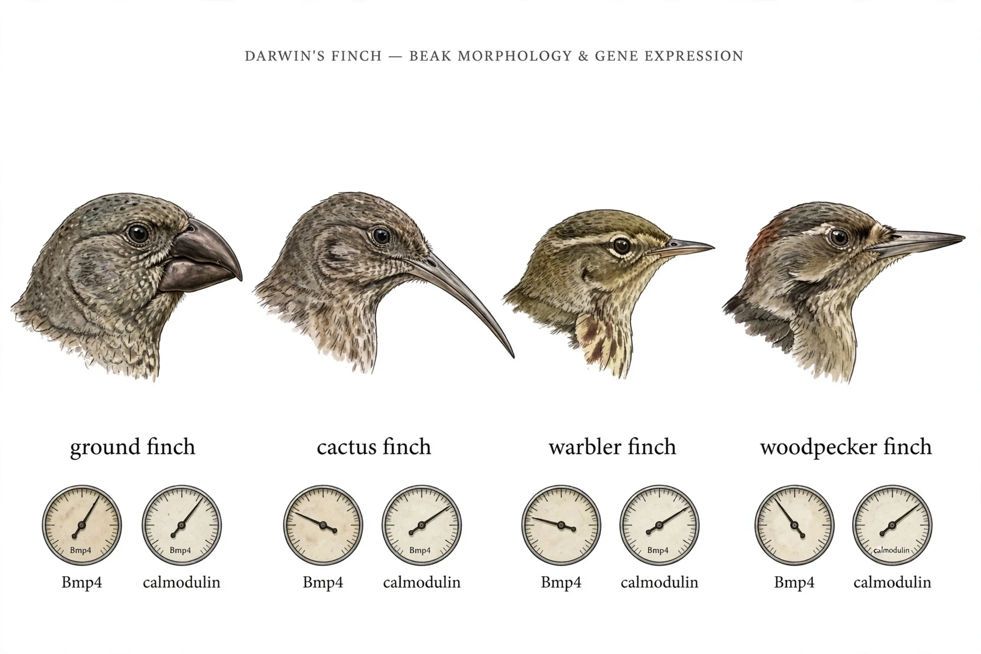
The three dials of evo-devo combine freely. A beak can get deeper (more Bmp4, acting longer) or longer (more calmodulin activity) independently — two dials, two dimensions, one beak. A limb can be lost by turning a gene off in one place (heterotopy) at one time (heterochrony) while the rest of the body keeps growing normally (allometry elsewhere). The finch case study in the next section shows the combination at its cleanest: two signals, two dimensions, and a whole radiation of beak shapes from the same toolkit.
Mechanism | What changes | Classic example | Result |
|---|---|---|---|
Heterochrony | Timing, rate, or duration of development | Axolotl neoteny | Adult keeps juvenile traits — gills, aquatic life |
Heterotopy | Place of gene expression | Antennapedia leg on the head | A structure appears in a new location |
Allometry | Relative growth rates of parts | Finch beak depth vs length | Shape changes by scaling, not by new parts |
Table 2: Heterochrony vs heterotopy (and allometry). Three dials, three kinds of change — when, where, and how much. Most evolutionary novelties use more than one at once.
Proportions are also signals in their own right — a long tail, a deep beak, a large crest can all carry meaning to mates and rivals. The companion article on sexual selection takes up that side of the story.
11. Case Study: Darwin's Finch Beaks — Two Dials, Two Dimensions
The Galápagos finches are the textbook example of adaptive radiation: about 14 closely related species in the classic count — now recognised as roughly 18, after taxonomic splits — descended from a single ancestor that reached the islands, now carrying beaks from crushing to probing — heavy seed-crushers, cactus-flower probers, insect-pickers, and woodpecker-like tool users. Darwin himself did not grasp their significance at the time. A century and a half later, evo-devo found the mechanism behind their diversity, and it turned out to be two dials, each controlled by one signalling pathway.
11.1 The birds
Fourteen species in the classic count, one ancestor, and a range of beaks that spans the functional spectrum. The large ground finch, Geospiza magnirostris, carries a massive, deep beak for crushing hard seeds. The cactus finch, Geospiza scandens, carries a long, pointed beak for probing cactus flowers. The warbler finch has a small, fine beak for picking insects. The woodpecker finch uses tools. The differences are not in the bones' basic plan — every finch has the same beak bones, in the same arrangement. The differences are in the proportions: how deep, how long, how wide.
That is exactly what evo-devo predicts. If beak shape is a matter of scaling — allometry — then the differences between species should be tunable by changing the amount and duration of growth signals in the developing beak. And that is what the research found.
11.2 Dial one — Bmp4: depth and breadth
The first dial is Bmp4, a signalling protein from the bone morphogenetic protein family. In the developing upper beak, Bmp4 is expressed in the mesenchyme — the embryonic tissue that will become bone and cartilage. Species with deep, broad beaks express more Bmp4, earlier and more strongly, than species with shallow beaks. The correlation is tight: measure Bmp4 in the embryo, and you can predict the adult beak's depth.
Correlation is not causation, so the researchers tested it directly. Arhat Abzhanov and colleagues took chick embryos — chickens are the laboratory stand-in for finches, whose embryos are harder to work with — and misexpressed Bmp4 in the developing beak. The result was a beak shaped like the large ground finch's: deeper, broader, built for crushing. One signal, turned up, and the beak changed shape in the predicted direction. The paper, published in Science in 2004, was one of the first to connect a specific developmental signal to a specific evolutionary difference in the wild.
11.3 Dial two — calmodulin: length
The second dial is calmodulin, a calcium-binding protein that acts through its signalling pathway in the developing beak. Species with long, pointed beaks — the cactus finches — show higher calmodulin-pathway activity in the beak mesenchyme than species with short beaks. And again, the experiment confirmed the correlation: replicating the higher calmodulin activity in a chick embryo elongated the upper beak, producing the long, pointed profile of a cactus finch.
Two dials, two dimensions. Bmp4 controls depth and breadth. Calmodulin controls length. Turn Bmp4 up and the beak deepens. Turn calmodulin up and the beak lengthens. Turn both, and the beak changes in both dimensions at once. The finch species are, in effect, one setting of two dials each — and the settings are heritable, selectable, and visible to natural selection.
11.4 The lesson: quantitatively tunable
The lesson for evo-devo is that beak shape is quantitatively tunable. The finches did not evolve new beak genes. They evolved new settings for old signals — more Bmp4 here, more calmodulin there — and the settings produce the full range of beak shapes from the same developmental toolkit. Later work showed the picture is even more precise: the variation is explained by scaling and shear transformations of the same underlying growth pattern, a mathematical description of how the two dials interact (Campàs et al., 2010).
That precision is what makes the finch study a landmark. It is not a story about a gene "for" a beak. It is a story about two signals, measured in embryos, manipulated in the laboratory, and matched to the beaks of wild birds. Selection acts on the beaks. Development builds them. Evo-devo connects the two.
The finches are also a reminder that selection and development are partners, not rivals. For the selection side of the story, see the companion article on natural selection, and for the long-running partnerships between species that shape beaks and flowers together, see coevolution.
12. Case Study: The Stickleback That Lost Its Pelvis — One Enhancer, Repeatedly
If the finches show evo-devo tuning a trait up and down, the stickleback shows it deleting a trait — repeatedly, in the wild, in real time, by breaking the same switch over and over again. It is the cleanest demonstration that regulatory evolution, not protein evolution, drives most morphological change.
12.1 The fish
The threespine stickleback is a small fish with a big defensive secret: a spiny pelvis, a set of bony spines on its underside that make it hard for a predator to swallow. Marine sticklebacks, living in the open ocean, all have the pelvis. But when sticklebacks colonised freshwater lakes after the last ice age — thousands of separate lakes, each colonised independently — many of those populations lost the pelvis. In lake after lake, the spines vanished, leaving a smooth underside.
The loss makes ecological sense. In lakes, the predators are different — dragonfly larvae and other insects that grab their prey, rather than fish that swallow it whole — and a spiny pelvis can be a liability, giving a predator something to grip. I have watched sticklebacks in a tank, the marine ones bristling with spines and the lake ones smooth as minnows, and the difference looks trivial until you remember what it means: each smooth fish is the product of a broken switch — the same switch, broken independently, in lake after lake. But the ecological story is not the remarkable part. The remarkable part is the genetics.
12.2 Shapiro et al. 2004: the protein is unchanged
In 2004, Michael Shapiro and colleagues mapped the pelvic loss to a single major genetic locus, plus four minor ones. The major locus was Pitx1, a transcription factor gene. But when they sequenced Pitx1 in pelvic-reduced fish, they found no change in the protein at all. The protein was identical to the marine version, letter for letter.
What had changed was expression. In pelvic-reduced fish, Pitx1 was switched off in the developing pelvis — but switched on normally everywhere else: in the jaw, in the pituitary gland, in all the other tissues where it works. The gene was intact. Its deployment in one place was gone — the evo-devo signature: same gene, different instructions. The fish had not lost a gene. It had lost a switch.
12.3 Chan et al. 2010: nine lakes, nine deletions, one switch
The mechanism arrived six years later. Yingguang Chan and colleagues found that the cause was the recurrent deletion of a single tissue-specific enhancer, PelA, sitting about 2.5 kilobases upstream of Pitx1. In nine different spineless populations, from nine different lakes, they found nine different deletions — all of them overlapping in a 488-base-pair region, all of them removing the same pelvic enhancer. Nine independent lakes, nine independent mutations, one switch.
The proof was completed by putting the enhancer back. When the researchers reintroduced a working PelA enhancer driving Pitx1 into a pelvic-reduced fish, the pelvis grew back. The gene had been there all along, waiting. Only the switch was missing.
A small footnote keeps the record honest: a 2006 corrigendum corrected two figures in the Shapiro paper, where the sense and antisense probes had been mislabelled. The main conclusion — that pelvic loss is a cis-regulatory change in Pitx1 — was unchanged. Science corrects itself in public, and the correction strengthens the result rather than weakening it.
12.4 Why an enhancer, not the gene
The reason evolution broke the enhancer rather than the gene is pleiotropy. One protein serves three organs: pelvis, jaw, and pituitary gland. Breaking the protein would damage all three — and a fish with a broken jaw and a broken pituitary does not survive to pass on its genes. Breaking the pelvic enhancer removes the pelvis and nothing else; the jaw and pituitary are unaffected, because their own enhancers still switch the gene on.
That is the surgical precision of regulatory evolution, and it is why evo-devo puts enhancers at the centre of morphological change. A protein is a blunt instrument: change it, and everything it touches changes. An enhancer is a scalpel: change it, and one deployment changes, in one tissue, at one time. Evolution, given the choice, uses the scalpel.
Three facts prove the stickleback story was regulatory, and they are worth listing plainly:
The Pitx1 protein is identical in pelvic and pelvic-reduced fish — no coding change.
Pitx1 expression is lost only in the pelvis; it is normal in the jaw and pituitary.
Nine separate lakes show nine independent deletions, all hitting the same 488-base-pair enhancer.
The repeated evolution is the clincher. If pelvic loss had happened once, it could have been a fluke. Nine times, in nine lakes, hitting the same switch — that is natural selection finding the same solution again and again, because it is the best solution available. For the timescale on which such repeated evolution plays out, see the companion articles on the tree of life and the molecular clock.
13. Case Study: Losing Legs — Snakes and Whales
The stickleback lost a pelvis. Snakes and whales lost whole limbs — and they lost them the same way: not by deleting the limb genes, but by switching them off in one place. Two independent stories, separated by hundreds of millions of years, converge on the same evo-devo mechanism. Keep the genes, change the switches.
13.1 Snakes: the thorax takes over
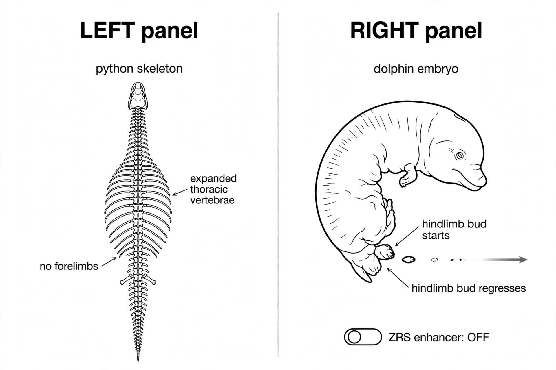
Snakes are tetrapods — four-limbed vertebrates — that lost their limbs. The python still carries the evidence in its skeleton: more than 300 vertebrae, most of them thoracic, where a lizard has a few dozen. In python embryos, the Hox expression domains that mark the thorax are expanded along the body axis, stretching thoracic identity over hundreds of segments. The forelimb field — the region where a front leg should form — is erased, because the Hox code that says "forelimb here" never gets expressed there. The snake did not lose its forelimbs by deleting a limb gene. It lost them by expanding the thorax until there was no room left for a forelimb.
That finding, published by Martin Cohn and Cheryll Tickle in Nature in 1999, was the first developmental explanation for snake limblessness. The Hox code sets the address. Change the address, and the limb never starts.
13.2 The hindlimb's slow fade
The hindlimb tells a slower story. Python embryos do start a hindlimb bud — a small swelling where the leg should be. The bud forms, and for a while it expresses the right signals. Then the signalling dies out. The apical ectodermal ridge, the signalling centre that drives limb outgrowth, stops expressing Fgf8. The zone of polarising activity, the centre that patterns the limb's digits, stops expressing Shh. Without those signals, the bud stalls and regresses, leaving only the tiny spurs that pythons still carry near their tails — the visible remnants of hindlimbs, complete with a claw.
The hindlimb was not deleted in one step. It faded, over evolutionary time, as its signals weakened and then vanished. The genes are still there. The switches that run them in the limb are not.
13.3 The smoking gun: a 17-letter deletion
The mechanism behind the fade was found in 2016, in the enhancer that controls Shh in the limb. Shh is switched on in the limb bud by a long-range enhancer called the ZRS, sitting about a million DNA letters away from the gene itself. In snakes, the ZRS carries a 17-base-pair deletion that destroys a binding site for ETS transcription factors — the proteins that switch the enhancer on.
The experiment that proved it was elegant. Kvon and colleagues replaced the mouse ZRS with the snake version, and the mouse's limbs were severely reduced. Then they restored a single binding site in the snake ZRS, and the mouse's limbs were restored. Seventeen letters of DNA, one binding site, and the difference between a limb and no limb. That is the precision of regulatory evolution — and the reason a snake can keep every limb gene it ever had, and still have no limbs.
13.4 Whales: the bud that starts and stops
Whales tell the same story from the other side of the vertebrate tree. Dolphin embryos start a hindlimb bud, just as python embryos do. The bud forms an apical ridge expressing Fgf8, and for a while it looks like a limb in the making. Then it arrests and degenerates, around the fifth week of gestation. The ridge is not maintained. Shh is never switched on, because its upstream regulator, Hand2, is never expressed in the hindlimb region. Without Hand2, there is no Shh. Without Shh, there is no limb.
The fossil record puts dates on the process. Thewissen and colleagues, combining the developmental evidence with fossils, estimated that Shh expression in the hindlimb was reduced around 41 million years ago, and that the distal elements of the limb were lost around 34 million years ago, near the origin of the modern whale groups. The limb shrank gradually, by small evolutionary steps, and then a regulatory switch turned it off for good.
13.5 The shared moral
The shared moral — the evo-devo moral — is the same in both stories: limb loss is loss of expression in one place, not loss of the genes. Every limb gene is still present in the snake genome and the whale genome, working elsewhere in the body — Shh patterns the brain and the gut, Hand2 works in the heart, the Hox genes pattern the spine. The genes were never deleted. Their limb switches were. And because the switches are modular, the rest of the animal kept working while the limbs faded away.
That is why snakes and whales are such powerful evidence for evo-devo. They are not stories about genes being lost. They are stories about genes being redeployed — or, in this case, un-deployed — one enhancer at a time. For the fossil evidence behind these transitions, see the companion articles on mass extinctions and the fossil record.
14. Case Study: Butterfly Eyespots — A New Trait From an Old Leg Gene
The finches tuned a trait. The stickleback deleted one. Snakes and whales lost limbs. But can evo-devo explain genuine novelty — a structure that never existed before, built from scratch? The butterfly eyespot says yes. And the gene that builds it is one we have already met: Distal-less, the leg-patterning gene.
14.1 The trait
Butterfly eyespots are concentric rings of colour on the wings — a dark centre, a pale ring, a dark outer ring — that look, at a glance, like eyes. They deflect predators: a bird pecks at the eyespot on the wing margin instead of the butterfly's body, and the butterfly escapes with a torn wing instead of a fatal wound. Eyespots vary enormously between species and within them: in number, in size, in colour, and in position on the wing. They are a recent evolutionary invention, about 90 million years old in the butterfly lineage, and one of evo-devo's showcase examples of novelty — built by a developmental process assembled from older parts.
14.2 The co-option: a leg gene on a wing
In 1994, Sean Carroll and colleagues showed that eyespots are marked, in the developing wing, by the expression of Distal-less — Dll, the same gene that patterns the ends of legs and antennae. Dll switches on at the centre of each future eyespot, the focus, and the rings of colour form around it. A leg gene, switched on in a new place on the wing, had been hired for a new job: marking the centre of an eyespot.
That is co-option in its purest form, and it is one of evo-devo's favourite explanations for novelty. The gene did not change. Its address changed. And the new address gave it a new function, because the wing tissue around the focus was competent to respond — it could read the Dll signal and build rings of pigment around it. The eyespot is not a leg, and it is not a wing. It is a new structure, built by an old gene in a new place.
14.3 The development: four stages
The eyespot develops in four stages, worked out by Paul Brakefield and colleagues in 1996: prepattern, in which the wing is divided into regions; focal determination, in which Dll marks the focus; focal signalling, in which the focus sends signals outward; and differentiation, in which the surrounding cells respond by producing the rings of colour. Each stage is controlled by its own set of genes, and each stage can be tuned independently — which is why eyespots vary so much between species.
14.4 The functional proof
Correlation is not causation, so the researchers tested Dll directly. In 2013, Antónia Monteiro and colleagues overexpressed Dll in the butterfly Bicyclus anynana, and the result was extra eyespots, and larger ones. Knocking Dll down produced smaller eyespots. Dll is a positive regulator of eyespot formation: more of it, more eyespot; less of it, less eyespot. The gene that patterns legs controls the size and number of eyespots on a wing — a direct, experimental demonstration that a co-opted gene does the job it was hired for.
Later CRISPR work (Connahs et al., 2019) found the outcome depends on which exon is hit: disrupting exon 2 of Dll produced missing and ectopic eyespots, while disrupting exon 3 produced only missing ones — and reaction-diffusion modelling of Dll as an activator reproduces these phenotypes. The eyespot is not just marked by Dll. It is made by it — and that is evo-devo's answer to where new traits come from.
14.5 Plasticity: the same genotype, different eyespots
The eyespot story has one more twist: plasticity. The same butterfly genotype produces different eyespots in the wet season and the dry season. Wet-season butterflies have large, conspicuous eyespots; dry-season butterflies have small ones, or none at all. The difference is developmental: temperature and day length during the pupal stage change the signalling at the focus, and the adult wing reflects the season it grew up in.
Plasticity is development as a responsive system, not a fixed blueprint. The genome does not specify a single eyespot size. It specifies a reaction norm — a range of possible outcomes, depending on conditions — and the environment selects among them. That responsiveness is itself an evolved trait, and it is part of why eyespots are so variable, and so successful, across the butterfly family.
For the defensive logic of eyespots and the wider world of visual deception, see the companion article on camouflage and mimicry. For how animals track the seasons that drive such plasticity, see animal migration.
15. From Fins to Fingers: Digits, Tiktaalik, and the Pentadactyl Puzzle
Your hands are the most personal evidence for evo-devo. Hold one up. Five fingers, each with its own identity — thumb, index, middle, ring, little — arranged in order, built by the same Hox machinery that patterns a fly's segments and a snake's spine. The story of how fins became fingers is the story of that machinery being rewired, one enhancer at a time, over hundreds of millions of years. And it begins with a fossil fish that had a neck.
15.1 Tiktaalik roseae: the fish with a neck
In 2004, on Ellesmere Island in the Canadian Arctic, a team led by Neil Shubin, Edward Daeschler, and Farish Jenkins found the fossil they had gone looking for: Tiktaalik roseae, a 375-million-year-old animal from the Late Devonian that was unmistakably a fish, and unmistakably on its way to becoming a tetrapod. It had scales and fins, like a fish. But it also had a neck — a head that could move independently of the body — ribs, and fin-limbs with a shoulder, a wrist, and finger-like bones inside them.
Tiktaalik was the fossil the theory predicted. If tetrapod limbs evolved from fish fins, there had to be an intermediate: a fish whose fins had begun to look like limbs. I have held a cast of a Tiktaalik fin in a museum shop, turning it over in my hands, and the wrist bones inside a fish's fin still feel impossible — until you remember that your own wrist is built from the same bones, rearranged. Shubin's team went to Ellesmere Island precisely because the rocks there were the right age, and they found Tiktaalik in the fourth summer of searching. The fossil was described in Nature in 2006, and it has become the most famous transitional fossil since Archaeopteryx — a fishapod, in Shubin's word, caught in the act of becoming a land animal.
15.2 The polydactyl past: eight fingers, then five
The earliest tetrapods did not have five digits. They had more. Acanthostega, one of the earliest known tetrapods, had eight digits on its forelimbs and hindlimbs. Ichthyostega, slightly later and more specialised, had seven on the hindlimb. The five-digit pattern — the pentadactyl ground state that almost every living tetrapod shares, from frogs to humans — came later, in the Carboniferous, and it has been conserved ever since.
That history corrects a common assumption. Five fingers is not the original plan, and it is not the only possible plan. It is the plan that survived. The earliest limbs were polydactylous — many-fingered — and the reduction to five was itself an evolutionary change, driven by the same developmental machinery that built the digits in the first place.
15.3 The digit dials: distal Hox genes
The digits are patterned by the distal Hox genes — Hoxa13 and Hoxd11 through Hoxd13 — acting in the tip of the limb bud. Their effect is dose-dependent: more Hox activity produces more digits, or larger ones; less produces fewer. In 2012, Sheth and colleagues showed that the Hox genes control digit number by tuning the wavelength of a Turing-type patterning mechanism — a self-organising system of activators and inhibitors that lays down repeating stripes in the limb bud. Change the Hox dose, and the wavelength changes, and the number of stripes — and therefore digits — changes with it.
That is a different kind of mechanism from the switches evo-devo has shown us so far. It is not a single gene turning a programme on or off. It is a quantitative dial, tuning a pattern that forms itself. The Hox genes set the parameters. The pattern does the rest.
15.4 The enhancer story: turning fin into limb
The fin-to-limb transition also involved new enhancers. Tetrapods carry digit-specific enhancers in the 5′ region of the Hoxd cluster — regulatory switches that drive Hoxd expression in the distal limb bud, where digits form. Fish lack those enhancers, or carry different versions of them. In 2012, Renata Freitas and colleagues tested the idea directly: they overexpressed hoxd13a in zebrafish fins, mimicking the effect of the tetrapod enhancers, and the fins responded with expanded distal cartilage and a reduced fin fold — changes that resemble the key steps in the fin-to-limb transition. The fin did not become a limb. But it moved in that direction, driven by a single regulatory change.
15.5 Shubin's point: your inner fish
Shubin's point, the title of his 2008 book Your Inner Fish, is that your hands are modified fins. The same Hox and Shh machinery that built a fin in a Devonian fish builds your hand today — the same genes, the same signals, the same patterning logic, rewired by new enhancers and new timing. Hold your hand up again. The thumb is the most anterior digit, the little finger the most posterior, and their identities are set by the same collinear Hox code that patterns a fly's segments. You are looking at 375 million years of regulatory evolution, written in your own fingers.
For the human side of that history, see the companion article on human evolution. For the fossil evidence, see the fossil record, and for the great diversification that made the fin-to-limb transition possible, see the Cambrian explosion.
16. Evo-Devo and the Cambrian Explosion: Did New Genes Make New Bodies?
The Cambrian explosion is the most dramatic event in the history of animal life: around 540 to 515 million years ago, the diversity of bilaterian animals — animals with a head, a tail, and a through-gut — increased rapidly, producing most of the animal body plans that exist today. It is natural to ask whether new genes caused it. Evo-devo's answer is careful, and it is worth getting right: new genes helped, but they did not do it alone, and the genes themselves were not the point. Their deployment was.
16.1 The event
The Cambrian explosion describes an apparently rapid increase in bilaterian diversity around 540–515 million years ago. Bilaterians are animals that explore the world in three dimensions: they have forward-facing sense organs, a brain, an anterior mouth, muscle blocks for crawling and burrowing, and an efficient through-gut with a separate mouth and anus. Before the Cambrian, the fossil record is dominated by simpler forms. After it, the seas are full of arthropods, molluscs, worms, and the ancestors of vertebrates. Something changed, and it changed fast.
16.2 Holland's argument: the patterning kit
In 2015, Peter Holland proposed that part of the explanation lies in the expansion of the ANTP class of homeobox genes — the family that includes the Hox genes. In the bilaterian stem lineage, these genes duplicated repeatedly, generating three distinct clusters: NK genes, Hox genes, and ParaHox genes. Comparative evidence suggests the three clusters took on different jobs: NK genes patterned the mesoderm, Hox genes coded position along the central nervous system, and ParaHox genes most likely specified the mouth, midgut, and anus of the newly evolved through-gut.
Together, the three clusters provided the patterning kit for an active, burrowing, three-dimensional animal: a nervous system with a front and a back, a gut with two ends, and muscle blocks to move between them. Without that kit, no combination of environmental factors could have produced a bilaterian radiation — because there would have been nothing to build the bodies with. The genes were permissive: they made the radiation possible, even if they did not cause it by themselves.
16.3 The key nuance: the genes predated the radiation
Here is the nuance that keeps the story honest. The full arthropod Hox set predated the arthropod radiation — the genes were already there, in place, before the diversity exploded. The radiation did not come from inventing new Hox genes. It came from rewiring the regulation of the Hox genes and their targets — new enhancers, new expression patterns, new combinations — using a toolkit that was already complete.
That is the same pattern evo-devo has shown throughout this article: the toolkit first, the deployment second. The genes are necessary but not sufficient. What matters is how they are used, and the Cambrian explosion was, above all, an explosion of new uses.
16.4 The balanced verdict
The balanced verdict is that gene duplications were permissive, not sufficient. Ecological and environmental factors mattered too — oxygen levels, the breakup of microbial mats, the opening of new habitats, the evolution of predation — and no single cause explains the explosion. Holland himself put the point in a sentence that deserves to stand on its own:
"It is not the birth of the genes that matters, but their deployment." — Peter W. H. Holland, 2015
Never write "Hox genes caused the Cambrian explosion". Write instead that homeobox duplications provided the patterning systems that made complex bilaterian bodies possible, and that the radiation itself was driven by the deployment of those systems in new combinations, alongside ecological change. That is a smaller claim, and it is the true one.
For the full story of the explosion itself — the fossils, the Burgess Shale, and the animals that emerged — see the companion article on the Cambrian explosion. For the beginning of the story, the origin of life, and for the extinctions that later pruned the tree, mass extinctions.
17. Common Mistakes: What Students (and Textbooks) Get Wrong About Evo-Devo
Evo-devo is easy to get wrong, because its central ideas sound simpler than they are. The mistakes below are the ones I see most often — in textbooks, in popular articles, and in exam answers. Each one takes a true idea and stretches it one step too far.
17.1 Mistake 1 — "Hox genes are the blueprint"
Hox genes are not a blueprint. A blueprint contains the whole design, in detail, ready to build from. Hox genes contain positional information: they tell a segment where it is, and which identity to adopt. The actual building — the cell divisions, the tissue movements, the differentiation — is done by hundreds of downstream genes, organised in networks, responding to signals from neighbouring tissues. The Hox gene issues the order. The network does the work. Calling Hox genes a blueprint gives them credit for a job they do not do, and it hides the real work of development.
17.2 Mistake 2 — "One gene, one structure"
No gene builds one structure and nothing else. Pitx1 is switched on in the pelvis, the jaw, and the pituitary gland. Pax6 works in the eye and the brain. Shh patterns the limb, the brain, and the gut. Pleiotropy — one gene doing many jobs — is the rule, not the exception, and it is the reason enhancers matter so much. Evolution escapes pleiotropy by changing enhancers, not genes: one switch per job, each changeable independently. If you read that a gene is "for" a structure, read it as shorthand for "this gene, switched on in this place, at this time, helps build this structure" — and remember the rest of the sentence.
17.3 Mistake 3 — "A mutant fly with legs on its head shows evolution in action"
A homeotic mutant is a laboratory re-decision, not a new species. The Antennapedia fly has legs on its head, but it is still a fruit fly, and its offspring are fruit flies. For a homeotic change to matter in evolution, it must be heritable — which it is — and it must be favoured, or at least tolerated, in the wild — which a leg on the head almost certainly would not be. The mutant shows that such changes are possible. It does not show that they are adaptive. The difference between possible and adaptive is the difference between development and evolution, and evo-devo is the study of both together, not of either one alone.
17.4 Mistake 4 — "Embryos replay evolution"
Haeckel's biogenetic law — "ontogeny recapitulates phylogeny" — claimed that an embryo passes through the adult stages of its ancestors: a human embryo, at some point, is a fish, then an amphibian, then a reptile. As a literal claim, it is wrong. Human embryos never have gills, and they never pass through an adult fish stage. What is true is von Baer's version: embryos diverge from a shared form, like branches from a trunk, and an embryo resembles another embryo — never another adult. The pharyngeal arches in a human embryo are homologous to the structures that become gills in a fish, but they are embryonic structures, and they become the jaw, the ear bones, and the throat. Resemblance is not replay.
17.5 Mistake 5 — "More genes means more complexity"
Humans have 39 Hox genes. Flies have 8. Zebrafish have 48. If more genes meant more complexity, the zebrafish would be the most complex animal of the three — and it plainly is not. The difference between a fly and a human is not the number of Hox genes. It is duplication plus rewiring: more copies, each one deployed in new combinations, in new places, at new times. Complexity comes from the network, not the count — a lesson evo-devo repeats in every case study. A small toolkit, well wired, builds more than a large toolkit wired badly.
"Hox genes are the blueprint": they are switches, not a blueprint; they give positional identity, and downstream networks do the building.
"One gene, one structure": pleiotropy is the rule; Pitx1 works in pelvis, jaw, and pituitary, and enhancers are how evolution escapes it.
"A mutant fly shows evolution in action": a homeotic mutant is a laboratory re-decision; evolution needs the change to be heritable and favoured or tolerated in the wild.
"Embryos replay evolution": Haeckel's recapitulation is wrong as a literal claim; von Baer's branching divergence is the correct picture.
"More genes means more complexity": the difference is duplication plus rewiring, and a zebrafish's extra clusters do not make it more complex than a human.
18. Myths About Evo-Devo
The mistakes in the last section were conceptual errors. The myths in this one are popular claims about evo-devo — the kind that circulate in articles, videos, and conversations, and sound convincing until you check them against the evidence.
"Evo-devo replaced natural selection." It did not. Evo-devo explains where variation comes from — how development produces new forms for selection to act on. Natural selection still decides which of those forms spread through a population. The two are complementary: development proposes, selection disposes — that partnership is the whole point of evo-devo. Anyone who tells you evo-devo overthrew Darwin has misunderstood both.
"Scientists found the gene 'for' the eye — or 'for' legs." There is no single gene for a structure. There are networks, and Pax6 and Hox genes are switches at the top of them. Saying Pax6 is "the gene for the eye" is like saying the light switch is "the generator for the house". The switch matters, and without it nothing happens — but the house is powered by the network behind it.
"Human embryos have gills." They do not. Human embryos have pharyngeal arches — embryonic structures homologous to the ones that become gills in fish. The arches are never gills in a human embryo: they never function in breathing, and they develop into the jaw, the middle-ear bones, and parts of the throat. The claim is Haeckel's recapitulation smuggled into a modern sentence, and it is wrong in the same way.
"Evo-devo proves evolution is directed or purposeful." It does not. Regulatory change is still random mutation filtered by selection. A mutation in an enhancer is a random event, like any other mutation. What makes evo-devo look directed is that the toolkit is conserved: the same switches have been there for hundreds of millions of years, so the same kinds of changes keep happening. That is history, not foresight. The toolkit's conservation is a record of what worked, not a plan for what comes next.
"If flies and humans share Hox genes, we are basically flies." Shared switches, different programmes. The same Hox gene can be wired into entirely different networks, switched on in different places and times, and produce entirely different animals. A fly and a human share the toolkit because they share an ancestor. They are not the same animal, because the wiring diverged hundreds of millions of years ago — and the wiring, not the switches, is what makes the animal.
19. Why Evo-Devo Matters — and What Comes Next
Evo-devo is the last chapter of this cluster, and it is the chapter that ties the rest together. From natural selection to the molecular clock to the tree of life, this cluster traced evolution from populations to genes. Evo-devo is the science that connects the two: the same toolkit, rewired, built every body plan on Earth, including yours. This section is about why that matters — and what comes next.
19.1 Medicine: where birth differences begin
Hox and Pax6 mutations cause real human conditions. A broken copy of PAX6 causes aniridia, the absence of the iris, and often other eye defects. Mutations in Hox genes cause limb malformations and congenital disorders of the spine and hands. These are not subtle biochemical defects. They are structural: a body built wrong in a visible way, because a switch failed at the right moment in development.
That is why evo-devo matters for medicine. Birth differences often begin in development, and development runs on the toolkit. Understanding how the switches work — and how enhancers control them — is how we start to understand the birth differences that begin in development. The work is early, and no one should promise cures. But the direction is clear: the genetics of development is the genetics of the body's construction, and construction errors are where many congenital conditions start.
19.2 The new frontier
Evo-devo is moving fast. Single-cell sequencing now lets researchers watch Hox genes switch on, cell by cell, in a developing embryo — a resolution the Heidelberg screen could only dream of. CRISPR gene editing lets them change a single enhancer and watch the result, testing evo-devo predictions directly in living animals. Ancient DNA is beginning to let researchers compare the regulatory regions of recently extinct relatives, such as Neanderthals, with our own, and ask when a particular switch changed.
These tools are turning evo-devo from a science of inference into a science of experiment. The old questions — how did this beak deepen, how did that pelvis vanish — can now be answered by rebuilding the change in the laboratory and watching it happen. The answers will refine the story, and some of them will surprise us. That is how science is supposed to work.
19.3 The closing thought
Here is the thought to carry away from this cluster, and from evo-devo. Every animal body — the fly with legs on its head, the finch with its deep beak, the snake with no legs, the butterfly with eyes on its wings, and you, reading this with five fingers on each hand — was built by the same ancient toolkit, rewired in different ways. The toolkit is small. The wiring is vast. And the wiring is what makes the difference between a fly and a human, between a fin and a hand, between a leg in the right place and a leg on a head.
Evo-devo does not diminish that diversity. It explains it — and in explaining it, it makes it more remarkable, not less. The same genes, used differently, built everything. That is the story of how bodies are made, and how they change. It is also, in the end, the story of how you came to be built the way you are.
19.4 What to read next
Three articles in this cluster pick up where this one leaves off. The tree of life shows where every body plan sits on the great branching diagram, and how scientists read it. The human genome explains the three billion letters that carry the toolkit, and what the rest of the genome does. And virus evolution shows the same evolutionary logic — mutation, selection, and change over time — running at a speed no animal can match.
FAQ
19.1 What is evo-devo?
Evo-devo is evolutionary developmental biology — the field that studies how changes in development produce evolutionary changes in body form. Its central finding is that new animal forms arise mostly not from new protein-coding genes, but from changes in when, where, and how much conserved regulatory genes — the toolkit, above all the Hox genes — are switched on.
19.2 What are Hox genes?
Hox genes are the master regulators at the heart of evo-devo — a family of transcription-factor genes that give cells their positional identity along the head-to-tail axis of the embryo. A fruit fly has 8 Hox genes, in two clusters; a mouse or a human has 39, in four clusters (HOXA–D) on four different chromosomes. Each Hox protein binds DNA and switches on a battery of downstream genes, telling a segment what structures to build. They are a subset of the wider homeobox gene family, defined by a shared 180-letter DNA sequence called the homeobox.
19.3 How do Hox genes control body plans?
Through collinearity: the order of Hox genes on the chromosome matches the order of their action in the embryo, from head to tail, and the order in which they switch on over time. Each Hox protein switches on a battery of downstream genes that build the structures that segment is supposed to make — a leg in the thorax, a haltere in the third thoracic segment, an abdomen further back. Change which Hox gene is active in a segment, and the segment builds a different structure.
19.4 What is deep homology?
Deep homology is homologous, deeply conserved genetic machinery running structures that are not themselves homologous. The classic example is Pax6: the same gene switches on eye development in flies, mice, and humans, yet insect eyes and vertebrate eyes are different organs that evolved independently. They share the switch, inherited from a common ancestor, but not the organ. The homology is in the machinery underneath, not in the structure on top.
19.5 Can the same genes really build a fly and a human?
Yes, at the level of the toolkit. Hox genes, Pax6, and their partners are conserved across animals, because they were present in the common ancestor of flies and humans — that shared toolkit is the basis of what evo-devo calls deep homology. But they are wired into different networks, switched on in different places and times, and at different strengths — so the outcomes differ completely. The genes are the same. The wiring is different. And the wiring is what makes the animal.
19.6 Do human embryos really have gills?
No. Human embryos have pharyngeal arches — embryonic structures that correspond to the gill-forming tissue in fish — but they never function as gills, and they become the jaw, the middle-ear bones, and parts of the throat. This is von Baer's branching divergence, not Haeckel's literal recapitulation: an embryo resembles another embryo, never another adult.
19.7 Why do snakes have no legs if they still have limb genes?
Because the genes are intact but their limb switches are off. In snakes, expanded Hox domains erase the forelimb field, and a 17-base-pair deletion in the ZRS enhancer of Shh destroys a binding site that switches the gene on in the limb bud — replacing the mouse ZRS with the snake version truncates the mouse's limbs. The limb genes are still there, working elsewhere in the body. Only their limb switches are off — loss of expression, not loss of genes.
Sources
Nobel Prize. The Nobel Prize in Physiology or Medicine 1995, press release, 9 October 1995
Carroll, S. B. (2005). Endless Forms Most Beautiful: The New Science of Evo Devo and the Making of the Animal Kingdom. W. W. Norton. ISBN 0393060160
Gould, S. J. (1977). Ontogeny and Phylogeny. Belknap Press / Harvard University Press
Shubin, N. (2008). Your Inner Fish: A Journey Into the 3.5-Billion-Year History of the Human Body. Pantheon
Von Baer's laws of embryology (1828) — Embryo Project Encyclopedia
Haeckel's biogenetic law (1866) — Embryo Project Encyclopedia