The word cancer can change the air in a room. I still remember the afternoon I first heard it said about someone in my own family — not shouted, not whispered exactly, but spoken in that careful, lowered voice people use when a single word carries more weight than the sentence around it. Nobody in the room needed an explanation of what the word meant. Everyone needed time to take in what it might mean next.
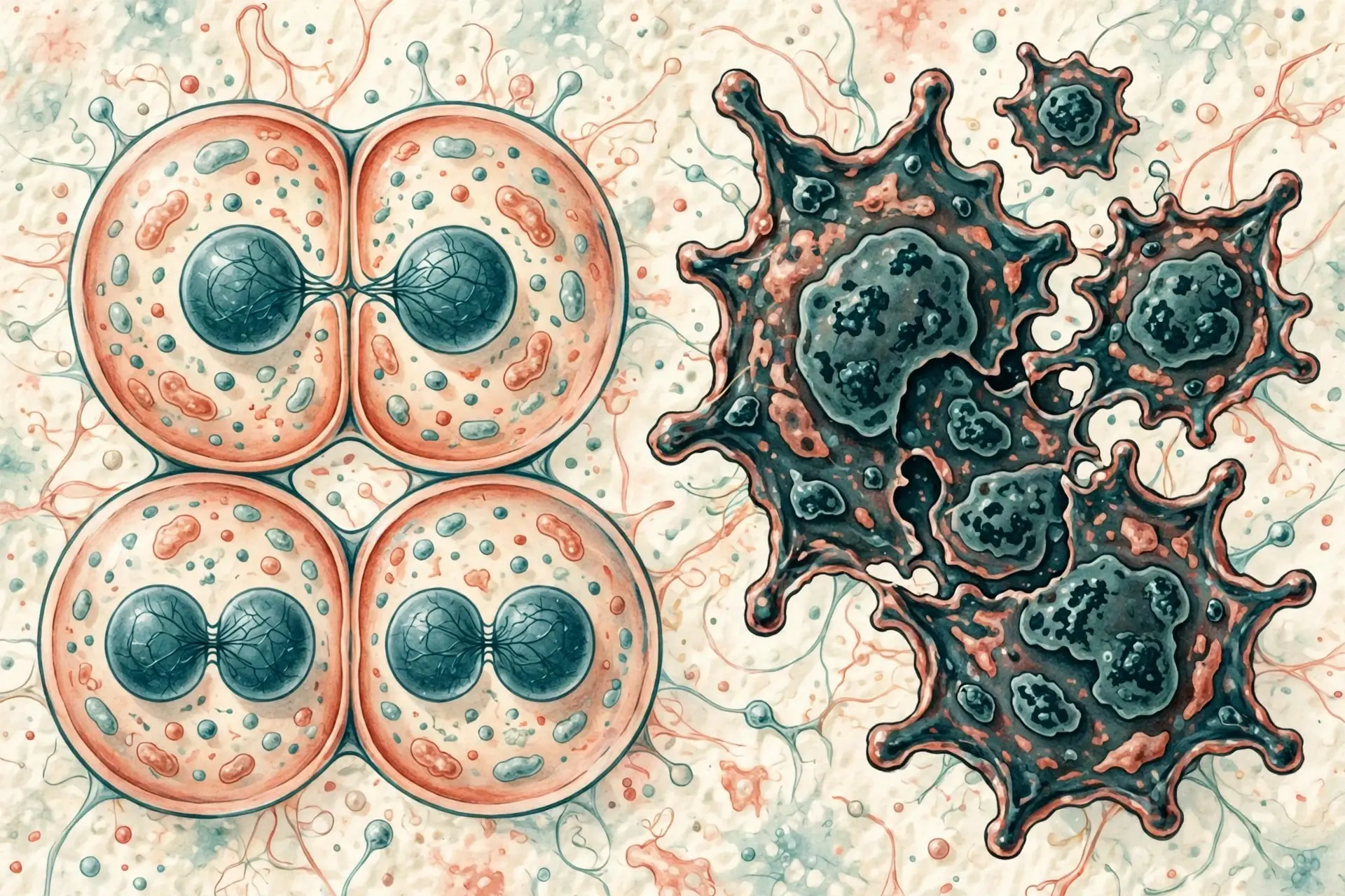
That is the strange power of this disease. Most illnesses arrive from outside: a virus, a bacterium, a broken bone. This one does not. It begins inside us, in cells that belong to us, built from our own DNA. Something in those instructions goes wrong, and a cell that was supposed to grow, divide and die on schedule stops listening. It divides when it should rest. It refuses to die when it should go. It wanders where it should stay put.
So what is it, really? In the plainest terms, it is what happens when some of the body's own cells start multiplying without restraint and travelling to places they do not belong. That definition, from the National Cancer Institute, carries the whole story — but the biology behind it is richer, stranger and, in places, more hopeful than most people expect. To understand the disease you first have to understand how a normal cell behaves, because every trick a tumour uses is a normal cellular skill bent out of shape.
This article follows that thread from start to finish: what a healthy cell does, how mutations rewrite its instructions, the fourteen hallmarks scientists use to recognise the disease, how a tumour feeds itself and travels, what raises risk and what does not, how doctors find and treat it, and where the science is heading. The numbers here are the latest available — the American Cancer Society's Cancer Statistics 2026 report and the global GLOBOCAN 2024 figures — and where the science is still arguing with itself, I will say so plainly. Nothing here is medical advice for any individual reader; it is an explanation of the biology, written so that the word in the room becomes a little less mysterious.
1. What Is Cancer?
1.1 A disease of our own cells
Every human body is a community of cells, each one running on instructions written in DNA. Most of the time the community works beautifully: skin cells replace themselves, gut lining renews, blood cells are made fresh every day. The disease begins when one cell in that community stops following the shared rules — and passes its rule-breaking on to its daughters.
"Cancer is a disease in which some of the body's cells grow uncontrollably and spread to other parts of the body." — National Cancer Institute
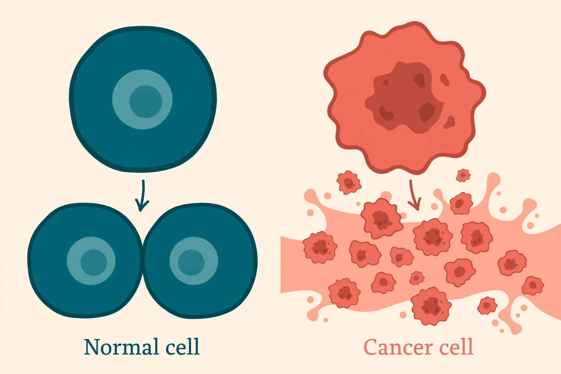
Two words in that definition do most of the work. "Uncontrollably" separates a tumour from ordinary growth: normal cells divide only when told to, and stop when told to stop. "Spread" separates a dangerous growth from a harmless one: a lump that stays where it started is a very different problem from one whose cells travel and set up colonies elsewhere. A single mutated cell is not yet a disease. It becomes one when its descendants keep dividing, ignore the signals that should restrain them, and learn to invade and travel.
1.2 Not one disease but more than a hundred
One of the first things to unlearn is the idea of a single illness. It is more than 100 different diseases that share a family resemblance, named in two parts: where they start, and what kind of cell they start in. A carcinoma begins in the skin or the tissue lining organs such as the breast, lung or colon. A sarcoma begins in bone, muscle or other connective tissue. Leukaemia begins in blood-forming tissue, so its cells circulate rather than forming a solid lump. Lymphoma begins in the lymph system, myeloma in plasma cells of the bone marrow, melanoma in the pigment-making cells of the skin, and brain and spinal cord tumours in the cells of the central nervous system.
That variety matters: a treatment that works against one type can be useless against another, because the two are driven by different broken genes.
1.3 Tumour vs cancer
People often use "tumour" and "cancer" as if they meant the same thing. They do not. A tumour is simply a mass of abnormal cells, and many tumours are benign: they grow slowly, stay in one place, and do not invade surrounding tissue or spread. A mole is a small benign tumour of pigment cells. Benign tumours can still cause trouble if they press on something important, but they do not behave like the disease this article describes.
A malignant tumour is different in kind, not just in degree. Its cells invade nearby tissue, break away, travel through blood or lymph, and start new growths far from home. That ability to invade and spread — metastasis — is what makes a tumour a cancer, and it is also what makes the disease so hard to treat. A lump that can be cut out is one problem; cells scattered through the body are another entirely.
1.4 The scale of it
Worldwide, about 20.6 million new cases were estimated for 2024, along with 9.8 million deaths, according to GLOBOCAN 2024, the global statistics compiled by the International Agency for Research on Cancer. Lung tumours were the most common, with almost 2.6 million cases — 12.8 percent of all cases — and the leading cause of death, at 1.9 million deaths, or 19.1 percent of the total. If current trends continue, the same project estimates about 34.4 million new cases a year by 2050, a rise of 67 percent from 2024, driven largely by population growth and ageing.
In the United States, the American Cancer Society's Cancer Statistics 2026 report projects 2,114,850 new cases in 2026 — about 5,800 every day — and 626,140 deaths. The same report carries the most hopeful headline in years: five-year relative survival for all types combined has reached 70 percent for people diagnosed between 2015 and 2021, the first time it has touched that mark, up from 49 percent in the mid-1970s. The global picture, from the GLOBOCAN 2024 estimates, is plain: about 1 in 5 people develop the disease in their lifetime, and about 1 in 9 men and 1 in 13 women die from it; the World Health Organization adds that about 400,000 children develop it each year worldwide.
1.5 A very old disease
The disease is often talked about as a modern one, a price of modern life. The written record says otherwise. The Edwin Smith Papyrus, an Egyptian medical text written around 1600 BCE and drawing on older material possibly dating to about 3000 BCE, describes breast tumours — among the earliest written records anywhere. Around 400 BCE, the Greek physician Hippocrates gave it the name that stuck: he called tumours "karkinos", the Greek word for crab, because the swollen veins around a tumour reminded him of a crab's legs. The Roman physician Celsus later translated the word into Latin as "cancer", and the name has followed the disease ever since. A crab grips and does not let go — and a malignant tumour does something similar. For most of human history the disease was rare in the records — not because it did not exist, but because people mostly died young of infections and other causes before the slow, age-linked failures behind it had time to appear.
2. How a Normal Cell Behaves
Before the rule-breaking makes sense, the rules have to be clear. A normal cell lives under a strict set of instructions. Four of those rules matter most for this story.
2.1 The cell cycle: grow, copy DNA, divide, rest
A cell that is going to divide moves through a cycle: it grows, copies its entire DNA, checks the copy, divides into two daughter cells, and then rests. At several points sit checkpoints — quality-control stops where the cell inspects its DNA for damage and either repairs it, pauses, or gives up dividing. Think of a printing press that stops the run, checks every page, and refuses to print a damaged plate. The stages of this cycle are walked through in our companion article on Mitosis vs Meiosis, and the broader picture in Cell Biology Explained.
I first watched this cycle in a school biology lab, hunched over a microscope at a slide of onion root tip cells. Most of the cells were just sitting there, quiet little boxes. A few were caught mid-division, chromosomes lined up like commuters on a platform. I remember the odd feeling that something so orderly could be happening inside something as ordinary as an onion — and the slow realisation, years later, that every tumour is what happens when that order comes apart.
2.2 Death on schedule: apoptosis
Healthy cells do not just divide on schedule; they die on schedule. The process is called apoptosis — programmed cell death — and it is as essential as division. Cells that are damaged, infected or simply no longer needed dismantle themselves neatly, and their neighbours clean up the remains. Your body replaces enormous numbers of cells this way every day: the lining of your gut renews itself constantly, skin cells flake away and are replaced, blood cells are made and retired in a constant cycle. One of the most reliable tricks a tumour cell learns is to switch this demolition crew off — to refuse the order to die, even when its DNA is badly damaged.
2.3 Talking to neighbours: contact inhibition, growth signals, sticking together
Cells are social. They listen for growth signals from their neighbours, they stop dividing when they touch each other on all sides — a behaviour called contact inhibition — and they stick together with molecular glue so that tissues hold their shape. That social life is a safety system. Break the listening — make the cell deaf to "stop" signals, or able to manufacture its own "go" signals — and the social order starts to fail.
2.4 The Hayflick limit: 40–60 divisions, telomeres shortening like a candle burning down
There is one more rule, and it is a strange one: normal human cells can divide only about 40 to 60 times before they stop. Leonard Hayflick and Paul Moorhead discovered this in 1961, and the limit now carries Hayflick's name. The reason sits at the ends of the chromosomes, in caps called telomeres. Every time a cell copies its DNA, the telomeres get a little shorter — between 50 and 200 base pairs per division — like a candle burning down a fraction with each lighting. When they get too short, the cell stops dividing for good.
That limit is a built-in safety brake against runaway growth. A tumour cell has to defeat it to keep growing, and most of them do it by switching back on an enzyme called telomerase, which rebuilds the telomeres. Telomerase is reactivated in about 85 to 90 percent of human cases; the rest use a different route called the ALT pathway, found in 10 to 15 percent. Either way, the candle stops burning down — and the cell becomes, in practical terms, immortal.
3. How Cancer Starts: Mutations
3.1 DNA, the instruction manual
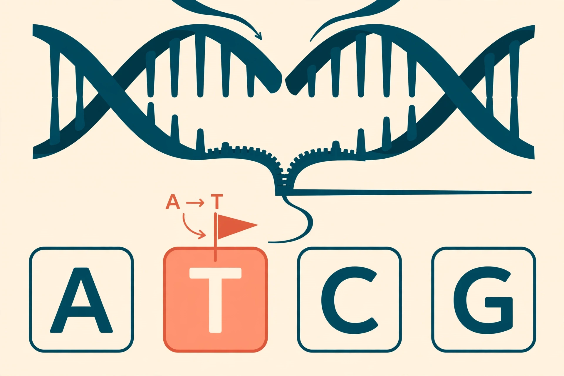
If a cell is a tiny factory, its DNA is the instruction manual — about 3 billion letters long, written in a four-letter alphabet, copied in full every time the cell divides. The cell has proofreading machinery that catches most errors, and repair crews that fix damage as it happens. Most days, the system works. Mistakes still slip through: a letter is swapped, a stretch is deleted, a whole paragraph is duplicated. Most of these changes are harmless. A few land in genes that control growth, division or death. Those are the changes that matter. The companion article on The Human Genome explains the manual itself, and DNA: Structure, Replication, and Role in Heredity covers the copying machinery.
3.2 Driver vs passenger mutations
A typical tumour carries between 2 and 8 driver mutations, drawn from a pool of about 140 known driver genes, according to the landmark analysis by Vogelstein and colleagues published in Science in 2013. Drivers are the mutations that actually push the cell toward uncontrolled growth. Around them sit many more passenger mutations — changes that happened to be present in the cell when it turned malignant, carried along for the ride, contributing little or nothing to the disease. Think of a car speeding down a hill with a broken brake: the driver mutations are the cut brake line and the jammed accelerator, while the passengers are the dent in the bumper and the coffee cup on the dashboard. Telling drivers from passengers is one of the central jobs of modern tumour genetics, because a drug aimed at a passenger will do nothing, while a drug aimed at a driver can change the course of the disease.
3.3 Accelerators: oncogenes
Genes that promote growth are called proto-oncogenes in their normal form. They are the accelerators of the cell: they send "divide now" signals when the body needs new cells, for example to heal a wound. When one of these genes is mutated, or switched on too strongly, or copied too many times, it becomes an oncogene — an accelerator pedal jammed to the floor. Three of these accelerators show up again and again. RAS genes, mutated in about 30 percent of human cases by the commonly cited estimate (newer incidence-weighted analyses put the figure lower, around 15 percent), normally relay growth signals from the cell surface inward; a mutated RAS relays the signal permanently, as if the phone were off the hook. MYC drives the machinery of growth and division, and HER2 is a surface receptor whose overactivity drives some breast tumours — and, usefully, can be targeted by a drug built to block it, as section 9.4 describes. A stuck accelerator alone is usually not enough to make a tumour. It needs the brakes to fail too.
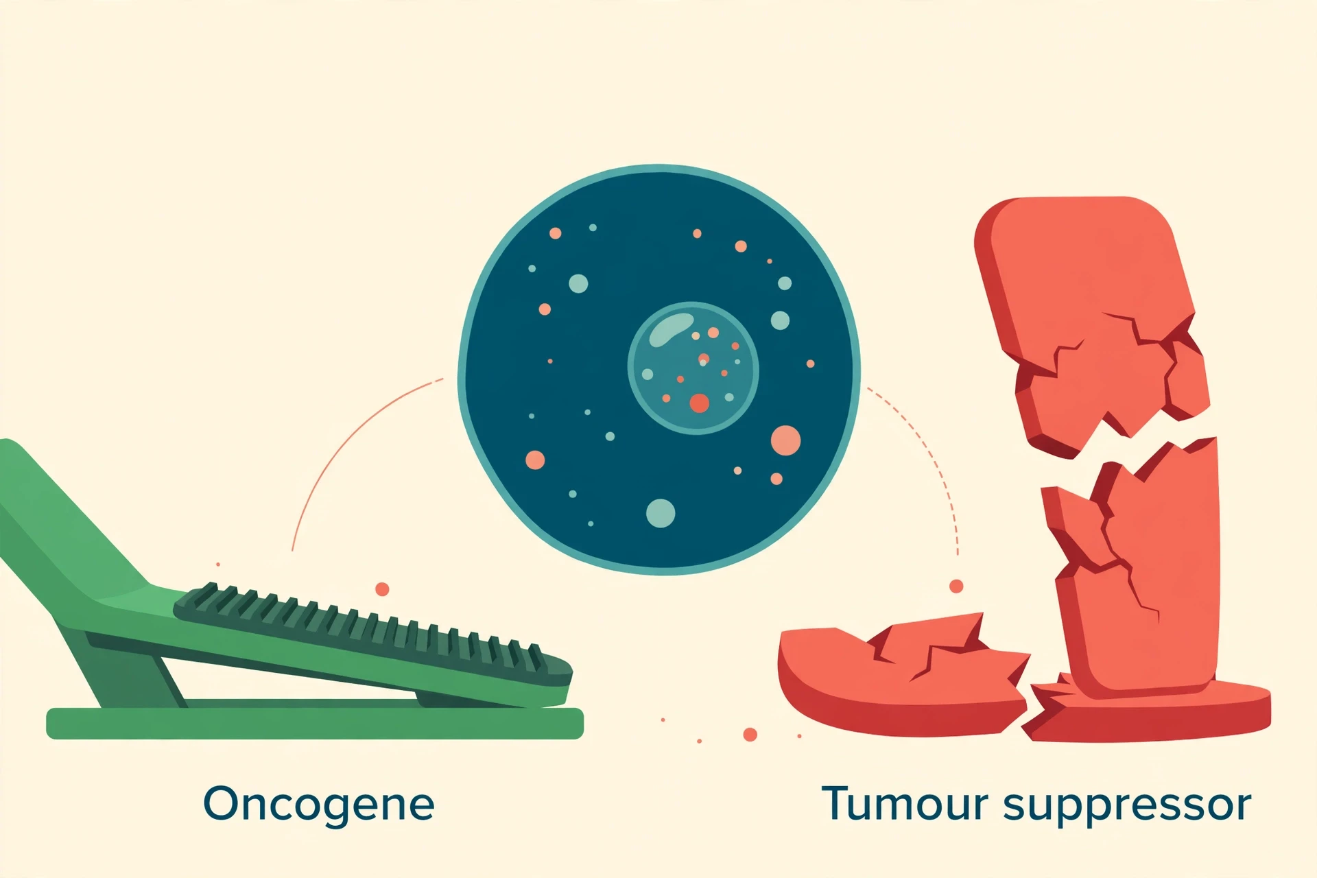
3.4 Broken brakes: tumour suppressor genes
If oncogenes are accelerators, tumour suppressor genes are brakes. They halt division when DNA is damaged, trigger cell suicide when the damage cannot be fixed, and generally keep growth honest. The most famous of them is TP53, which encodes the p53 protein — often called the "guardian of the genome". When DNA is damaged, p53 halts the cell cycle so repairs can happen, or orders apoptosis if the damage is too great. TP53 is the most commonly mutated gene in human cancer, altered in about half of all tumours by the commonly cited figure (incidence-weighted estimates are lower, around a third). Lose it, and a cell with broken DNA keeps dividing anyway. Two other brakes matter here: RB1, another tumour suppressor gene, controls the gateway into cell division, and BRCA1 and BRCA2 are repair genes that help fix the most dangerous kind of DNA damage, breaks across both strands. A classic idea from Alfred Knudson's work on retinoblastoma, a childhood eye tumour, explains why these brakes are hard to lose completely: the "two-hit" hypothesis says both copies of a tumour suppressor gene usually have to be knocked out before control fails. One working copy is often enough to hold the line — which is why inherited risk and acquired damage so often combine, one hit inherited and the second arriving during life.
3.5 Inherited vs acquired
Most people assume the disease runs in families. The numbers say something subtler. Only about 5 to 10 percent of cases are linked to inherited mutations — changes present in every cell from birth. The large majority are acquired during life, in one cell and its descendants, from copying errors and exposures. Inherited mutations still matter enormously for the people who carry them. The best-studied examples are BRCA1 and BRCA2: carriers of a harmful BRCA1 mutation face about a 72 percent lifetime risk of breast cancer by age 80, and about 44 percent for ovarian cancer; for BRCA2 the figures are about 69 percent for breast and 17 percent for ovarian, according to a large 2017 study in JAMA by Kuchenbaecker and colleagues. Those are population-level risks for carriers, not predictions for any individual. How inheritance itself works is covered in our Genetics Explained article.
3.6 Epigenetics: the same genes, switched off
Mutations change the letters of the manual. Epigenetics changes which pages are open. Chemical marks on DNA — most famously methylation, the addition of small methyl groups — and chemical changes to the histone proteins DNA wraps around can switch genes off without altering a single letter of the sequence. A tumour suppressor gene can be perfectly intact and still silent, because methylation has padlocked its promoter shut. That matters for two reasons. First, a cell can lose its brakes without any mutation at all. Second, epigenetic marks are in principle reversible, which is one reason drugs that strip away methylation or loosen histones are an active area of research. The full story is told in Epigenetics Explained: How Your Environment Changes Your Genes, and it is directly relevant here: the 2022 update to the hallmarks of the disease, described in the next section, added nonmutational epigenetic reprogramming as a hallmark in its own right.
3.7 Where mutations come from
Mutations arrive from several directions at once. The first is the copying process itself: every division is a chance for a proofreading slip, and a body that makes billions of cell divisions across a lifetime will accumulate them. Then there are the outside agents — tobacco smoke, ultraviolet light, ionising radiation, certain chemicals, chronic inflammation, and infections that damage DNA or force cells to divide more often. DNA repair systems fix thousands of lesions every day; when repair genes themselves fail — as with BRCA1 and BRCA2, or with the mismatch-repair genes MLH1 and MSH2 in Lynch syndrome — risk rises sharply. How much of this is simply chance? In 2015, Tomasetti and Vogelstein published a much-discussed analysis in Science arguing that random copying errors — "bad luck" — explain a large share of the variation in risk between tissue types. The claim has been debated ever since: other researchers argue it underplays the role of environment and prevention, and the exact proportion remains unsettled. The honest summary is that copying errors contribute to many cases, they are only part of the picture, and the balance between chance, inheritance and exposure differs from one type to the next. Our article on Natural Selection Explained gives useful background on how random mutation and selection work together — the same two forces, at work inside a single body.
4. The Hallmarks of Cancer: Ten Rules, Broken
4.1 The 2000 list: six hallmarks
In 2000, Douglas Hanahan and Robert Weinberg published a paper in Cell that gave the whole field a shared vocabulary. They proposed that, for all their variety, malignant cells share six essential abilities — six rules of normal cell life, broken in a characteristic pattern. The paper became one of the most cited in biology, because it turned a bewildering zoo of diseases into a checklist a researcher could hold in one hand.
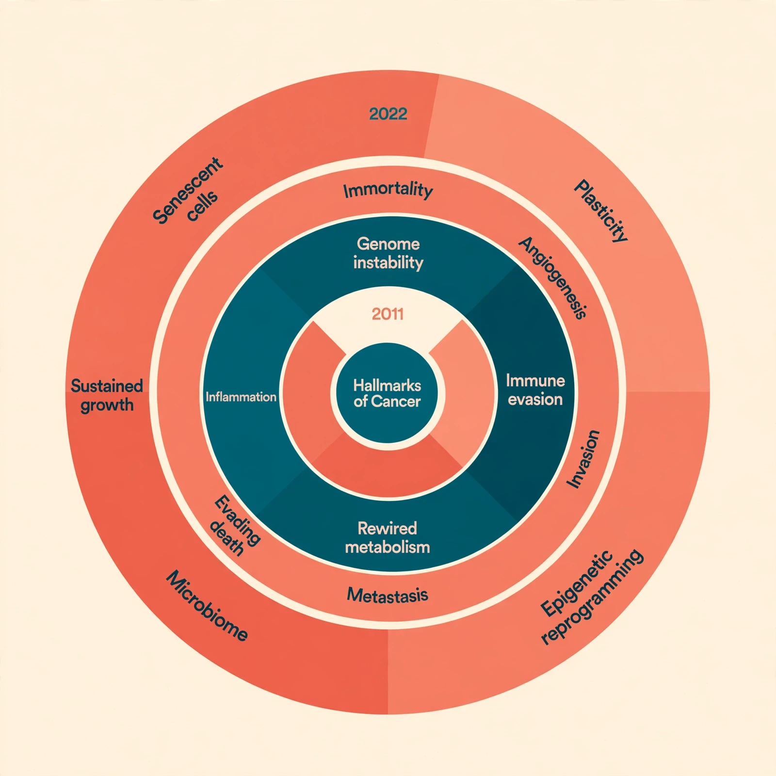
The six were: self-sufficiency in growth signals (making their own "go" signals); insensitivity to growth-inhibitory signals (ignoring "stop"); evasion of programmed cell death (refusing apoptosis); limitless replicative potential (defeating the Hayflick limit); sustained angiogenesis (building their own blood supply); and tissue invasion and metastasis (moving and colonising). Each maps directly onto a rule from section 2 — and each, on its own, is not enough. A cell that divides without permission but still dies on schedule is not a tumour. A cell that refuses to die but cannot feed itself will not grow far.
4.2 The 2011 expansion: ten hallmarks
Eleven years later, Hanahan and Weinberg returned with an update. Two enabling characteristics joined the list — genome instability and mutation, which generate the genetic diversity tumours evolve from, and tumour-promoting inflammation, in which immune cells that should attack the tumour are co-opted into helping it grow. Two emerging hallmarks were added as well: reprogramming of energy metabolism, the Warburg effect described in section 5.2, and evading immune destruction, the tumour's ability to hide from the immune system. That brought the total to ten. The 2011 paper also made an important shift: the hallmarks are not a ladder the cell climbs one rung at a time, but capabilities acquired in different orders, at different times, in different tumours.
4.3 The 2022 update: fourteen
In 2022, Hanahan published a further update in Cancer Discovery, adding four more hallmarks and bringing the total to fourteen: unlocking phenotypic plasticity (cells changing their identity, sliding backwards to a more primitive state); nonmutational epigenetic reprogramming (the silencing described in section 3.6); polymorphic microbiomes (the bacteria and other microbes living in and around a tumour, influencing its behaviour); and senescent cells (aged, non-dividing cells that linger and secrete signals promoting growth and inflammation). The list keeps growing because the science keeps looking closer — and each new hallmark is also a new place to aim a treatment.
4.4 A working summary
Fourteen hallmarks are a lot to hold in mind, so here is the whole set compressed into a plain-language table, with one concrete example for each. If you remember only five ideas from this article, make them these: growth without permission, death refused, immortality, a private blood supply, and invasion. Everything else is commentary.
Hallmark | What it means in plain words | One example |
|---|---|---|
Self-sufficiency in growth signals | The cell makes its own "divide" signals instead of waiting for them | Mutated RAS signalling permanently, as if the phone were off the hook |
Insensitivity to growth-inhibitory signals | The cell ignores "stop" signals from neighbours | Loss of RB1 lets the cell cycle run past its checkpoint |
Evasion of programmed cell death | The cell refuses apoptosis even when badly damaged | Loss of p53 removes the order to self-destruct |
Limitless replicative potential | The cell defeats the Hayflick limit and divides indefinitely | Telomerase reactivated in 85 to 90 percent of cases |
Sustained angiogenesis | The tumour builds its own blood supply | VEGF signals summon new vessels to feed the growth |
Tissue invasion and metastasis | Cells break away, travel and colonise distant organs | An estimated 90 percent of deaths involve metastasis |
Genome instability and mutation | The genome becomes unstable, generating new variants fast | Failure of DNA repair genes such as MLH1 and MSH2 |
Tumour-promoting inflammation | Immune cells are recruited to help the tumour grow | Chronic inflammation feeding growth signals |
Reprogramming of energy metabolism | Cells rewire how they make energy | The Warburg effect: glycolysis even when oxygen is available |
Evading immune destruction | The tumour hides from or switches off immune attack | Checkpoint proteins that tell T cells to stand down |
Unlocking phenotypic plasticity | Cells slide back to a more primitive, flexible state | Dedifferentiation that resists treatment |
Nonmutational epigenetic reprogramming | Genes are silenced by chemical marks, not mutations | Methylation switching off tumour suppressor genes |
Polymorphic microbiomes | Microbes in and around the tumour shape its behaviour | Gut and tumour bacteria influencing growth and treatment response |
Senescent cells | Aged, non-dividing cells linger and secrete growth signals | Senescent cells promoting inflammation around a tumour |
5. How Cancer Grows: Feeding the Tumour
5.1 Angiogenesis: building its own blood supply
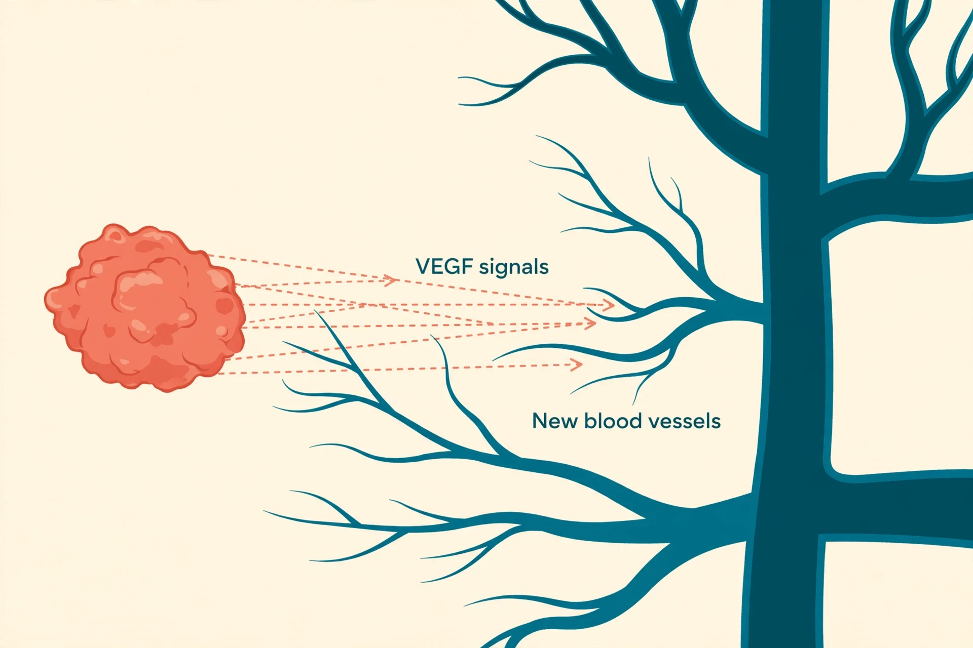
A small clump of abnormal cells can survive on whatever oxygen and nutrients diffuse in from nearby tissue — but only up to a point. Past a tiny size, the cells in the middle begin to starve. To grow further, a tumour has to solve a plumbing problem, and it does so by recruiting its own blood vessels, a process called angiogenesis. In 1971, Judah Folkman proposed that tumours could not grow beyond a tiny size without this step, an idea that was controversial at the time and is now textbook biology. The main signal the tumour uses is VEGF, vascular endothelial growth factor, which tells nearby blood vessels to sprout new branches toward it — a supply line for oxygen and nutrients, and, as a side effect, a highway out.
5.2 Rewired metabolism: the Warburg effect
Normal cells, when oxygen is available, burn glucose efficiently in their mitochondria, extracting a large amount of energy from each molecule. Tumour cells often do something that looks wasteful: they favour glycolysis — a fast, inefficient way of burning glucose — even when oxygen is plentiful. This is the Warburg effect, named after Otto Warburg, who described it in the 1920s and won the Nobel Prize in 1931 for his work on respiration. Why would a cell choose the inefficient route? Speed and raw materials, most likely. Glycolysis is fast, and its byproducts feed into the building blocks a dividing cell needs — nucleotides, amino acids, lipids. A tumour cell is not trying to be efficient; it is trying to build two cells out of one as quickly as possible.
5.3 The tumour microenvironment: a tumour is not alone
It is tempting to picture a tumour as a lonely lump of rogue cells. In reality it is a whole distorted neighbourhood. Around the malignant cells sit fibroblasts that have been recruited to lay down scaffolding, blood vessels feeding the mass, and immune cells that have been corrupted or exhausted into helping rather than attacking. Scientists call this the tumour microenvironment, and it often decides whether a treatment works: drugs must cross it, and the signals it produces can protect the tumour.
5.4 Heterogeneity: a tumour is a crowd, not a clone
A tumour begins from a single cell, but it does not stay uniform. As it grows, its cells keep mutating, and different branches of the family tree pick up different changes. The result is heterogeneity: a single tumour contains many subpopulations of cells, some sensitive to a given drug and some already resistant to it. This is the same evolutionary logic — random variation plus selection — that shapes whole species, and it is why a treatment can shrink a tumour dramatically and still leave behind the cells that will regrow it. Antibiotic Resistance Explained tells the parallel story in bacteria, where the same dynamic produces drug-resistant superbugs.
6. How Cancer Spreads: Metastasis
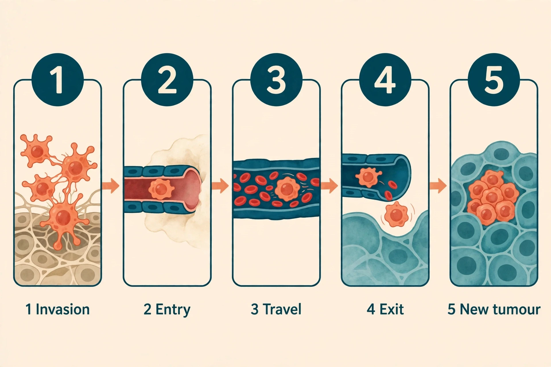
6.1 The journey: invasion, travel, landing, colonisation
Metastasis is a journey with five stages, and a tumour cell has to survive all five to succeed. First comes local invasion: cells break through the basement membrane that contains their tissue and push into surrounding structures. Then intravasation: they squeeze into a blood vessel or a lymph vessel. Then survival in circulation, carried along in the bloodstream while being battered by shear forces and hunted by immune cells. Then extravasation: exiting the vessel at a distant site. And finally colonisation: growing into a new tumour in foreign tissue. Stephen Paget, a London surgeon, described the pattern in 1889 with an analogy that still holds: the travelling cell is the "seed", and the organ it lands in is the "soil". A seed only grows where the soil suits it, which is why the disease spreads in patterns — some types favour the lungs, others the liver, the bones or the brain — rather than scattering at random.
6.2 Why it is so deadly
About 90 percent of deaths from the disease are caused by metastasis, according to a 2011 review in Science by Chaffer and Weinberg — not by the original tumour, but by the colonies it founds elsewhere. A primary tumour can often be removed or irradiated. Secondary growths scattered through the lungs, liver, bones and brain are far harder to reach, and they are what damage vital organs beyond repair. That is also why early detection matters so much, and why so much research focuses on the travelling cell: what lets it survive in the blood, what tells it where to land, and what wakes a dormant cell years after it arrived.
6.3 Why most travelling cells die — and why a few succeed
The metastatic journey is extraordinarily wasteful. Many cells are shed into the bloodstream, and the overwhelming majority die: torn apart by the flow, starved, or killed by immune cells. Only a tiny fraction complete the journey. The ones that succeed tend to be the most adaptable — cells that can change shape to squeeze through vessel walls, cluster together with platelets for protection, and switch their identity to survive in a new tissue. As a student I was stuck for a long time on a simple question: how could a tumour that began in the lung appear in the brain? The organs are not connected by any tube I could picture. The penny dropped when I finally mapped the route properly — the bloodstream is not a local road system but a single loop that visits everywhere, including the brain. Once that picture clicked, the patterns of spread stopped looking mysterious and started looking inevitable: anywhere the blood goes, a travelling cell can go too.
6.4 Staging: TNM in plain words
When doctors describe how far the disease has spread, they use a system called TNM. T stands for the size and extent of the primary tumour. N stands for whether nearby lymph nodes — the small filters of the lymph system, often the first stop for travelling cells — contain malignant cells, and how many. M stands for metastasis: whether the disease has spread to distant parts of the body. The three letters combine into a stage, usually from stage 0 to stage IV, with higher stages meaning further spread. Staging is not a verdict on a person; it is a description of geography, telling the care team how far the disease has travelled and which treatments are in play.
7. What Causes Cancer?
There is no single cause. For most types, the honest answer is a combination: inherited susceptibility, exposures across a lifetime, and random copying errors, in proportions that differ from person to person and type to type. What follows is the population-level picture — the factors that raise risk across large groups of people — described in general terms, never as personal advice.
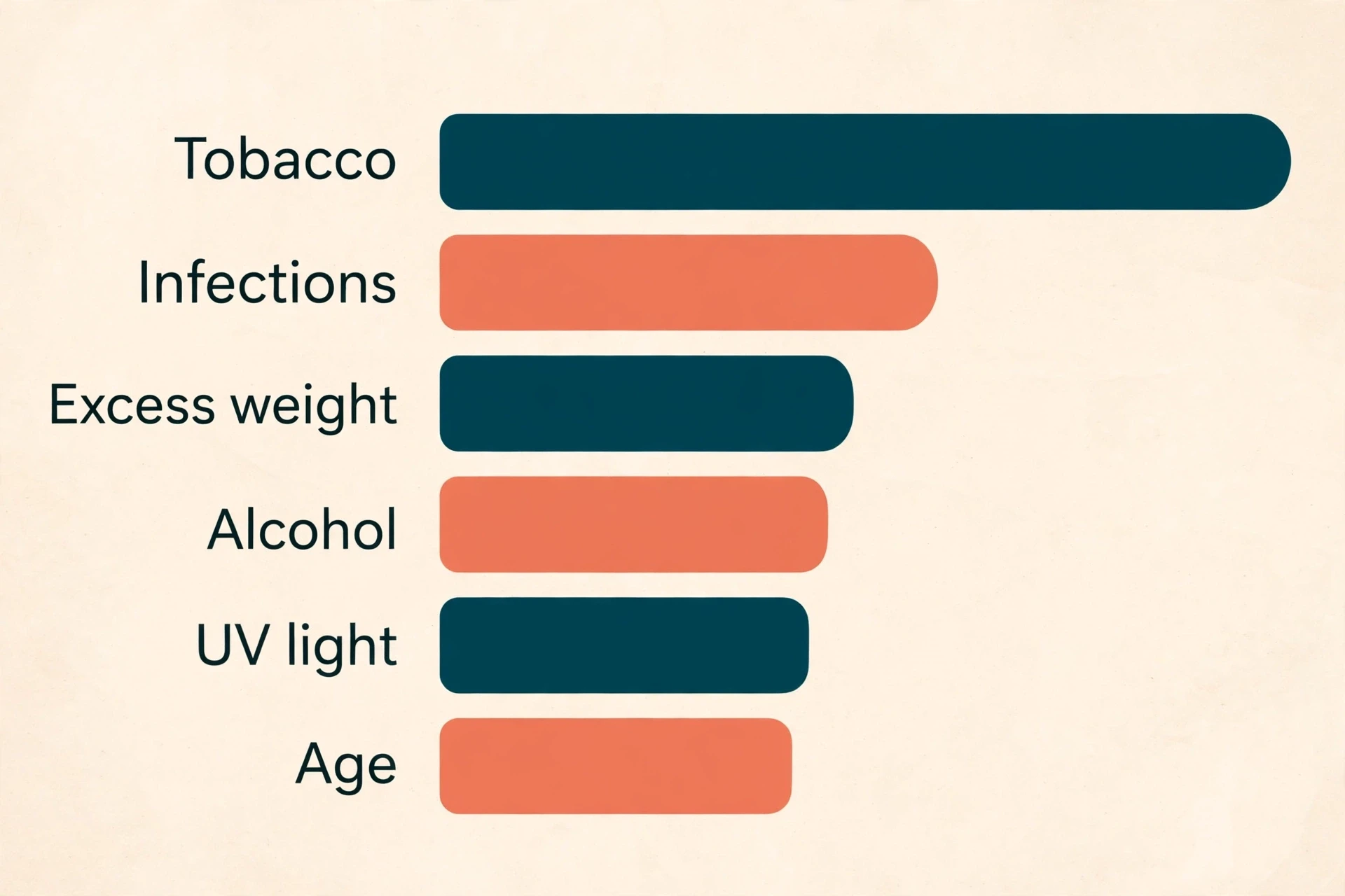
7.1 Tobacco
Tobacco is the single largest preventable contributor. Smoking accounts for about 30 percent of all deaths from the disease in the United States, and about 80 percent of lung cancer deaths, according to the American Cancer Society — and about 1 in 5 of all new cancer cases in the US. Tobacco smoke carries dozens of chemicals that damage DNA directly, and it forces the cells lining the airways to divide more often to repair the damage, giving copying errors more chances to occur. The link extends well beyond the lungs: smoking raises the risk of tumours of the mouth, throat, oesophagus, bladder, kidney, pancreas and several other sites.
7.2 Infections
About 13 percent of cases worldwide are linked to infections, according to the International Agency for Research on Cancer's 2018 estimate (the 2024 update puts the share at about 12 percent). The human papillomavirus, HPV, causes about 95 percent of cervical cancers, according to the World Health Organization; hepatitis B and C viruses cause most liver cancers; and the bacterium Helicobacter pylori causes most stomach cancers. These infections do not make the disease contagious — the infections can spread between people, but the disease itself cannot be caught from another person, as the FAQ below explains. How viruses change over time, including the ones that cause these infections, is covered in Virus Evolution Explained: How Viruses Mutate and Adapt.
7.3 Sun and radiation
Ultraviolet radiation from sunlight is the main cause of skin cancer, and melanoma — the tumour of pigment cells — is the deadliest of the skin tumours. UV light damages DNA directly, and sunburn is a visible sign of that damage. Other radiation matters too: radon gas seeping from the ground is a recognised cause of lung cancer, and occupational exposures such as asbestos and benzene, along with air pollution, are classified by the International Agency for Research on Cancer in its highest category, Group 1, meaning they are known to cause cancer in humans. The story of UV light itself — why it damages skin, and how the ozone layer filters it — is told in The Ozone Layer Explained.
7.4 Food, weight, alcohol, movement
Excess body weight is linked to at least 13 types, including tumours of the breast (after the menopause), colon, kidney, liver and pancreas. Alcohol is a known carcinogen: any amount raises risk, and risk rises with the amount consumed. Physical inactivity contributes too, partly through its effect on weight and partly on its own. These are population-level patterns, not personal verdicts: many people with none of these risk factors develop the disease, and many people with several never do.
7.5 Age
The biggest single risk factor is simply age. The median age at diagnosis in the United States is 67, according to the National Cancer Institute's SEER programme. That is not because older bodies are somehow weaker in a general sense, but because the disease is a disease of accumulated damage: decades of copying errors, decades of exposures, and decades of gradual decline in DNA repair. A cell needs several driver mutations to turn malignant, and gathering them takes time. This is also why the number of cases is projected to rise so sharply by 2050: the world's population is both growing and ageing. The evolutionary side of ageing — why selection cannot fully protect us late in life — is explored in Why Do We Age? The Evolutionary Theory of Aging Explained.
7.6 Chance and the genes you inherit
Put the pieces together and the balance looks like this: a minority of risk is inherited, a large share is linked to exposures and lifestyle at population level, and a meaningful share comes from random copying errors that no one chose and no one could have prevented. That last part is worth saying plainly, because it cuts against a cruel reflex — the idea that a diagnosis must be somebody's fault. Sometimes it follows a lifetime of smoking; sometimes it follows nothing at all that anyone could have changed.
7.7 What does NOT cause cancer
A few claims circulate so widely that they deserve a direct answer. Mobile phones: large studies so far have not found a consistent link between mobile phone use and brain tumours, and the radio waves they emit do not carry enough energy to damage DNA directly. Sugar: it is true that tumour cells consume glucose eagerly, as the Warburg effect describes — but that does not mean eating sugar feeds a tumour directly, and cutting sugar out of the diet has not been shown to starve one. Deodorant and antiperspirant: no reliable evidence links them to breast cancer, despite years of claims online. Each of these is a case where a plausible-sounding mechanism outran the evidence, and the evidence, checked against the National Cancer Institute's summary of common cancer myths, has not confirmed the claim.
8. How Cancer Is Found
8.1 Screening
Screening means looking for the disease in people who feel well, before symptoms appear — and it works because earlier is, in general, better. The established programmes are mammography for breast cancer, cervical screening and HPV testing, colorectal screening, and low-dose CT scanning for people with a heavy smoking history. Each has been shown in large trials to catch disease earlier, when treatment is more likely to help, and each is offered to defined groups at defined intervals rather than to everyone at all times. Screening is a population-level programme with real benefits and real trade-offs — false alarms, and findings that would never have caused harm.
8.2 Diagnosis
When screening or symptoms raise a suspicion, diagnosis proceeds in steps. Imaging — X-rays, CT, MRI, ultrasound, PET — shows where a mass is and how far it reaches. A biopsy takes a small piece of tissue so a pathologist can examine the cells under a microscope and confirm what kind of cells they are. Blood tests can support the picture, and genetic testing of the tumour itself has become routine: knowing which driver mutations a particular tumour carries can determine which drug is likely to work. The tumour's genetic profile is now as much a part of the diagnosis as its location.
8.3 Staging and grading
Once a diagnosis is confirmed, two more descriptions are added. Staging, the TNM system from section 6.4, describes how far the disease has spread. Grading describes how abnormal the cells look under the microscope — how far they have drifted from the tissue they came from, and how quickly they appear to be dividing. A low-grade tumour looks relatively orderly and slow; a high-grade tumour looks chaotic and fast. Stage and grade are measurements, not prophecies: they guide the choice of treatment, and they say nothing about the person beyond the disease.
9. How Cancer Is Treated
What follows is an educational overview of the treatment families — how each one works, where it came from, and what it can and cannot do. It is not guidance for any individual; treatment decisions depend on the type, the stage, the tumour's genetics and the person's overall health, and they belong to the person and their care team.
9.1 Surgery
Surgery is the oldest treatment, and still the most common route to a cure when a tumour is localised. The idea is simple: remove the tumour and a margin of healthy tissue around it. The practice is anything but simple — modern surgery is guided by imaging, by frozen sections examined during the operation, and by a careful weighing of what can be removed against what the organ still needs to do. For a tumour that has not spread, an operation can remove the whole problem at once. For one that has, surgery can still help: relieving a blockage, reducing the bulk of disease, or obtaining tissue for diagnosis.
9.2 Chemotherapy
Chemotherapy uses drugs that kill fast-dividing cells. Its origins are wartime: nitrogen mustard, a chemical weapon, was observed to damage rapidly dividing bone marrow cells, and in the 1940s researchers turned that observation into the first chemotherapy drugs. In 1948, Sidney Farber achieved the first remissions in childhood leukaemia with an antifolate drug, aminopterin — the first time a drug had ever driven the disease into remission. The logic has not changed since: malignant cells divide fast, so drugs that damage dividing cells hit them harder than most healthy tissue. The side effects follow from the same logic: hair follicles, gut lining and bone marrow also divide fast, so they are hit too.
9.3 Radiation
Radiation therapy uses high-energy rays to damage the DNA of tumour cells, so badly that they cannot divide again. About half of all patients receive radiation at some point in their care. It can be aimed from outside the body, with beams shaped to match the tumour, or delivered from inside, with radioactive sources placed near it. The skill lies in the targeting: delivering a lethal dose to the tumour while sparing as much healthy tissue as possible. Like surgery, radiation is mainly a local treatment — it treats the place it is aimed at — which is why it is often combined with drugs that travel through the whole body.
9.4 Targeted therapy
Targeted therapy aims at a specific broken molecule rather than at dividing cells in general. The proof that this could work came in 1998, when trastuzumab (Herceptin) was approved for breast cancers that overproduce the HER2 receptor, and in 2001, when imatinib (Gleevec) was approved for chronic myeloid leukaemia, where it blocks the abnormal BCR-ABL protein that drives the disease. These drugs work only when the tumour carries the target they were built for, which is why genetic testing of the tumour has become part of diagnosis. When the match is right, the results can be dramatic, with fewer of the broad side effects of chemotherapy. When the target is absent, the drug does nothing — a reminder that a tumour's genetics, not just its location, decides the treatment.
9.5 Immunotherapy
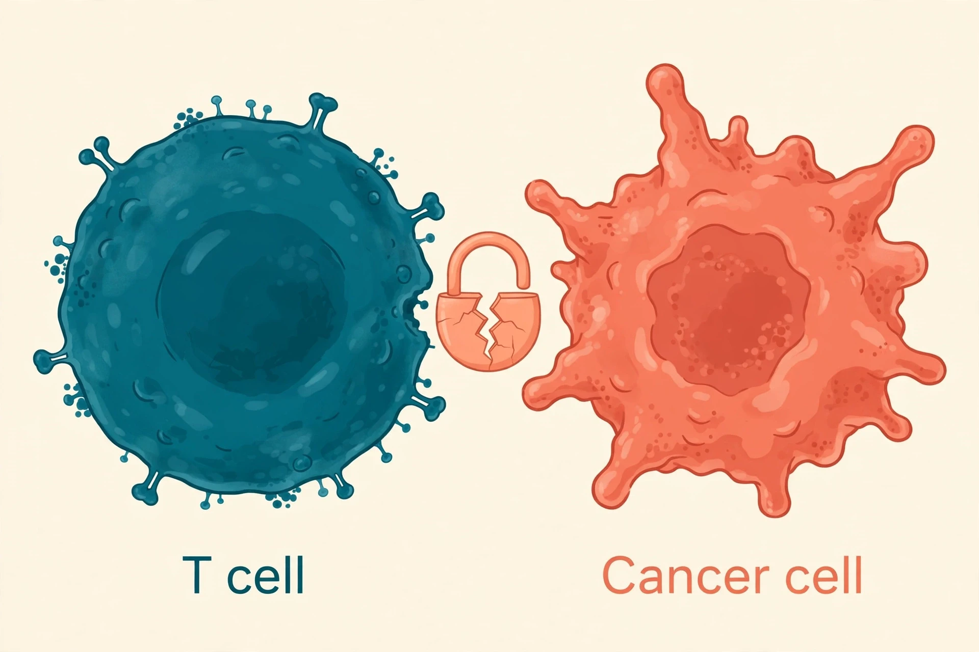
The immune system already knows how to kill abnormal cells — it does so every day — so the idea behind immunotherapy is to remove the brakes the tumour has put on it. The breakthrough came from understanding checkpoints: proteins such as CTLA-4 and PD-1 that tell T cells to stand down. James Allison worked out the role of CTLA-4, and Tasuku Honjo discovered PD-1 in 1992. In 2018 the two men shared the Nobel Prize in Physiology or Medicine:
"for their discovery of cancer therapy by inhibition of negative immune regulation" — The Nobel Prize in Physiology or Medicine 2018, awarded to James P. Allison and Tasuku Honjo
The first checkpoint inhibitor drug, ipilimumab, was approved in 2011 for melanoma, and checkpoint drugs are now used across many types. A second branch of immunotherapy takes the patient's own T cells, engineers them to recognise the tumour, and returns them: CAR T-cell therapy. The first approval came on 30 August 2017, for tisagenlecleucel (Kymriah), for relapsed or refractory B-cell acute lymphoblastic leukaemia in patients up to age 25. In the ELIANA trial, 83 percent of patients — 52 of 63 — achieved overall remission. The first patient treated, Emily Whitehead, was six years old when she received the therapy in 2012. How the immune system normally tells friend from foe is explained in How the Human Immune System Works, and the gene-editing tools that make engineered T cells possible are covered in CRISPR Gene Editing Explained.
9.6 Why treatment is hard
Three problems recur, and they explain most treatment failures. The first is heterogeneity, from section 5.4: a tumour is a crowd of different cells, and a drug that kills 99 percent of them leaves the resistant 1 percent to regrow. The second is evolution under pressure: treatment itself selects for resistant cells, the same way antibiotics select for resistant bacteria. The third is concealment: tumours hide from the immune system, build a protective microenvironment, and recruit normal cells to defend them. Beating the disease, in most cases, means combining treatments that attack from different directions at once — surgery plus drugs, drugs plus radiation, or several drugs together — so that no single escape route saves the tumour.
9.7 Survival today
The overall trend is genuinely encouraging, and it is worth stating plainly. Five-year relative survival for all types combined in the United States has reached 70 percent for people diagnosed between 2015 and 2021, according to the American Cancer Society's Cancer Statistics 2026 report — up from 49 percent in the mid-1970s. As the report's lead author Rebecca Siegel put it, seven in 10 people now survive five years or more, up from only half in the mid-1970s. The gains have been striking for some types: survival in myeloma rose from 32 percent to 62 percent since the mid-1990s, liver cancer from 7 to 22 percent, and lung cancer from 15 to 28 percent. Even for distant-stage disease — disease that has already spread — five-year survival has doubled since the mid-1990s, from 17 percent to 35 percent. And the mortality rate has continued to decline through 2023, with about 4.8 million deaths averted since 1991, mainly through reductions in smoking, earlier detection and better treatment. Those are population figures, not promises for any individual — survival varies enormously by type and stage — but they measure real, hard-won progress.
10. Can Cancer Be Prevented?
10.1 The share that is modifiable
About 40 percent of new cases in the United States — and about 44 percent of deaths — among adults aged 30 and older in 2019, excluding non-melanoma skin cancers, are attributable to potentially modifiable risk factors, according to a 2024 analysis by Islami and colleagues published in CA: A Cancer Journal for Clinicians. That figure is a population-level estimate, not a personal score: it means that, across the whole country, a large share of cases is associated with factors that can in principle be changed, such as smoking, excess weight, alcohol, inactivity, diet, sun exposure and infections. It also means the majority of cases are not linked to those factors — a reminder that prevention, however valuable, is not a guarantee and never a matter of blame.
10.2 The big, boring, effective things
The measures with the strongest evidence are, frankly, unglamorous. At population level, the list is short: not smoking, and helping others to stop; vaccination against HPV and hepatitis B; taking part in screening programmes when invited; maintaining a healthy weight; limiting alcohol; and protecting skin from the sun. None of these is a promise. Each of them shifts the odds across a whole population, and across a population of millions, a small shift in the odds is a large number of lives.
10.3 The HPV story
The HPV story shows what prevention can look like when the biology is understood well enough to act on. HPV types 16 and 18 cause around 76 percent of cervical cancers, according to the World Health Organization, and vaccination programmes against the virus began in 2006, with England's school programme following in 2008. In England, researchers reported no cervical cancer deaths among women aged 20 to 24 between 2020 and 2024 — the first generation to have been offered the vaccine as schoolgirls. The finding, from Sasieni and Falcaro in The Lancet, is an early signal rather than a final verdict, and it needs to be read alongside screening, which remains important for older age groups. Still, it is a rare thing in medicine: a line on a graph that bends because a vaccine was given to children years earlier.
11. What Nature Already Knows: Peto's Paradox
11.1 The paradox: elephants and whales should get more cancer — they don't
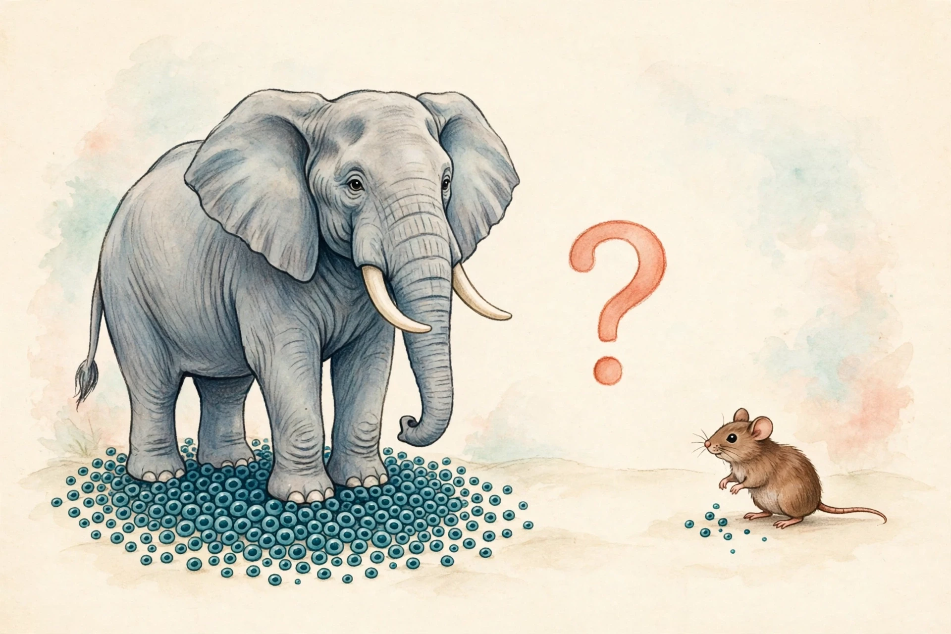
In the 1970s, the epidemiologist Richard Peto noticed something that should not be true. Every cell division is a chance for a mutation, so an animal with vastly more cells, dividing over a much longer life, should develop the disease far more often than a small, short-lived animal. An elephant has far more cells than a mouse and lives far longer. By the simple arithmetic of mutations, elephants should be riddled with tumours. They are not. Whales, the largest animals on Earth, show the same pattern. This is Peto's paradox, and it has puzzled biologists for half a century.
I first met the paradox in a lecture hall, and I remember the small jolt of it: a graph that went the wrong way, a piece of arithmetic that refused to add up. The lecturer put the numbers on the board and asked the room why the elephant was not the sickest animal alive. Nobody had an answer. A good paradox is a doorway, and this one opens onto the question of what defences evolution can build when it has millions of years to work.
11.2 Elephants and TP53: ~20 copies, cells twice as likely to self-destruct after radiation
Part of the answer, for elephants at least, seems to lie in TP53 — the guardian gene from section 3.4. Elephants carry about 20 copies of TP53: one canonical copy plus 19 retrogenes, extra copies created when the gene was copied back into the genome during evolution. Their cells respond to DNA damage by undergoing apoptosis about twice as readily as human cells do after radiation, according to a 2015 study in JAMA by Abegglen and colleagues — a hair-trigger self-destruct response that removes damaged cells before they can turn malignant.
That story comes with an honest caveat, and it matters. A 2022 re-evaluation published in Evolutionary Applications found that most of the elephant TP53 retrogenes are truncated, and questioned whether they spread through the elephant genome by natural selection at all — the extra copies may be a byproduct of other processes rather than an anti-cancer adaptation. The elephant's resistance is real, and the hair-trigger apoptosis is real, but the simple story — "more copies of the guardian gene, therefore less of the disease" — is debated, not settled. It is a good example of how science revises its favourite stories when it looks closer.
11.3 Naked mole-rats and bowhead whales — other resistance stories under study
Elephants are not the only animals under study. Naked mole-rats, small rodents that live for decades underground, almost never develop tumours, and researchers are studying their unusual cell biology for clues. Bowhead whales, which can live for more than 200 years, also show remarkably low rates of disease for their size, and their DNA repair is under study too. None of these animals offers a ready-made therapy. What they offer is proof of principle: evolution has solved this problem more than once, in different ways, and each solution is a clue about what defences a body can build.
12. Where the Science Is Heading
12.1 Liquid biopsies and multi-cancer early detection
One of the most active frontiers is finding the disease from a blood sample. Tumours shed fragments of their DNA into the bloodstream — circulating tumour DNA — and new tests can read those fragments for the mutations that mark a tumour. The bigger ambition is multi-cancer early detection: a single blood test that screens for signals from many types at once, years before symptoms. These tests are still being validated in large trials, and a positive signal still needs to be followed by imaging and diagnosis. A blood test that finds disease early is only useful if what it finds can be acted on.
12.2 Cancer vaccines and personalised neoantigen vaccines
Vaccines against the disease come in two very different kinds, and the distinction matters. Preventive vaccines, like the HPV and hepatitis B vaccines, stop the infections that cause disease. Therapeutic vaccines try to teach the immune system to recognise a tumour that already exists. The most personalised version of this idea is the neoantigen vaccine: scientists sequence a person's tumour, identify the mutated proteins unique to it, and build a vaccine — often using mRNA technology — that trains the immune system to attack exactly those targets. Early trials have shown that these vaccines can trigger immune responses against the tumour. The gene-editing and mRNA platforms behind these vaccines are the same ones described in CRISPR Gene Editing Explained. Like everything in this section, these are research directions with real data behind them, not promises.
12.3 AI in pathology and imaging
A third frontier is pattern-spotting at scale. Machine-learning systems trained on thousands of tissue slides and scans can help pathologists spot subtle patterns, grade tumours more consistently, and flag cases for a second look. AI tools are also being tested for reading mammograms and CT scans, and for predicting which mutations a tumour carries from the shape of its cells alone. The aim is not to replace the pathologist but to give them a second pair of eyes that never tires. The same technology raises familiar questions about validation, bias in training data and accountability when a machine misses something.
12.4 The honest close: progress is real and measurable, and the disease is still formidable
It is worth ending this section where the numbers began. Survival is up, deaths averted are counted in the millions, and treatments exist now — checkpoint inhibitors, targeted drugs, engineered T cells — that were science fiction a generation ago. At the same time, the disease still kills about 9.8 million people a year worldwide, cases are projected to rise to about 34.4 million a year by 2050, and many types remain very hard to treat once they have spread. There is no "cure around the corner" in this article, because there is no single cure to be around a corner: there are more than 100 diseases, each with its own genetics, and progress comes type by type, drug by drug, patient by patient. That is slower than a headline, and it is also the reason the progress is real.
Conclusion
Back in that room, the word hung in the air and nobody knew what to say next. I think about that room whenever I return to this topic — and I keep returning to it, out of curiosity and out of respect for the people living with the disease, because understanding a thing is the only honest way I know to make it less frightening. The biology does not make the word easy. Nobody who has heard it in a doctor's office would say it did. But it does make the word less mysterious, and less mysterious is a genuine kind of help.
The through-line of this article is simple enough to carry out of it: cancer is our own biology with the brakes cut. Every cell already knows how to divide, how to move, how to build blood vessels and how to hide — the disease is what happens when those normal abilities are switched on at the wrong time, in the wrong place, without permission. And that is also why every advance in treatment is, at root, an advance in understanding normal cells. The checkpoints, the telomeres, the immune brakes, the guardian gene — all of them were discovered by studying healthy biology first, and each one became a treatment only after someone understood what it did when it worked properly.
The numbers give the story its shape: 20.6 million new cases a year worldwide, 2,114,850 in the United States in 2026, and against them a survival figure that has reached 70 percent for the first time, with 4.8 million deaths averted since 1991. The disease is common, formidable and ancient — and it is also increasingly understood, increasingly treatable, and increasingly survivable. The word in the room is still heavy. It is no longer a mystery.
Frequently Asked Questions
What is cancer?
In the National Cancer Institute's definition, it is a disease in which some of the body's cells multiply out of control and spread elsewhere in the body. It is not one disease but more than 100 related diseases, named for the tissue they start in and the cell type involved. A tumour becomes cancer when its cells invade nearby tissue and travel to distant organs.
What causes cancer?
Cancer starts when mutations accumulate in a cell's DNA, damaging genes that control growth, division and death. Those mutations come from copying errors, inherited changes, and exposures such as tobacco smoke, ultraviolet light, radiation and certain infections. Age is the biggest single risk factor. Most cases have no single cause — several factors combine over a lifetime.
How do genes affect cancer growth?
Genes control when a cell divides, when it stops and when it dies. Mutations can turn growth-promoting genes called oncogenes into stuck accelerators, or disable tumour suppressor genes such as TP53, the brakes. Only a small share — around 5 to 10 percent — of cases trace back to inherited mutations; most are acquired during life. Tumours typically carry 2 to 8 driver mutations.
How does cancer spread?
Spread, or metastasis, begins when cells break away from a tumour, invade nearby tissue and enter blood or lymph vessels. Most travelling cells die in transit, but a few survive, exit the vessel at a distant organ and grow into a new tumour. An estimated 90 percent of deaths involve metastasis, which is why finding the disease before it spreads matters so much.
Is cancer contagious?
No. Cancer itself cannot be caught from another person through touch, kissing, sharing food or any other contact. Some infections that raise risk can spread, though — for example, the human papillomavirus, hepatitis B and C, and Helicobacter pylori. Preventing or treating those infections lowers the risk of the cancers they are linked to.
Can cancer be prevented?
Not entirely, but a large share of cases is linked to modifiable factors — about 40 percent of new cases in the United States among adults aged 30 and older, according to a 2024 analysis. Population-level measures with the strongest evidence are not smoking, HPV and hepatitis B vaccination, taking part in screening, keeping a healthy weight, limiting alcohol and protecting skin from the sun. No measure guarantees prevention.
What is the survival rate for cancer?
Five-year relative survival for all cancers combined in the United States stands at 70 percent for diagnoses in 2015–2021, per the American Cancer Society's Cancer Statistics 2026 report — against 49 percent back in the mid-1970s. Survival varies enormously by type and stage, from above 90 percent for some early-stage diseases to very low for others.
Sources
American Cancer Society, Cancer Statistics 2026 press release — pressroom.cancer.org
Siegel, R. L. et al., Cancer Statistics, 2026, CA: A Cancer Journal for Clinicians — doi.org
Siegel, R. L. et al., Cancer Statistics, 2025, CA: A Cancer Journal for Clinicians — doi.org
Sung, H. et al., Global cancer statistics 2024: GLOBOCAN estimates of incidence and mortality worldwide for 34 cancers in 186 countries, CA: A Cancer Journal for Clinicians, 2026 — doi.org
Bray, F. et al., Global cancer statistics 2022: GLOBOCAN estimates of incidence and mortality worldwide (historical reference; superseded by GLOBOCAN 2024) — doi.org
World Health Organization, Cancer fact sheet — who.int
National Cancer Institute, What Is Cancer? — cancer.gov
Hanahan, D. & Weinberg, R. A., The Hallmarks of Cancer, Cell, 2000 — doi.org
Hanahan, D. & Weinberg, R. A., Hallmarks of Cancer: The Next Generation, Cell, 2011 — doi.org
Hanahan, D., Hallmarks of Cancer: New Dimensions, Cancer Discovery, 2022 — doi.org
Vogelstein, B. et al., Cancer Genome Landscapes, Science, 2013 — doi.org
Kuchenbaecker, K. B. et al., Risks of Breast, Ovarian, and Contralateral Breast Cancer for BRCA1 and BRCA2 Mutation Carriers, JAMA, 2017 — doi.org
Chaffer, C. L. & Weinberg, R. A., A Perspective on Cancer Cell Metastasis, Science, 2011 — doi.org
Abegglen, L. M. et al., Potential Mechanisms for Cancer Resistance in Elephants and Comparative Cellular Response to DNA Damage in Humans, JAMA, 2015 — doi.org
Sulak, M. et al., TP53 Copy Number Expansion Is Associated with the Evolution of Increased Body Size and an Enhanced DNA Damage Response in Elephants, eLife, 2016 — doi.org
Nunney, L., Cancer suppression and the evolution of multiple retrogene copies of TP53 in elephants: A re-evaluation, Evolutionary Applications, 2022 — doi.org
The Nobel Prize, Press release: The Nobel Prize in Physiology or Medicine 2018 — nobelprize.org
Kymriah development overview (journal version) — doi.org
Centers for Disease Control and Prevention, Tobacco and Cancer — cdc.gov
National Cancer Institute, Common Cancer Myths and Misconceptions — cancer.gov
World Health Organization, Cervical Cancer fact sheet — who.int
Sasieni, P. & Falcaro, M., Cervical cancer mortality trends following HPV vaccination in England, 2001–24, The Lancet, 2026;408:162–168 — doi.org
Tomasetti, C. & Vogelstein, B., Variation in cancer risk among tissues can be explained by the number of stem cell divisions, Science, 2015 — doi.org
Islami, F. et al., Proportion and number of cancer cases and deaths attributable to potentially modifiable risk factors in the United States, 2019, CA: A Cancer Journal for Clinicians, 2024 — doi.org
American Cancer Society, Cancer Facts & Statistics — cancer.org