
Introduction
The first time I held a tube of purified DNA, I was underwhelmed. After years of hearing about the molecule of life, I expected something dramatic. What I saw was a small, clear, slightly stringy drop at the bottom of a plastic tube. It looked like nothing. It looked like water that had gone slightly wrong.
Then I loaded it onto a gel, ran the current, and watched the bands appear. That clear drop resolved into a pattern — sharp, repeatable, readable. A pattern that could tell me which organism it came from and whether my experiment had worked or failed. I have never forgotten the gap between how ordinary DNA looks and how much information it holds. That gap is the subject of this guide to DNA structure and replication.
Every cell in your body carries about two metres of DNA, packed into a nucleus a few micrometres across. That DNA holds the instructions for building proteins, for running a cell, and for passing traits from one generation to the next. It does this with an alphabet of just four letters, a shape like a twisted ladder, and a copying machine so accurate that it makes only about one mistake per billion letters.
1. What DNA Actually Is
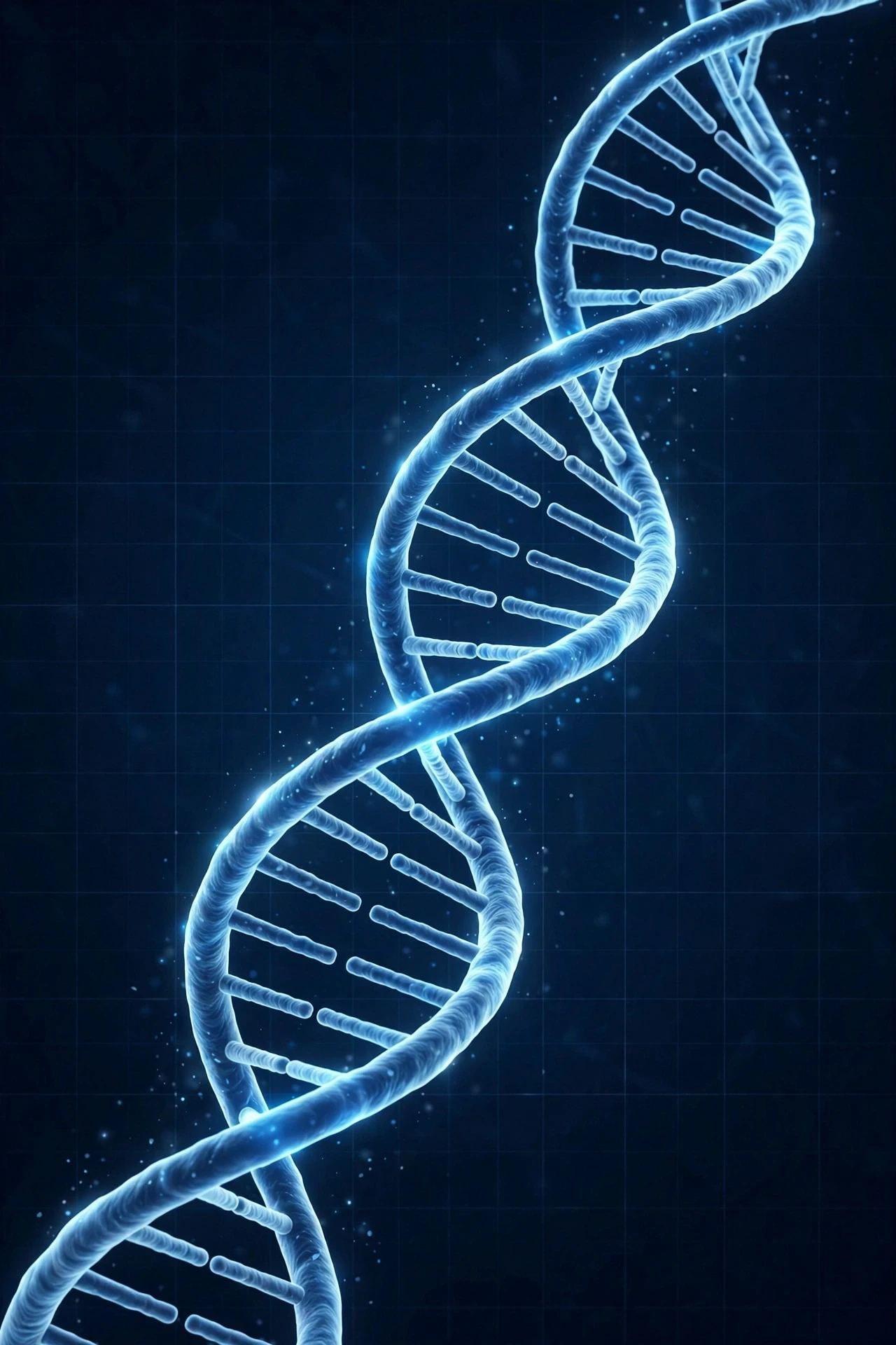
DNA is a molecule that stores information in chemical form. A recipe book stores information in ink. A hard drive stores it in magnetic patterns. DNA stores it in the order of chemical units along a long chain. Change the order, and you change the message.
1.1 Nucleotides: the four-letter alphabet
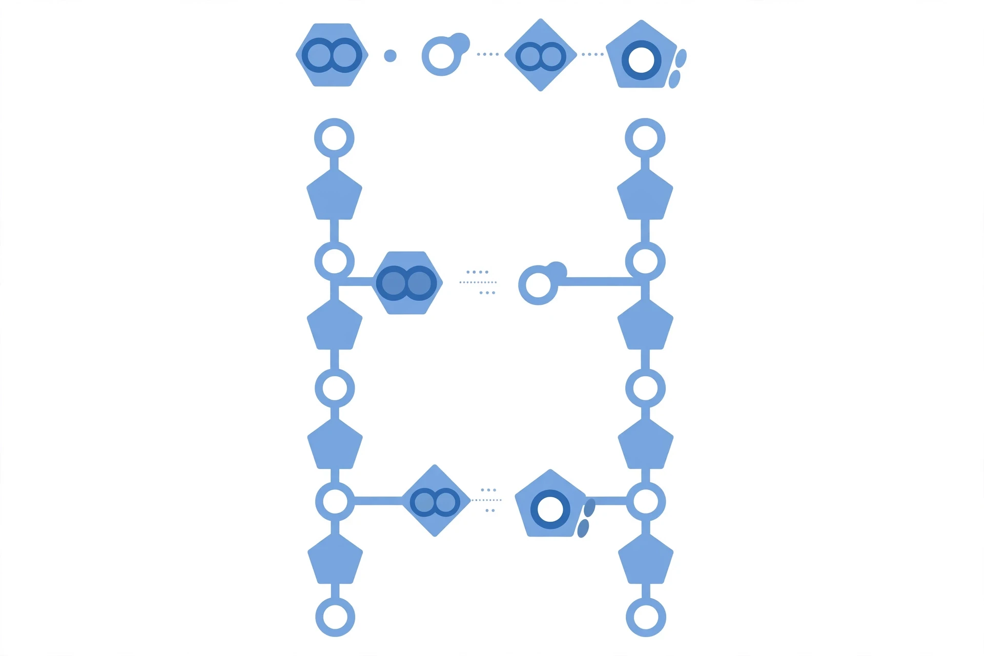
DNA is a polymer, a long chain made of repeating units called nucleotides. Each nucleotide has three parts: a phosphate group, a sugar (deoxyribose), and one of four nitrogen-containing bases. The phosphate and sugar form the backbone of the chain — the rails of the ladder. The bases stick out sideways, like rungs, and carry the information.
The four bases are adenine (A), thymine (T), guanine (G), and cytosine (C). A and G are purines, built from a double ring of atoms. T and C are pyrimidines, built from a single ring. A purine always pairs with a pyrimidine, so the width of the ladder stays constant.
The sugar in each nucleotide is deoxyribose, a five-carbon sugar. Its carbons are numbered 1' to 5', and those numbers give DNA its direction. One end of a strand is the 5' end, where a phosphate group sits on the 5' carbon. The other end is the 3' end, where a hydroxyl group sits on the 3' carbon. DNA is always read and built in the 5' to 3' direction.
I learned how much direction matters the hard way. Early in my bench work, I set up a reaction with a short DNA primer and waited for a product that never came. I checked the enzyme, the temperature, the reagents. Hours later, a colleague glanced at my notes and pointed at one line: I had written the primer sequence backwards. The letters were right. The direction was wrong. DNA does not forgive that mistake, because its enzymes read in one direction only.
1.2 How nucleotides link into a strand
Nucleotides join through phosphodiester bonds: the phosphate of one nucleotide links to the sugar of the next. The result is a sugar-phosphate backbone with bases hanging off it in a row. The backbone is the same all along the chain. The bases vary.
1.3 Chargaff's rules: the first clue to the shape
In the late 1940s, Erwin Chargaff measured the base composition of DNA from different organisms. He found a pattern that looked like a coincidence until it turned out to be a key. In any DNA sample, the amount of adenine roughly equals the amount of thymine, and the amount of guanine roughly equals the amount of cytosine. A = T, and G = C.
2. The Double Helix: DNA Structure in Three Dimensions
2.1 Watson and Crick, 1953
In 1953, James Watson and Francis Crick proposed the double helix model of DNA. They built on Chargaff's rules, chemical data on the bases, and X-ray diffraction images of DNA fibres — including the famous Photo 51, produced by Rosalind Franklin and Raymond Gosling, which showed the clear cross pattern of a helix. The model was simple, elegant, and it explained the biology in one stroke: two strands, wound around each other, with complementary bases facing inward.
2.2 Base pairing: A with T, G with C
The two strands are held together by hydrogen bonds between facing bases. Adenine pairs with thymine through two hydrogen bonds. Guanine pairs with cytosine through three. That difference makes G-C pairs slightly stronger than A-T pairs.
Base pairing is also the logic of copying. If you know one strand, you know the other: every A on one strand faces a T on the other, every G faces a C. Each strand is a template for its partner. That complementarity is the reason DNA can be copied so faithfully.
Base pairing is the whole trick. A with T, G with C — and from that one rule, copying, heredity, and the reading of genes all follow.
2.3 Antiparallel strands and the grooves
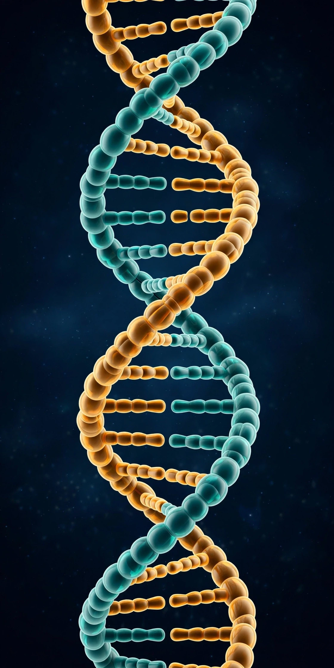
The two strands run in opposite directions — antiparallel. One runs 5' to 3' in one direction, the other runs 5' to 3' in the opposite direction. This is not a design flourish. It is a consequence of how the bases pair, and it has a direct consequence for replication: the two new strands are built in opposite directions.
The helix also has two grooves running along its surface: a wide major groove and a narrow minor groove. Proteins that read DNA reach into the major groove to "feel" the sequence of bases without unwinding the helix. DNA is read by touch as much as by sight.
3. From DNA to Chromosomes: How Two Metres Fit in a Nucleus
3.1 Histones, nucleosomes, and chromatin
The first layer of packaging is the nucleosome. About 147 base pairs of DNA wrap nearly twice around a core of eight histone proteins, like thread around a spool.
Nucleosomes coil into a thicker fibre, and that fibre loops and folds further, forming chromatin. Chromatin comes in two broad states. Euchromatin is loosely packed and generally accessible, so its genes can be read. Heterochromatin is tightly packed and generally silent.
3.2 Chromosomes: the final package
At cell division, chromatin condenses further into chromosomes — the compact, rod-shaped structures you see in textbook pictures. Humans have 46 chromosomes in most cells: 22 pairs of autosomes, plus two sex chromosomes, XX in most females and XY in most males. Each chromosome is a single, very long DNA molecule, plus its proteins.
3.3 Genes and the rest: what is non-coding DNA?
A gene is a stretch of DNA that carries instructions for a functional product — most often a protein, sometimes a functional RNA molecule.
Only a small fraction of the human genome — around one to two percent — directly codes for proteins. The rest is non-coding DNA. Some of it regulates genes. Some of it forms structural features, such as the centromere that holds sister chromatids together, or the telomeres that cap chromosome ends. Some of it has no known function yet — which is not the same as having no function.
The elements that make up DNA also connect it to the rest of chemistry. The bases are built from carbon, hydrogen, nitrogen, and oxygen, and the backbone adds phosphorus — the same elements whose patterns Sciory's Periodic Table of Elements: Structure & Trends lays out. DNA is not separate from chemistry. It is chemistry, organised into information.
4. DNA Structure and Replication: How DNA Copies Itself
4.1 Semi-conservative replication and the Meselson-Stahl experiment
When Watson and Crick proposed the double helix, they also proposed how it might copy itself: the two strands separate, and each serves as a template for a new partner strand. Each new double helix would then contain one old strand and one new strand. This is semi-conservative replication.
That was a beautiful idea, but it needed a test. In 1958, Matthew Meselson and Franklin Stahl provided it. They grew bacteria in a medium containing a heavy isotope of nitrogen, so the bacterial DNA became heavy, then switched the bacteria to normal, light nitrogen and let them divide. After one round of division, the DNA was of intermediate weight — exactly what semi-conservative copying predicts, with one heavy old strand and one light new strand in each molecule. The result ruled out the rival ideas: fully conservative copying, where the old double helix stays intact, and dispersive copying, where old and new material mix randomly along each strand.
I still use that experiment when I teach replication, because it shows something about good science. Meselson and Stahl did not argue from elegance. They designed a measurement that could distinguish between three clear predictions, and the data picked one.
4.2 The replication fork and the enzyme team
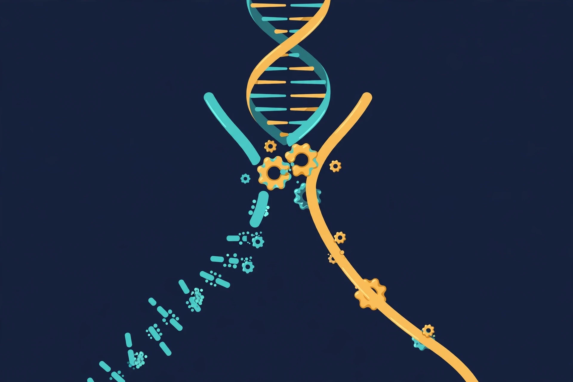
Replication begins at specific sites called origins of replication. At each origin, the two strands separate and a replication fork forms — a Y-shaped opening where the copying happens. In bacteria, there is usually a single origin per chromosome. In human cells, there are tens of thousands.
The enzymes at the fork each have a job:
Helicase unwinds the double helix, breaking the hydrogen bonds between base pairs and opening the fork. Topoisomerase relieves the strain ahead by cutting, swivelling, and rejoining the DNA.
Single-strand binding proteins coat the separated strands and keep them from snapping back together.
Primase lays down a short RNA primer, a starting point, because the main copying enzyme cannot start from nothing.
DNA polymerase builds the new strand, adding nucleotides one by one in the 5' to 3' direction, choosing each new base by complementarity.
DNA ligase joins fragments together, sealing the gaps between them.
4.3 How DNA polymerase works — and why it needs a primer
DNA polymerase is the workhorse of replication. It reads the template strand, picks the complementary nucleotide, and adds it to the growing strand. It also proofreads its own work, as Section 6 explains.
One limitation shapes the whole process: DNA polymerase can only add nucleotides to an existing 3' end. It cannot start a new strand from scratch. That is why primase lays down a short RNA primer first — a temporary starting handle, usually about ten nucleotides long, that polymerase can extend. The primer is later removed and replaced with DNA. Each incoming nucleotide arrives as a triphosphate, carrying energy in its phosphate bonds, and adding it to the chain releases that energy, driving the reaction forward.
4.4 The steps, in order
Replication follows a clear sequence at each origin:
Initiation: initiator proteins recognise the origin and open a small bubble in the helix.
Unwinding: helicase extends the bubble into two forks moving in opposite directions, with topoisomerase relieving the strain ahead.
Priming: primase lays down RNA primers on each template strand.
Elongation: DNA polymerase extends the primers, building new strands base by base.
Joining: ligase seals the gaps, and the primers are removed and replaced with DNA.
Termination: the forks meet, the two new double helices separate, and the cell checks the result before dividing.
5. Leading and Lagging Strands: The Problem of Direction
5.1 The lagging strand and Okazaki fragments
The cell solves this with a workaround. On the lagging strand, polymerase builds the new DNA in short pieces, each one built 5' to 3' away from the fork, starting from a fresh RNA primer each time. As the fork opens further, a new piece begins. These short pieces are Okazaki fragments, named after Reiji Okazaki, who discovered them. In human cells, they are roughly 100 to 200 nucleotides long.
Once the fragments are in place, the RNA primers are removed, the gaps are filled with DNA, and DNA ligase joins the fragments into one continuous strand. The finished lagging strand is identical in quality to the leading strand. It just takes more steps to get there.
Feature | Leading strand | Lagging strand |
|---|---|---|
Direction of synthesis | Continuous, following the fork | Discontinuous, in short fragments |
Number of primers | One per origin | Many, one per fragment |
Fragmentation | None | Okazaki fragments, joined by ligase |
Speed of completion | Faster to finish | Slower, more steps required |
Final product | One continuous new strand | One continuous new strand after joining |
5.2 Why this matters for accuracy
The lagging-strand workaround shows how biology trades elegance for reliability. A continuous copy would be simpler. But the chemistry of DNA polymerase does not allow it, so the cell accepts a more complicated process — fragments, primers, joining — and still achieves the same accuracy. The cost is time and energy. The benefit is that the copying rule, 5' to 3' only, is never broken.
6. Proofreading, Repair, and Mutations
6.1 Proofreading by DNA polymerase
The first layer is built into DNA polymerase itself. As it adds each nucleotide, it checks the fit. When it detects a mistake, it pauses, removes the wrong nucleotide, and tries again.
6.2 Mismatch repair and other repair systems
The second layer is mismatch repair. After replication, a separate team of proteins scans the new DNA for mismatches that proofreading missed. They can tell the new strand from the old one and cut out the wrong segment from the new strand, replacing it using the old strand as the template.
Beyond mismatch repair, the cell runs several other repair systems. Base excision repair fixes small, damaged bases. Nucleotide excision repair removes larger damaged stretches, such as the distortions caused by ultraviolet light. Double-strand break repair rejoins broken chromosomes. Peer-reviewed research has consistently shown that these systems are essential: when they fail, mutation rates rise, and the risk of disease rises with them.
6.3 What mutations are — and what they are not
A mutation is a change in the DNA sequence. Mutations can be tiny — a single base swapped for another — or large, involving the loss, duplication, or rearrangement of whole stretches of DNA. They can arise from copying errors that escape repair, from chemical damage, from radiation, or from the movement of mobile DNA elements.
Most mutations have no noticeable effect. They land in non-coding regions, or they change a codon to another codon for the same amino acid. Some mutations are harmful, disrupting a protein or its regulation. A few are beneficial, in a particular environment, and those are the raw material of evolution.
One more point matters here. Mutation is random in the sense that it does not happen because the organism needs it. It happens, and then selection acts on the results. That distinction — random mutation, non-random selection — is the core of Darwin's theory, and Sciory's Darwin's Theory of Evolution: Evidence Explained lays out the evidence for how variation and selection together shape species over time.
7. Prokaryotes vs Eukaryotes, and the Telomere Problem
7.1 Replication in bacteria
Bacteria usually carry a single circular chromosome. Replication starts at one origin and proceeds in both directions around the circle, with two forks meeting on the far side. Because the chromosome is circular, there is no "end" to copy.
7.2 Replication in eukaryotes
Eukaryotic cells — including human cells — carry multiple linear chromosomes, each with many origins of replication. Thousands of forks work at once, which is the only way to copy three billion base pairs in a few hours. Replication is licensed to happen once per cycle, and only once.
Feature | Prokaryotes (e.g. bacteria) | Eukaryotes (e.g. humans) |
|---|---|---|
Chromosome shape | Usually one circular chromosome | Multiple linear chromosomes |
Origins of replication | Usually one per chromosome | Many per chromosome |
Speed | Very fast, one origin suffices | Slower per fork, many forks in parallel |
Polymerases | Fewer types, mainly one for copying | Several, with specialised roles |
End problem | None, because the chromosome is circular | Present, solved by telomerase |
Cell-cycle control | Looser, linked to growth rate | Strict, once per cycle only |
7.3 Telomeres: the ends of the line
Linear chromosomes have a problem that circular ones do not. DNA polymerase needs a primer to start, and when the last primer at the very end of a chromosome is removed, there is nothing for polymerase to extend from. A short stretch of DNA is lost from the end with each round of replication.
The cell's answer is the telomere: a stretch of repeated DNA at each chromosome end that acts as a buffer. Telomeres shorten with each division, but the lost DNA is expendable — it is the buffer, not a gene. When telomeres become critically short, the cell usually stops dividing, a state called senescence.
Some cells solve the problem differently. Telomerase, an enzyme with its own RNA template, extends telomeres, adding back the repeats that replication loses. Telomerase is active in stem cells and germ cells, which need to divide many times. It is also active in most cancer cells, which is one reason they can keep dividing without limit. Sciory's Longevity and Healthy Aging: A Science-Based Guide covers how DNA damage and telomere shortening fit into the wider science of ageing.
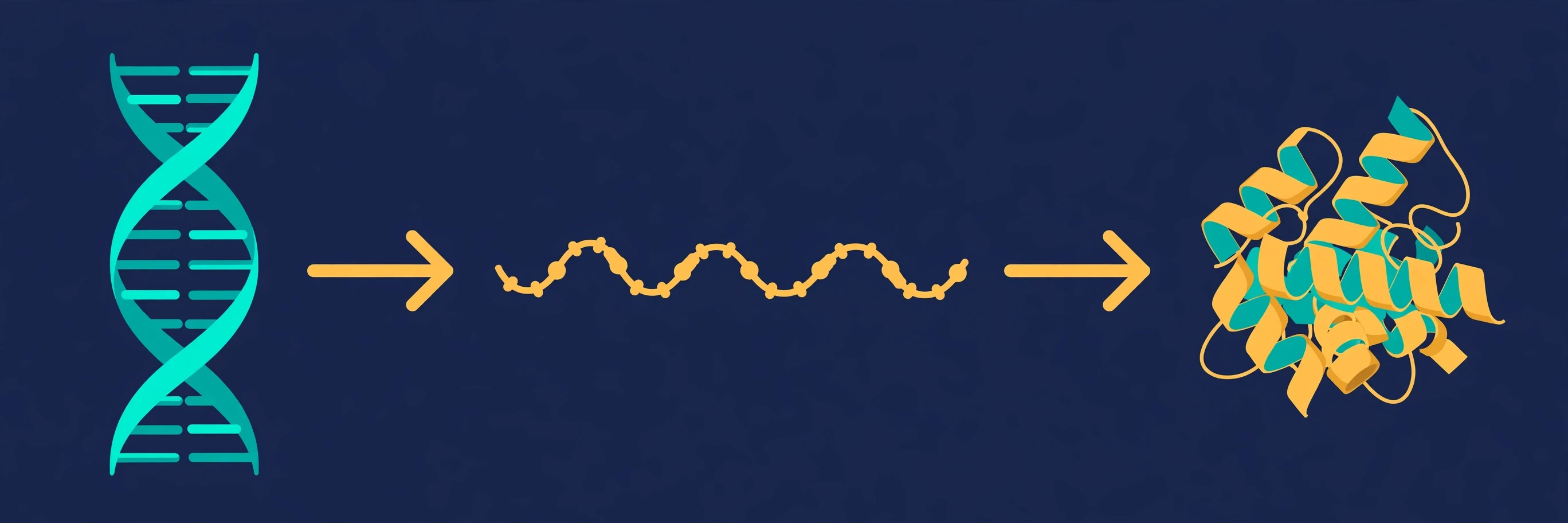
8. From DNA to Protein: The Central Dogma in Brief
8.1 Transcription: DNA to RNA
Transcription is the copying of a gene's sequence into a messenger RNA (mRNA) molecule. An enzyme called RNA polymerase unwinds a short stretch of DNA, reads one strand, and builds a complementary RNA strand. RNA uses the sugar ribose instead of deoxyribose, it is usually single-stranded, and it uses the base uracil (U) in place of thymine (T).
8.2 Translation: RNA to protein
Translation is the reading of mRNA to build a protein. It happens on ribosomes, molecular machines that move along the mRNA, reading it in groups of three bases called codons. Each codon specifies one amino acid, or a signal to start or stop. Transfer RNA (tRNA) molecules bring the right amino acid to each codon, and the ribosome links the amino acids into a chain.
The genetic code is nearly universal: the same codons mean the same amino acids in almost every organism on Earth, from bacteria to humans. That universality is strong evidence for a common origin of life.
8.3 Why DNA is not the whole story
The central dogma is a useful map, but it is not the whole territory. Some RNA molecules never become proteins — they act as regulators, or as parts of the ribosome itself. Some proteins modify DNA or RNA after they are made. And information can flow in other directions: some viruses copy RNA back into DNA, using an enzyme called reverse transcriptase. The dogma describes the main road, not every side street.
The same flow runs in every living thing, including plants. In photosynthesis, the proteins that capture sunlight are built from genes in the plant's DNA — and from a small separate genome inside the chloroplast itself, a relic of the ancient bacterium that became the chloroplast. Sciory's Photosynthesis Explained: Process & Equation covers the chemistry of that process.
9. Heredity: How DNA Moves from Parent to Child
9.1 Mendel, genes, and alleles
Mendel crossed pea plants that differed in single traits — seed colour, flower colour, plant height — and counted the results. Some traits disappeared in one generation and reappeared in the next; some appeared in a predictable three-to-one ratio. He proposed that traits are carried by discrete units, which he called factors, and that each organism carries two copies of each factor, one from each parent.
Today we call those factors genes, and the different versions of a gene alleles. You carry two alleles of each gene, one on each chromosome of a pair. If the two alleles are the same, you are homozygous for that gene. If they differ, you are heterozygous.
9.2 Dominant and recessive: what the words really mean
A dominant allele is one whose effect shows when it is present in a single copy. A recessive allele shows its effect only when two copies are present. Dominant does not mean common, and recessive does not mean rare or weak. It is a statement about how alleles interact in a heterozygote, nothing more.
9.3 Meiosis: halving the genome
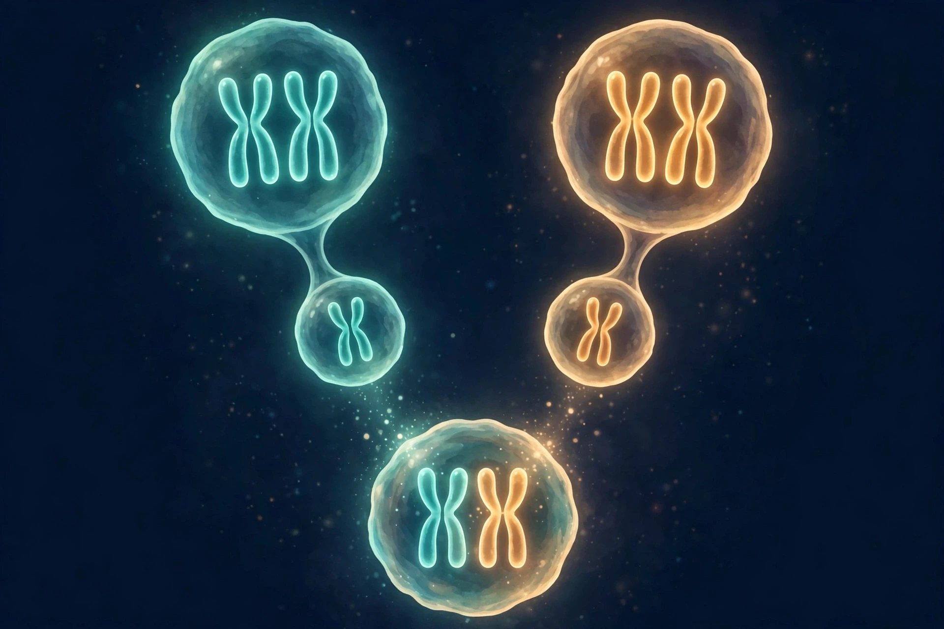
Ordinary cell division, mitosis, produces two identical daughter cells. Meiosis is different: it produces four cells, each with half the usual number of chromosomes. In the first division, homologous chromosomes separate. In the second, sister chromatids separate. The result is a gamete: a sperm or egg cell carrying one copy of each chromosome.
Two features of meiosis create genetic variation. First, independent assortment: which member of each chromosome pair goes to a given gamete is random. Second, crossing over: homologous chromosomes exchange segments before they separate, mixing alleles between the two parental chromosomes.
I once extracted DNA in a kitchen, with a group of students, using dish soap, salt, and cold alcohol on mashed strawberries. The DNA came out as white, stringy clumps that we could spool onto a wooden stick. One student stared at the clump and said, "That is in me?" It is. The same molecule, the same four letters, the same copying — running in a strawberry, in a student, and in every living thing we know.
9.4 Inheritance patterns
Different traits follow different inheritance patterns:
Autosomal dominant: one copy of the variant allele is enough to show the trait. Each child of an affected parent has a roughly one-in-two chance of inheriting it.
Autosomal recessive: two copies are needed. Two carriers can have an affected child, even though neither parent shows the trait.
X-linked: the gene sits on the X chromosome, so inheritance differs between sons and daughters.
Mitochondrial: the DNA in mitochondria comes almost entirely from the mother, so mitochondrial traits pass through the maternal line.
Polygenic: many genes contribute, each with a small effect, as with height or blood pressure.
9.5 DNA in medicine and forensics
DNA has practical uses beyond the textbook. In medicine, genetic testing can identify variants linked to inherited conditions and guide drug choices. In forensics, DNA profiling compares short repeated sequences that vary between people, producing a pattern that can link a sample to a person with very high confidence.
10. Common Misconceptions About DNA — Corrected
"DNA is destiny." Genes influence traits, but they rarely determine them alone. Environment, behaviour, and chance all shape the outcome. Identical twins share a genome, yet they differ.
"One gene equals one trait." Most traits involve many genes. The one-gene-one-trait idea is a teaching shortcut, not a law of nature.
"Mutations are always harmful." Most are neutral. Some are harmful. A few are helpful in a given environment, and those drive evolution.
"DNA never changes during your life." Your inherited DNA sequence is largely stable, but mutations accumulate in individual cells over time, and gene regulation changes constantly.
"Genes are the same as chromosomes." A chromosome is a long DNA molecule plus proteins. A gene is a stretch of DNA on that molecule. One chromosome carries many genes.
"Replication is perfect." It is extremely accurate, but not perfect. Proofreading and repair catch most errors, and the few that remain are the source of new variation.
10.1 Current research and open questions
Another open area is the brain. Genes shape brain development, and differences in gene regulation help explain why brain cells differ from other cells despite sharing the same genome. Sciory's Human Brain Anatomy and Functions: A Complete Guide describes the structures those genes build, from neurons to the networks that produce thought.
Conclusion: One Molecule, Two Jobs
DNA has two jobs, and its structure serves both. It stores information in a sequence of four bases, protected inside a double helix. And it copies that information, using each strand as a template for a new partner, with a team of enzymes that unwind, prime, build, proofread, and join.
The next time you hear that someone "has their mother's eyes," you will know what is actually being passed on: not a trait, but a sequence. A sequence that is read, regulated, and copied — three billion letters at a time, in every cell, for a lifetime.
Frequently Asked Questions
What is DNA structure and replication?
DNA structure and replication are two sides of the same process. DNA is a double helix of two antiparallel strands, held together by base pairing — A with T, G with C. Replication copies it by separating the strands and using each one as a template for a new partner strand, producing two identical double helices.
What are the four bases of DNA?
The four bases are adenine (A), thymine (T), guanine (G), and cytosine (C). A pairs with T through two hydrogen bonds, and G pairs with C through three.
How does DNA replication work?
Replication starts at origins, where helicase unwinds the helix and a replication fork forms. Primase lays down RNA primers, DNA polymerase builds new strands in the 5' to 3' direction, and ligase joins the fragments. The result is two double helices.
What is semi-conservative replication?
Semi-conservative replication means each new DNA molecule keeps one strand from the parent molecule and one newly made strand. The Meselson-Stahl experiment in 1958 confirmed this by showing that after one round of copying, DNA was of intermediate weight — one heavy old strand, one light new strand.
What is the difference between the leading and lagging strands?
The leading strand is built continuously, following the replication fork. The lagging strand is built in short Okazaki fragments, because DNA polymerase only works in the 5' to 3' direction and the lagging template runs the other way. Ligase later joins the fragments.
What are Okazaki fragments?
Okazaki fragments are short pieces of newly made DNA on the lagging strand, each starting from its own RNA primer. In human cells they are roughly 100 to 200 nucleotides long. After the primers are removed, DNA ligase joins the fragments together.
What is the role of DNA in heredity?
DNA carries hereditary information from parent to child. Meiosis halves the genome into gametes, fertilisation combines two gametes, and the resulting child carries a unique combination of alleles. Mendel's rules describe how those alleles are passed on.
What are telomeres and why do they matter?
Telomeres are repeated DNA sequences at the ends of chromosomes that act as a buffer against the loss of DNA during replication. They shorten with each division. Telomerase can rebuild them in stem cells, germ cells, and most cancer cells.
How accurate is DNA replication?
Replication is extremely accurate, with roughly one mistake per billion bases after proofreading and repair. DNA polymerase proofreads as it works, and mismatch repair fixes most remaining errors afterwards.
What is the central dogma of molecular biology?
The central dogma describes the flow of genetic information: DNA is transcribed into RNA, and RNA is translated into protein. It is the main route from gene to working molecule.