
1. Two Ways to Divide: A Cut Finger and a New Life
A few years ago I watched a student press a paper cut against her thumb and ask a question that stopped the whole class: "How does my skin know how to rebuild itself — and how is that different from how a baby gets half its chromosomes from each parent?" She had put her finger, quite literally, on the two great jobs of cell division.
Your body runs both jobs every day. When you cut your skin, scrape a knee, or simply grow, your cells copy themselves exactly. That is mitosis. When your body makes eggs or sperm, it does something far stranger: it deliberately halves the chromosome number and shuffles the genetic deck. That is meiosis. Mix the two up and the whole picture of heredity falls apart — yet students, and plenty of adults, do mix them up, because both processes use the same stage names: prophase, metaphase, anaphase, telophase.
This mitosis vs meiosis comparison untangles them properly. You will see each process step by step, count chromosomes the way a biologist counts them, compare the two side by side in a full table, and learn what happens when the machinery slips — from Down syndrome to cancer. By the end, you should be able to look at any diagram of dividing cells and say, with confidence, which process it shows and why.
The vocabulary is small, and that is the good news. A chromosome is a packaged length of DNA. A sister chromatid is one of the two identical copies of a chromosome after DNA replication, joined at a centromere. Homologous chromosomes are the matching pair — one inherited from each parent — carrying the same genes in the same order, but often different versions of those genes. Diploid (2n) means two sets of chromosomes; haploid (n) means one set. In humans, 2n = 46 and n = 23. Keep those four terms in your pocket and the rest follows.
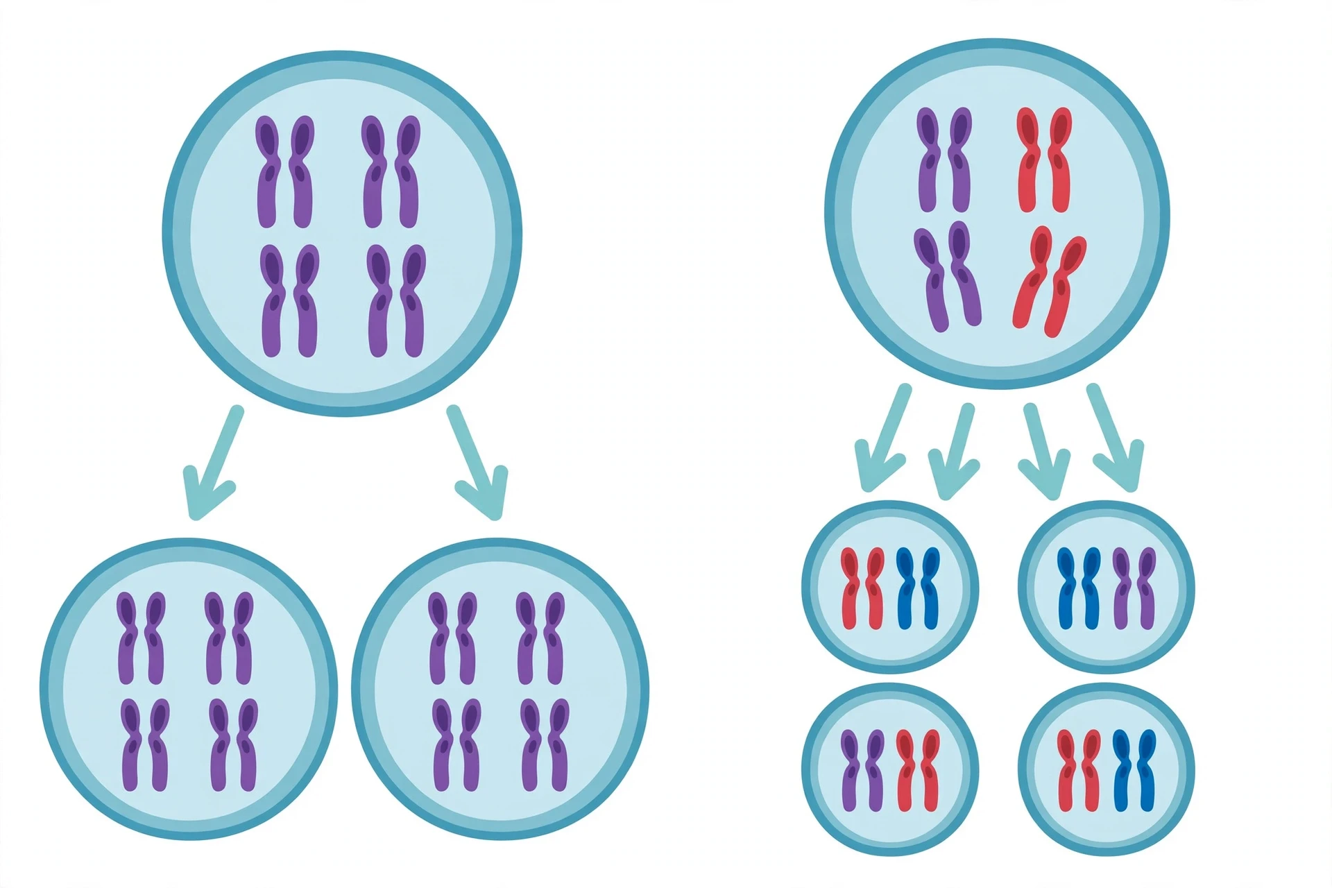
The one-sentence version: Mitosis copies a cell into two identical cells with the same chromosome number; meiosis halves the chromosome number and shuffles genes to make four genetically different sex cells.
If you want the background on what a cell actually contains before it divides — nucleus, cytoplasm, organelles — our guide to cell biology covers it in full. Here we will take only what division needs and move on.
2. Before Either Division: The Cell Cycle and the Copying Step
Neither mitosis nor meiosis starts from nothing. Both are preceded by interphase, the long working life of the cell, divided into G1 (growth), S (DNA synthesis), and G2 (final preparation). During the S phase, every chromosome is copied, so each one now consists of two identical sister chromatids held together at the centromere.
Here is the first trap, and I have watched bright students fall into it for years: after DNA replication, the amount of DNA has doubled, but the number of chromosomes has not. A replicated chromosome still counts as one chromosome, because we count chromosomes by centromeres, not by arms. A human cell in G2 has 46 chromosomes, each made of two chromatids — 92 chromatids in total, but still 46 chromosomes. Only when the sister chromatids separate does the count of chromosomes change. Hold on to that rule; it is the key to every chromosome-counting question you will ever meet.
Both processes also share the same basic machinery: the mitotic spindle, built from microtubules, which attaches to chromosomes at protein structures called kinetochores and pulls them apart. The spindle is the same tool used in two very different jobs, like a crane that can either lift an identical copy of a bridge into place or dismantle half a bridge and ship it away.
The cell cycle itself is tightly policed. Checkpoints at the end of G1, at the G2/M boundary, and during metaphase ask whether the DNA is intact, fully copied, and properly attached to the spindle. If the answer is no, the cell pauses or, in serious cases, destroys itself. As the OpenStax textbook Biology 2e explains, the cell cycle is "an ordered series of events involving cell growth and cell division that produces two new daughter cells," and those checkpoints are what keep the order from becoming chaos.
3. Mitosis: The Faithful Copy
3.1 What mitosis is
Mitosis is the division of a cell's nucleus into two nuclei with identical sets of chromosomes, usually followed by cytokinesis, the division of the cytoplasm. One diploid parent cell becomes two diploid daughter cells, each with the same chromosome number as the parent — in humans, 46 stays 46. The National Human Genome Research Institute puts it with a lovely image: think of mitosis as copying an instruction manual, page by page, and handing one complete copy to each of two people. Every page must arrive, exactly once, with no misspellings.
Mitosis is the workhorse of your body. It builds you from a single fertilized egg into trillions of cells. It replaces the lining of your gut every few days, renews your skin roughly every month, and repairs a cut by filling the gap with fresh, identical cells. It is also how many organisms reproduce asexually — a single-celled yeast budding, a strawberry plant sending out a runner, a starfish regrowing an arm.
3.2 The stages of mitosis: PMAT
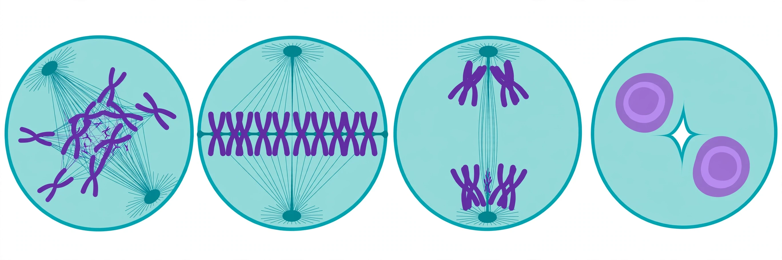
Mitosis runs through four named stages, remembered by the mnemonic PMAT: prophase, metaphase, anaphase, telophase. In textbooks they look like neat boxes; in a real cell they flow into one another, and the boundaries are a teaching convenience. I learned this the hard way in my first microscopy lab, staring at an onion root tip slide and trying to sort a hundred cells into four tidy piles. Half of them refused to cooperate. The cells in between stages were the lesson: division is a continuous process, and the names are signposts, not walls.
Prophase. The replicated chromosomes condense from loose chromatin into compact, visible X-shaped structures. The nucleolus disappears, the nuclear envelope begins to break down, and the centrosomes move to opposite poles, building the spindle between them.
Prometaphase. The nuclear envelope fragments fully. Kinetochores form at each centromere, and spindle microtubules from opposite poles capture them. Each chromosome is tugged until its two sister chromatids face opposite poles.
Metaphase. All the chromosomes line up along the metaphase plate, the cell's equator, each sister chromatid attached to opposite poles. This is the moment of maximum tension and maximum order — and the moment when a cell's chromosomes are easiest to photograph, which is why karyotypes, the chromosome portraits used in genetic testing, are made from cells arrested in metaphase.
Anaphase. The glue holding sister chromatids together, a protein complex called cohesin, is cut. The chromatids — now each a chromosome in its own right — are pulled rapidly toward opposite poles. Watching anaphase under a microscope for the first time is genuinely thrilling: after minutes of slow lining-up, everything suddenly moves at once.
Telophase and cytokinesis. The chromosomes arrive at the poles and decondense. Nuclear envelopes reform around each set, the spindle disassembles, and the cytoplasm divides — by a cleavage furrow pinching inward in animal cells, or by a cell plate built across the middle in plant cells. Two daughter cells, each genetically identical to the parent, go their separate ways.
3.3 Where mitosis happens, and where it does not
Mitosis happens in somatic cells — the body cells — wherever growth or replacement is needed: skin, gut lining, bone marrow, plant root tips and shoot tips. It does not happen in the production of gametes, and it does not happen in cells that have permanently left the cycle. Mature nerve cells and heart muscle cells sit in a resting state called G0 and rarely divide at all, which is one reason brain and heart injuries heal so poorly. The same machinery that builds you can also betray you: when the checkpoints that control mitosis fail, cells divide without permission, and that uncontrolled division is the core of cancer.
4. Meiosis: The Halving and the Shuffle
4.1 What meiosis is
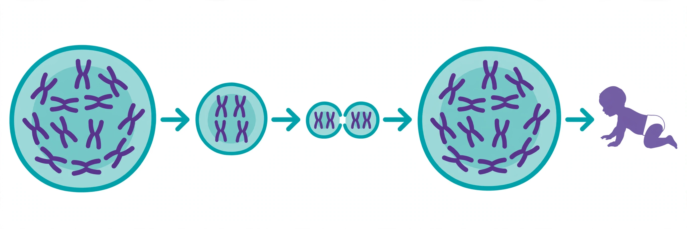
Meiosis is a specialized form of cell division that produces sex cells — gametes — with half the chromosome number of the parent cell. One diploid cell (46 chromosomes in humans) goes through one round of DNA replication and then two rounds of division, producing four haploid cells (23 chromosomes each). When two gametes fuse at fertilization, the diploid number is restored: 23 + 23 = 46.
Without meiosis, the numbers would double every generation — 46, then 92, then 184 — and heredity would collapse within a few generations. Meiosis is the accounting trick that keeps the chromosome number constant across generations while shuffling the genes within it. The National Human Genome Research Institute describes it plainly: during meiosis, "each diploid cell undergoes two rounds of division to yield four haploid daughter cells — the gametes."
Meiosis happens only in the reproductive organs — the testes and ovaries in animals, the anthers and ovaries in flowering plants. A cell that enters meiosis has chosen a different career from its mitotic neighbours, and it will never go back.
4.2 Meiosis I: the reduction division
Meiosis I is where meiosis earns its reputation as the strange one. It begins with an interphase identical in outline to the one before mitosis, then runs through its own prophase, metaphase, anaphase, and telophase — but the events inside those stages are different in the ways that matter most.
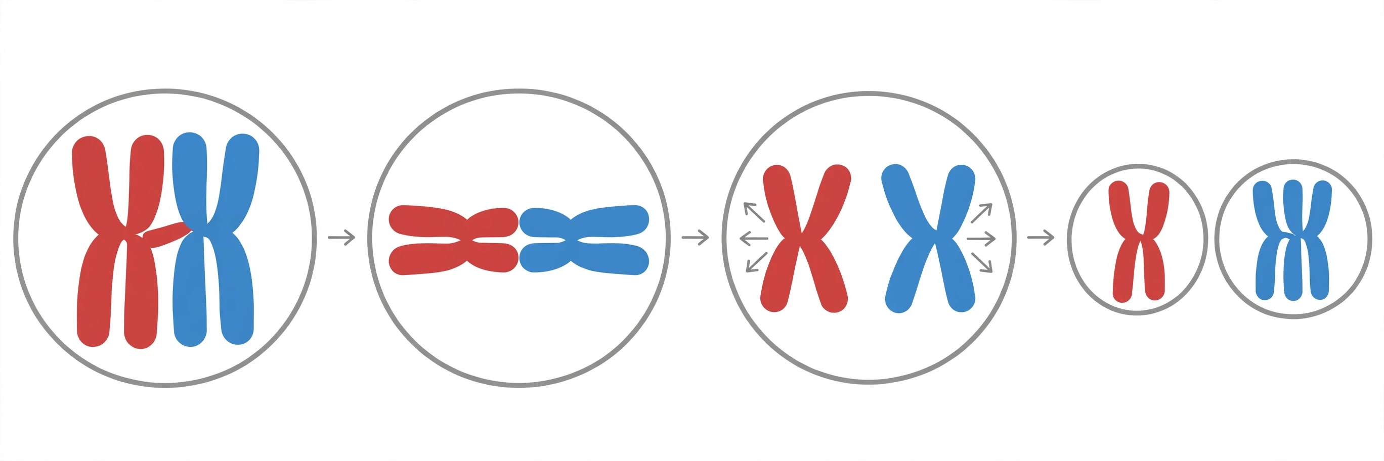
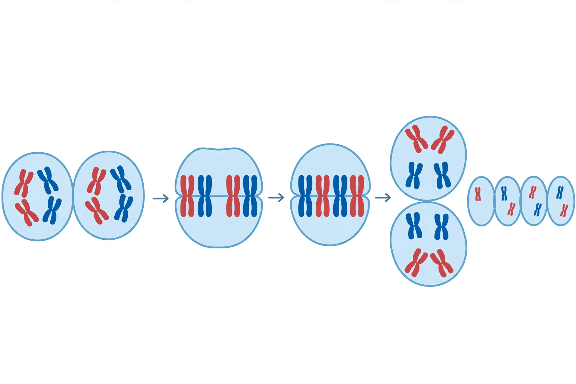
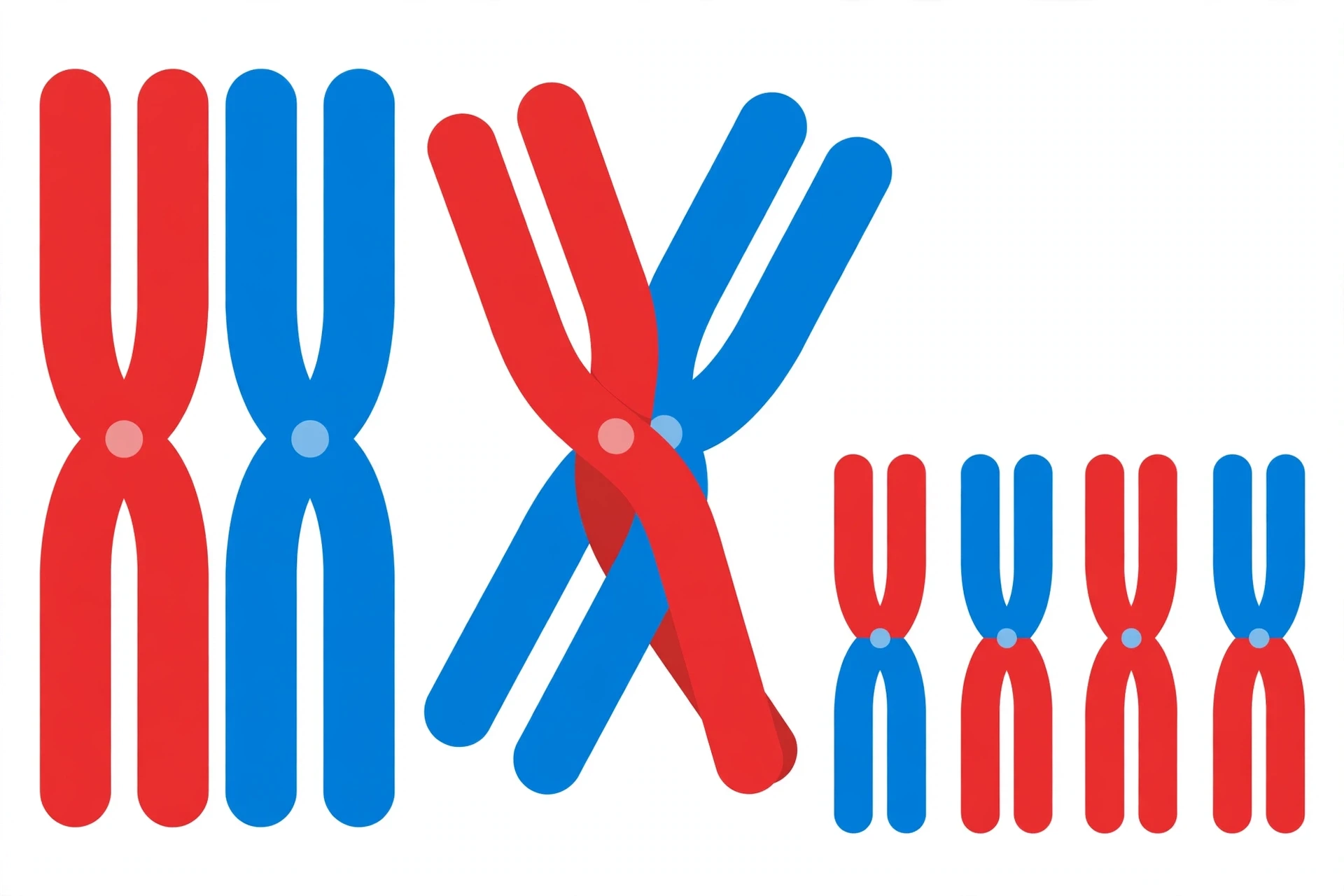
Prophase I is the longest and most eventful phase of either process, and it has five sub-stages of its own: leptotene, zygotene, pachytene, diplotene, and diakinesis. Two things happen here that never happen in mitosis. First, synapsis: homologous chromosomes find each other and pair up along their full length, held together by a protein lattice called the synaptonemal complex, forming a four-chromatid structure called a tetrad or bivalent. Second, crossing over: non-sister chromatids of the paired homologs swap segments of DNA at points called chiasmata. The swapped segments carry different versions of genes, so the chromatids that emerge are new combinations that never existed before — part maternal, part paternal.
I still remember the first time a student asked me why crossing over matters if the genes are "the same genes in the same order." It matters because the versions differ. Your mother and father each gave you a copy of the gene for, say, eye colour regulation, but the copies are not identical. Crossing over stitches a maternal version and a paternal version onto the same chromatid, creating a combination no ancestor ever carried. That is new genetic material in the most literal sense.
Metaphase I. The tetrads line up at the metaphase plate — but as pairs, not as individual chromosomes. Each pair orients randomly: the maternal homolog may face one pole or the other, independently of every other pair. This is independent assortment, the second great shuffling mechanism. With 23 pairs in humans, there are 2^23 — over eight million — possible combinations from assortment alone, before crossing over adds more.
Anaphase I. The homologous chromosomes separate and move to opposite poles. Crucially, the sister chromatids stay together — the centromeres do not split yet. This is the reduction step: each pole now receives one set of chromosomes, so the cell has gone from diploid to haploid in terms of chromosome sets, even though each chromosome still consists of two chromatids.
Telophase I and cytokinesis. Two haploid cells form. There may be a brief pause, called interkinesis, but there is no DNA replication between meiosis I and meiosis II. That absence is the whole point: the second division will separate chromatids without copying them first.
4.3 Meiosis II: the equational division
Meiosis II looks like mitosis, and that resemblance is a trap for the unwary. The two haploid cells from meiosis I each divide again, through prophase II, metaphase II, anaphase II, and telophase II. In metaphase II the chromosomes line up individually at the equator, and in anaphase II the centromeres finally split and the sister chromatids separate to opposite poles. Telophase II and cytokinesis produce four haploid cells.
The difference from mitosis is the starting material: meiosis II begins with haploid cells and no new DNA replication, so it ends with four haploid cells instead of two diploid ones. Think of meiosis I as sorting the pairs into two piles, and meiosis II as splitting each item in the pile into its two copies. The first division halves the sets; the second separates the copies.
4.4 How meiosis creates genetic variation
Three mechanisms, working together, make it almost certain that no two gametes from one person are genetically identical:
Crossing over in prophase I swaps segments between homologous chromosomes, creating recombinant chromatids.
Independent assortment in metaphase I orients each tetrad randomly, producing over eight million possible chromosome combinations in humans.
Random fertilization — which of the millions of possible sperm meets which of the millions of possible eggs — multiplies the variation further.
OpenStax states the result directly: given crossing over and independent assortment, "it is highly unlikely that any two haploid cells resulting from meiosis will have the same genetic composition." That variation is the raw material of evolution, and it is also why siblings, who share the same parents, are not identical. The one exception proves the rule: identical twins come from a single fertilized egg dividing by mitosis, not from two separate meiotic products.
4.5 From gametes to a new individual
In human males, meiosis produces four functional sperm from each starting cell, continuously from puberty onward. In human females, the story is different and more poignant: meiosis begins before birth, pauses in prophase I for years or decades, resumes at ovulation, and pauses again at metaphase II until fertilization. A woman is born with her lifetime supply of eggs already partway through meiosis. The long pause is one reason the risk of chromosome segregation errors rises with maternal age — a thread we pick up in Section 8.
Fertilization restores the diploid number, and the resulting zygote divides by mitosis to build the new individual. The two processes hand off to each other like relay runners: meiosis halves and shuffles, fertilization restores, mitosis builds. For the genetics behind what those shuffled genes actually do, see our guide to genetics, and for the molecule being copied and shuffled, our guide to DNA.
5. Mitosis vs Meiosis: The Complete Comparison Table
This is the table students ask for most, so here it is in full. Read it row by row, and notice how many rows trace back to a single decision: one division or two.
Feature | Mitosis | Meiosis |
|---|---|---|
Purpose | Growth, repair, replacement of cells, asexual reproduction | Production of gametes (sex cells) for sexual reproduction |
Where it happens | Somatic (body) cells throughout the body | Germ cells in reproductive organs only (testes, ovaries) |
Number of divisions | One | Two (meiosis I and meiosis II) |
DNA replication | Once, before division | Once, before meiosis I; none between meiosis I and II |
Daughter cells produced | 2 | 4 |
Chromosome number in daughter cells | Same as parent: diploid (2n); 46 in humans | Half the parent: haploid (n); 23 in humans |
Genetic identity of daughter cells | Genetically identical to the parent (barring mutation) | Genetically different from the parent and from each other |
Pairing of homologous chromosomes | No pairing (synapsis does not occur) | Yes — pairing (synapsis) in prophase I, forming tetrads |
Crossing over | Does not normally occur | Occurs in prophase I between non-sister chromatids |
What lines up at the metaphase plate | Individual duplicated chromosomes | Pairs of homologous chromosomes (metaphase I); individual chromosomes (metaphase II) |
What separates in the first anaphase | Sister chromatids | Homologous chromosomes (anaphase I); sister chromatids separate only in anaphase II |
Genetic variation created | Essentially none (except new mutations) | Extensive — crossing over, independent assortment, random fertilization |
Length of prophase | Relatively short | Prophase I is long and complex, with five sub-stages |
Role in the life cycle | Builds and maintains the body | Produces gametes; keeps chromosome number constant across generations |
Errors can lead to | Uncontrolled division (cancer), mosaicism, tissue defects | Aneuploidy — e.g. Down syndrome, Turner syndrome, Klinefelter syndrome |
6. What Mitosis and Meiosis Share
The differences get the attention, but the similarities are real and worth naming, because they explain why the two processes were confused for so long after their discovery. Both begin with a single round of DNA replication in the S phase. Both use the same spindle machinery, the same kinetochores, and the same stage names — prophase, metaphase, anaphase, telophase. Both end with cytokinesis dividing the cytoplasm. Both are policed by checkpoints that can halt division if something is wrong.
Most people assume the two processes are opposites, but what we actually see is a shared toolkit with one crucial modification: meiosis runs the toolkit twice, with a special first division that separates homologs instead of chromatids. Meiosis is not a different machine; it is the same machine run in a different program. That is why meiosis II looks so much like mitosis — because, mechanically, it nearly is mitosis, performed on haploid cells.
7. Counting Chromosomes: The Skill That Unlocks Every Diagram
Chromosome-counting questions cause more lost exam marks than any other part of this topic, and the fix is a single rule repeated until it is automatic: count centromeres, not chromatids. A chromosome with two sister chromatids still counts as one chromosome. Two separate chromatids after separation count as two.
Walk through the human numbers:
G1 (before replication): 46 chromosomes, 46 chromatids, DNA content 2C.
G2 (after replication): 46 chromosomes, 92 chromatids, DNA content 4C. The DNA doubled; the chromosome number did not.
After mitosis: two cells, each 46 chromosomes, 46 chromatids, 2C.
After meiosis I: two cells, each 23 chromosomes, 46 chromatids, 2C — haploid in sets, but each chromosome still duplicated.
After meiosis II: four cells, each 23 chromosomes, 23 chromatids, 1C.
The most common error is to say a G2 cell has 92 chromosomes. It has 92 chromatids and 46 chromosomes. The second most common error is to say meiosis produces cells with "half the DNA" immediately after meiosis I. It produces cells with half the chromosome sets — the DNA per cell is still 2C until meiosis II splits the chromatids. Keep the two quantities separate — chromosome number and DNA content — and the diagrams stop being frightening.
8. When Division Goes Wrong: Errors and Disease
8.1 Nondisjunction: the failure to separate
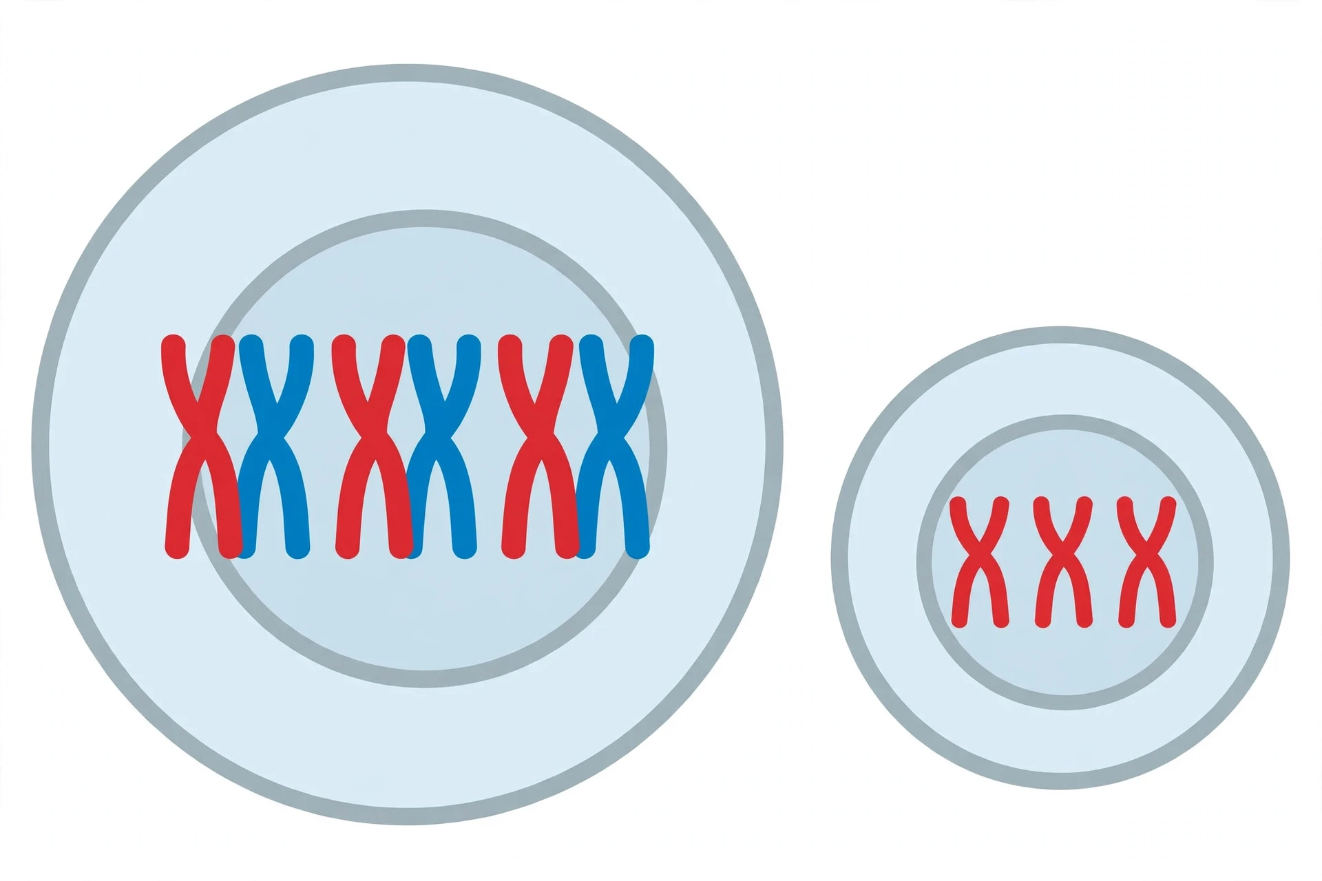
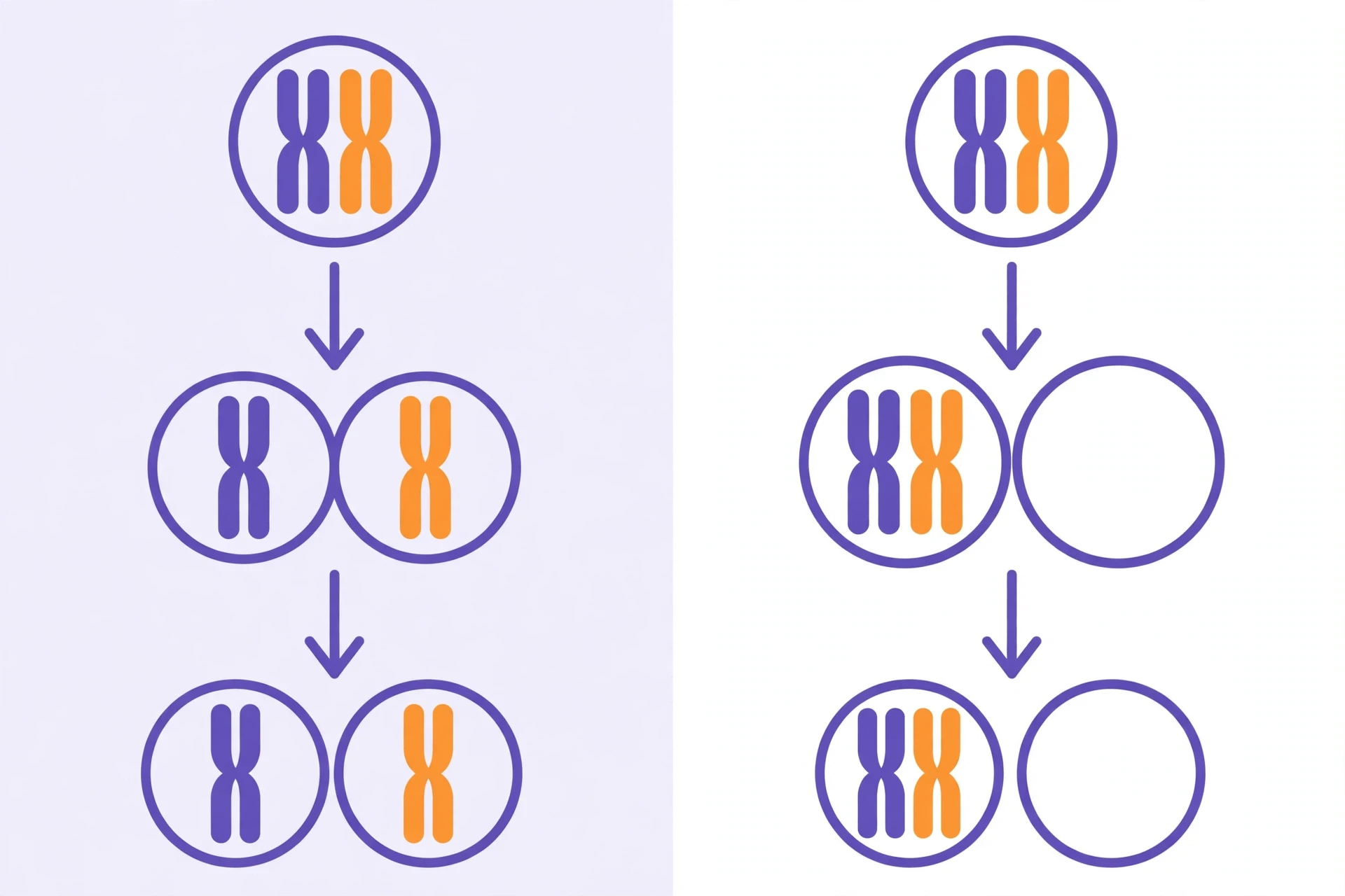
The most consequential error in either process is nondisjunction — the failure of chromosomes to separate properly. If homologous chromosomes fail to separate in meiosis I, or sister chromatids fail to separate in meiosis II (or in mitosis), the resulting cells get too many or too few chromosomes. The condition of having an abnormal chromosome number is called aneuploidy.
The best-known result is Down syndrome (trisomy 21): three copies of chromosome 21 instead of two, usually caused by nondisjunction of chromosome 21 during meiosis. The CDC reports that Down syndrome is the most common chromosomal condition diagnosed in the United States, with about 5,775 babies born with it each year, and that about 95% of cases are trisomy 21. The risk rises with maternal age, which is linked to the long pause in prophase I described earlier — the segregation machinery, assembled decades before it is used, becomes more error-prone with time.
Other aneuploidies follow the same logic: Turner syndrome (a single X chromosome, 45,X), Klinefelter syndrome (XXY), Edwards syndrome (trisomy 18), and Patau syndrome (trisomy 13). Most aneuploidies are not compatible with life, and many end in early miscarriage — a sobering reminder of how precise segregation has to be. Only a few, involving small chromosomes or the sex chromosomes, are compatible with survival.
8.2 Mitotic errors and cancer
Errors in mitosis are quieter but no less serious. If the spindle attaches incorrectly or the metaphase checkpoint fails, a daughter cell can gain or lose a chromosome. A single such error rarely causes cancer by itself, but the loss of a chromosome carrying a tumour-suppressor gene, or the gain of one carrying a growth-promoting gene, can push a cell toward uncontrolled division. Cancer is, at its root, mitosis without brakes: the checkpoints that should stop a damaged cell from dividing are broken, and the cell copies itself again and again. That is why so many cancer drugs target dividing cells, and why they carry such heavy side effects — they also strike the healthy cells that divide often, like those in bone marrow and gut lining.
8.3 Mosaicism and other mistakes
When nondisjunction happens in mitosis during early embryonic development, the result is mosaicism: some cells of the body carry the error and some do not. Mosaic Down syndrome, in which some cells have three copies of chromosome 21 and others have two, accounts for about 2% of Down syndrome cases and often produces milder features. Errors can also occur at the DNA level rather than the chromosome level — a copying mistake during S phase that escapes repair becomes a mutation, passed on to every descendant of that cell by mitosis, or to offspring if it occurs in the germline during meiosis.
9. Common Misconceptions — and What the Evidence Actually Shows
"Meiosis is just mitosis done twice." It is not. Meiosis I is fundamentally different from mitosis: homologs pair, cross over, and separate, and there is no second round of DNA replication. Only meiosis II resembles mitosis.
"Mitosis produces genetically identical cells, always." Almost always — but mutations during DNA replication mean daughter cells can differ slightly. "Identical" is a useful expectation, not a guarantee. The difference matters in cancer, where a mutation acquired in one cell is inherited by all its mitotic descendants.
"Crossing over happens in mitosis too." It does not, in any routine sense. The pairing of homologs that makes crossing over possible is a meiosis-only event.
"Meiosis happens in all your cells." No — only in germ cells in the reproductive organs. Your skin, liver, and bone marrow cells divide by mitosis alone. A student once told me she thought every cell in her body was "doing meiosis right now." I asked her what her skin cells would produce if they did. She laughed, and the point landed: if skin cells halved their chromosomes, the tissue would collapse within days.
"Humans have 46 chromosomes because 46 is somehow the right number." There is nothing universal about 46. Dogs have 78, fruit flies have 8, and some ferns have over a thousand. The number matters only in that it must stay constant within a species — and meiosis is the mechanism that keeps it constant.
10. Why the Difference Matters in Real Life
10.1 Medicine and fertility
Fertility medicine lives inside this topic. In vitro fertilization (IVF) clinics screen embryos for aneuploidy before implantation, because a wrong chromosome number is a leading cause of failed implantation and miscarriage. Karyotyping — photographing a person's chromosomes from cells arrested in metaphase — remains a front-line diagnostic test for suspected chromosomal conditions. And cancer treatment, as noted above, is largely a fight over the control of mitosis.
10.2 Agriculture and breeding
Plant breeders exploit meiosis deliberately. Crossing over and independent assortment are the engines of variation that breeders select from; polyploid crops such as wheat, which carries multiple chromosome sets, exist because of meiotic quirks harnessed over thousands of years. Understanding meiosis is also how breeders produce seedless fruit: a triploid plant, with three sets of chromosomes, cannot complete meiosis properly and so produces no viable seeds.
10.3 Forensics, ancestry, and the microbiome of ideas
DNA fingerprinting and ancestry testing both depend on meiotic variation — the shuffling that makes your genome a unique combination of your parents' genomes. Even microbiology connects here: many single-celled organisms reproduce by mitosis-like division alone, and the absence of meiotic shuffling shapes how quickly they can adapt — and how quickly antibiotic resistance spreads through a population of identical clones.
11. Latest Research: What Scientists Are Still Learning
The story is not finished. In the last few years, researchers have been mapping the machinery of meiosis at a resolution that would have astonished its discoverers. A few directions worth knowing:
The synaptonemal complex in atomic detail. Structural biologists have resolved the protein lattice that holds homologous chromosomes together during prophase I, revealing how it assembles, how it positions crossover sites, and why at least one crossover per chromosome pair is needed for proper separation.
Why eggs make more errors with age. Research on the long prophase I arrest in human eggs has traced age-related aneuploidy to the gradual weakening of cohesin — the protein glue holding sister chromatids together — over decades, and to the deterioration of the spindle machinery. This is an active area of fertility research.
Meiosis in the lab. Scientists have produced functional mouse eggs and sperm from stem cells by guiding them through meiosis in vitro, a breakthrough with profound implications for fertility treatment and for understanding the basic rules of the process. Human applications remain distant and heavily regulated, but the principle is established.
Cancer and chromosome instability. Work on chromosomal instability in tumours has shown that many cancers do not just divide too much — they mis-segregate chromosomes at an elevated rate, generating the genetic diversity that lets tumours evolve resistance to treatment. Drugs that target the mitotic spindle or the spindle checkpoint are a major research frontier.
The field agrees on the core story — one division that copies, two divisions that halve and shuffle — and argues, productively, about the machinery in between. That is how science is supposed to work.
12. How to Remember It: A Study Guide
If you take one memory aid from this article, take this one: mitosis maintains, meiosis halves. One division, two identical cells, same number. Two divisions, four different cells, half the number. Everything else is detail that hangs off that frame.
A few more practical tricks:
PMAT for the mitotic stages: Prophase, Metaphase, Anaphase, Telophase. For meiosis, the same letters with a I or II after them.
"Homologs separate in meiosis I, sisters separate in meiosis II — and in mitosis, sisters separate." Ask "what is separating?" at every stage, and most diagrams solve themselves.
Draw it once. Take a cell with two chromosome pairs (2n = 4), draw it in colour — one parent's chromosomes red, the other's blue — and walk it through mitosis and through meiosis on the same page. You will understand more from that one drawing than from a week of rereading notes. I make my students do exactly this, and the ones who do it stop confusing the two processes.
Count centromeres, not chromatids. Say it aloud every time you count.
13. The Takeaway
Mitosis and meiosis are the two answers to the same problem: how to pass genetic instructions onward without losing them or doubling them. Mitosis answers by copying faithfully — one cell becomes two identical cells, and the body grows, heals, and renews itself. Meiosis answers by halving and shuffling — one cell becomes four different cells, and a new generation begins with the right chromosome number and a genome that has never existed before.
A cut finger healing and a child resembling both parents and neither exactly are the same story told twice: careful copying on the one hand, careful variation on the other. Once you can tell the two apart — one division or two, sisters separating or homologs separating, identical or different — the whole of heredity opens up. That is a good trade for a single afternoon of reading.
Frequently Asked Questions
1. What is the main difference between mitosis and meiosis?
Mitosis produces two genetically identical diploid daughter cells for growth and repair through one division, while meiosis produces four genetically different haploid daughter cells for sexual reproduction through two divisions. Mitosis maintains the chromosome number; meiosis halves it.
2. How many daughter cells do mitosis and meiosis produce?
Mitosis produces two daughter cells. Meiosis produces four daughter cells. In humans, the two cells from mitosis each have 46 chromosomes, while the four cells from meiosis each have 23 chromosomes.
3. Do mitosis and meiosis have the same stages?
Both use the same stage names — prophase, metaphase, anaphase, and telophase — but the events differ. Meiosis runs through those stages twice (meiosis I and meiosis II), and prophase I includes synapsis and crossing over, which never happen in mitosis.
4. Where do mitosis and meiosis occur in the human body?
Mitosis occurs in somatic (body) cells throughout the body — skin, gut lining, bone marrow, and anywhere growth or repair is needed. Meiosis occurs only in the reproductive organs: the testes in males and the ovaries in females, where it produces sperm and eggs.
5. Why does meiosis have two divisions?
The first division (meiosis I) separates homologous chromosomes and halves the chromosome number. The second division (meiosis II) separates sister chromatids. Two divisions are needed because the cell must both reduce the chromosome number and distribute the copied chromatids, with only one round of DNA replication before them.
6. What is crossing over, and when does it happen?
Crossing over is the exchange of DNA segments between non-sister chromatids of homologous chromosomes. It happens during prophase I of meiosis, and it creates recombinant chromatids with new combinations of maternal and paternal genes. It does not normally occur in mitosis.
7. What is the chromosome number after mitosis and meiosis in humans?
After mitosis, each daughter cell has 46 chromosomes (diploid, 2n), the same as the parent. After meiosis, each daughter cell has 23 chromosomes (haploid, n), half the parent's number. Fertilization restores 46.
8. What happens if chromosomes fail to separate during meiosis?
The failure, called nondisjunction, produces gametes with too many or too few chromosomes. If such a gamete is fertilized, the result is aneuploidy — for example, trisomy 21 (Down syndrome), in which there are three copies of chromosome 21 instead of two.
9. Is meiosis II the same as mitosis?
Very similar, but not identical. Meiosis II resembles mitosis because individual chromosomes line up and sister chromatids separate, but it starts with haploid cells and involves no new DNA replication, so it ends with haploid cells rather than diploid ones.
10. Which is longer, mitosis or meiosis?
Meiosis takes much longer, mainly because prophase I is a long and complex stage with five sub-stages (leptotene, zygotene, pachytene, diplotene, diakinesis). In human females, meiosis can even pause in prophase I for decades before completing.