
Introduction
The first cell I ever saw, in my first cell biology class, was not impressive. It was a thin slice of onion skin on a school microscope slide, and for a long minute all I could see was my own eyelash reflected in the glass. Then the focus knob caught, the blur sharpened, and a grid of little boxes snapped into view — neat, empty-looking rooms, wall to wall. I remember thinking: that is it? That is what living things are made of?
Years later, in a university teaching lab, I watched a student have the same moment with a drop of her own cheek cells, stained pale blue. She laughed out loud. "They look like fried eggs," she said. And then, as the lecturer talked her through what she was seeing — a nucleus holding two metres of DNA folded into a space six micrometres wide, a membrane deciding what came in and what stayed out, tiny power stations burning sugar for fuel — her face changed. The fried egg turned into a city.
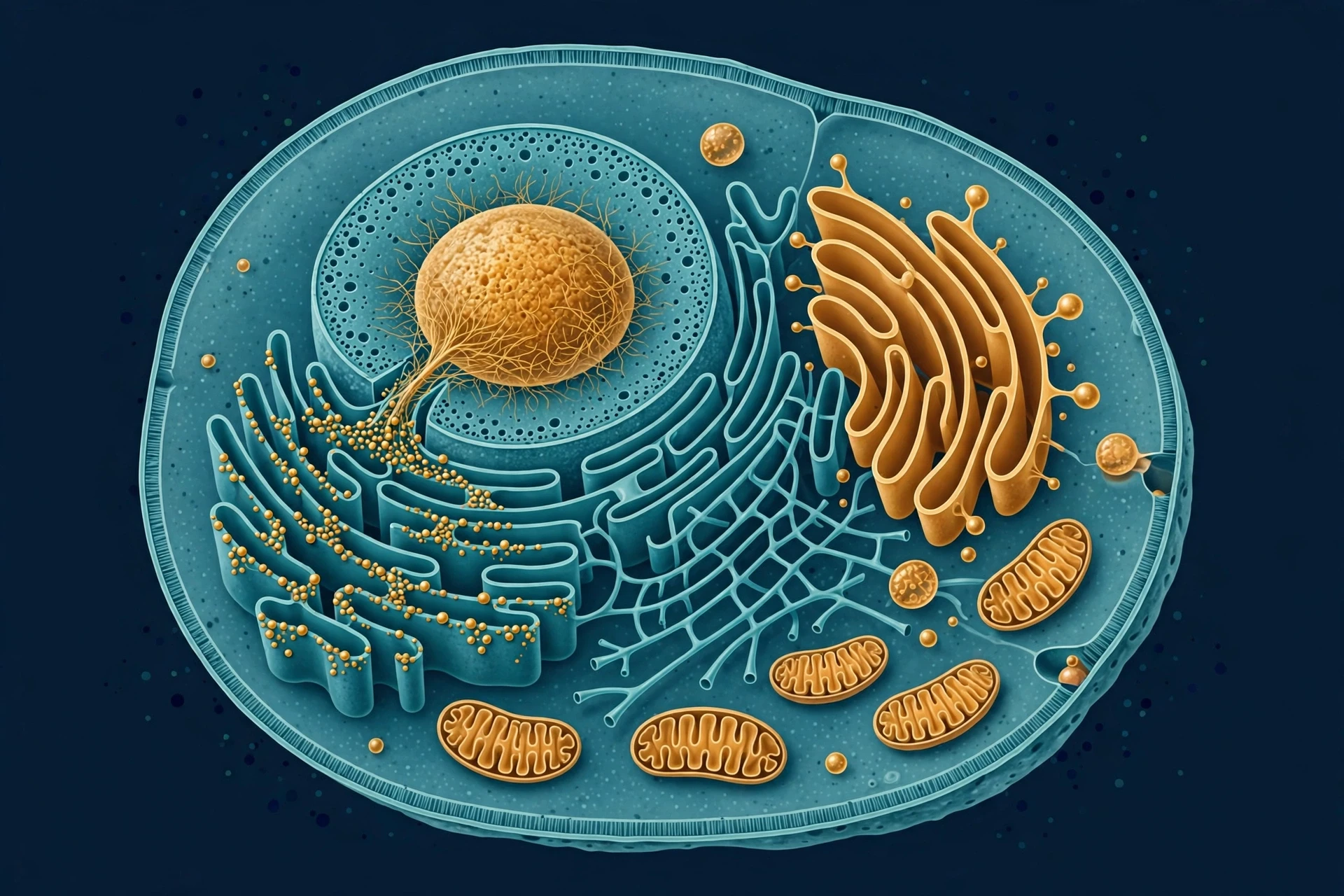
That is what cell biology does. It takes the smallest unit of life and shows you that it is not small at all. A single cell eats, breathes, builds, repairs, talks to its neighbours, and, when the time comes, divides or dies on schedule. Your body contains around 37 trillion of them, in more than 200 types, and every breath you take, every memory you keep, every wound that heals, is cell biology in action. This guide follows the story from the first microscopes to the newest research — the structure of cells, the organelles inside them, the processes that keep them alive, and what happens when those processes go wrong.
1. What Cell Biology Is — and Why One Cell Matters
Cell biology is the study of cells: their structure, their chemistry, how they work, and how they interact. It sits at the crossroads of almost every other branch of biology. Genetics asks what the DNA says; cell biology asks how the cell reads it. Medicine asks why a drug works; cell biology asks what it does inside the cell. Evolution asks how life changed over billions of years; cell biology shows the machinery that made that change possible.
1.1 The cell theory: three sentences that changed biology
By the late 1830s, two German scientists, Matthias Schleiden (a botanist) and Theodor Schwann (a zoologist), had compared notes and reached a startling conclusion: plants and animals, different as they look, are both built from cells. A few years later, Rudolf Virchow added the third piece — cells come only from other cells. Together, those ideas became cell theory, and it still stands today:
All living things are made of one or more cells. A bacterium is one cell. You are about 37 trillion.
The cell is the smallest unit of life. Nothing smaller than a cell is alive on its own. A virus, for example, cannot copy itself without hijacking a cell.
All cells come from pre-existing cells. Every cell in your body traces back, through an unbroken chain of divisions, to a single fertilised egg — and beyond that, through your parents, to the first cells on Earth.
I have taught cell theory to first-year students for years, and the third point is the one that always lands hardest. Sit with it for a moment: the cells dividing in your bone marrow right now are the direct descendants, by division, of cells that divided in your mother, and in her mother, in a chain that has never once been broken in roughly four billion years. You are not just made of cells. You are the latest link in the longest unbroken chain on the planet.
1.2 What every cell has in common
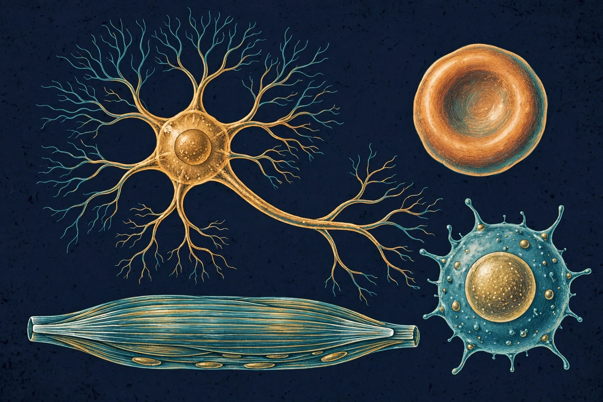
Cells come in bewildering variety — a neuron with branches a metre long, a red blood cell with no nucleus at all, a plant cell stiffened by a wall — yet all of them share four features. OpenStax Biology puts it plainly: cells from organisms as different as bacteria, onion, and human share certain fundamental characteristics, including a membrane, genetic material, ribosomes, and cytoplasm (OpenStax Biology 2e, Chapter 4).
A plasma membrane — a thin, flexible boundary that separates inside from outside and controls what crosses.
Cytoplasm — the gel-like interior where most chemical reactions happen, crowded with proteins, sugars, salts, and water.
Genetic material — DNA carrying the instructions for building proteins and running the cell.
Ribosomes — the molecular machines that read those instructions and build proteins.
Everything else — nucleus, mitochondria, chloroplasts, the whole cast of organelles — is variation on this theme. That shared toolkit is one of the strongest pieces of evidence that all life on Earth descends from a common ancestor. The machinery is too similar, down to the same genetic code, to be coincidence. For the story of how that shared ancestry shaped all life, see our guide to Darwin's Theory of Evolution: Evidence Explained.
1.3 A misconception worth correcting early
Most people assume cells are mostly empty space with a few neat parts floating in it, the way textbook diagrams show them. What we actually see in the data is the opposite. The inside of a cell is packed — so crowded with proteins, filaments, and membranes that a water molecule bumps into something every few nanometres. Biologists call this "macromolecular crowding," and it changes how chemistry works: reactions happen faster, proteins fold differently, and the cell's organisation matters as much as its ingredients. The tidy diagram is a map, not the territory. The territory is a rush-hour city at microscopic scale, and the fact that it works at all is one of the wonders of cell biology.
"People always think of them as sort of the building blocks of the body, but I really like to think of them almost individually as their own little kind of nanoparticles, or little nanobots, that have their own individual activities, and together they form things like tissues, organs." — National Human Genome Research Institute, on what a cell is (NHGRI Genetics Glossary: Cell)
2. How We Learned to See Cells
Nobody knew cells existed until someone built a tool to see them. The history of cell biology is, in large part, the history of the microscope — and of people looking at ordinary things and refusing to believe their eyes.
2.1 Hooke's cork and Leeuwenhoek's "animalcules"
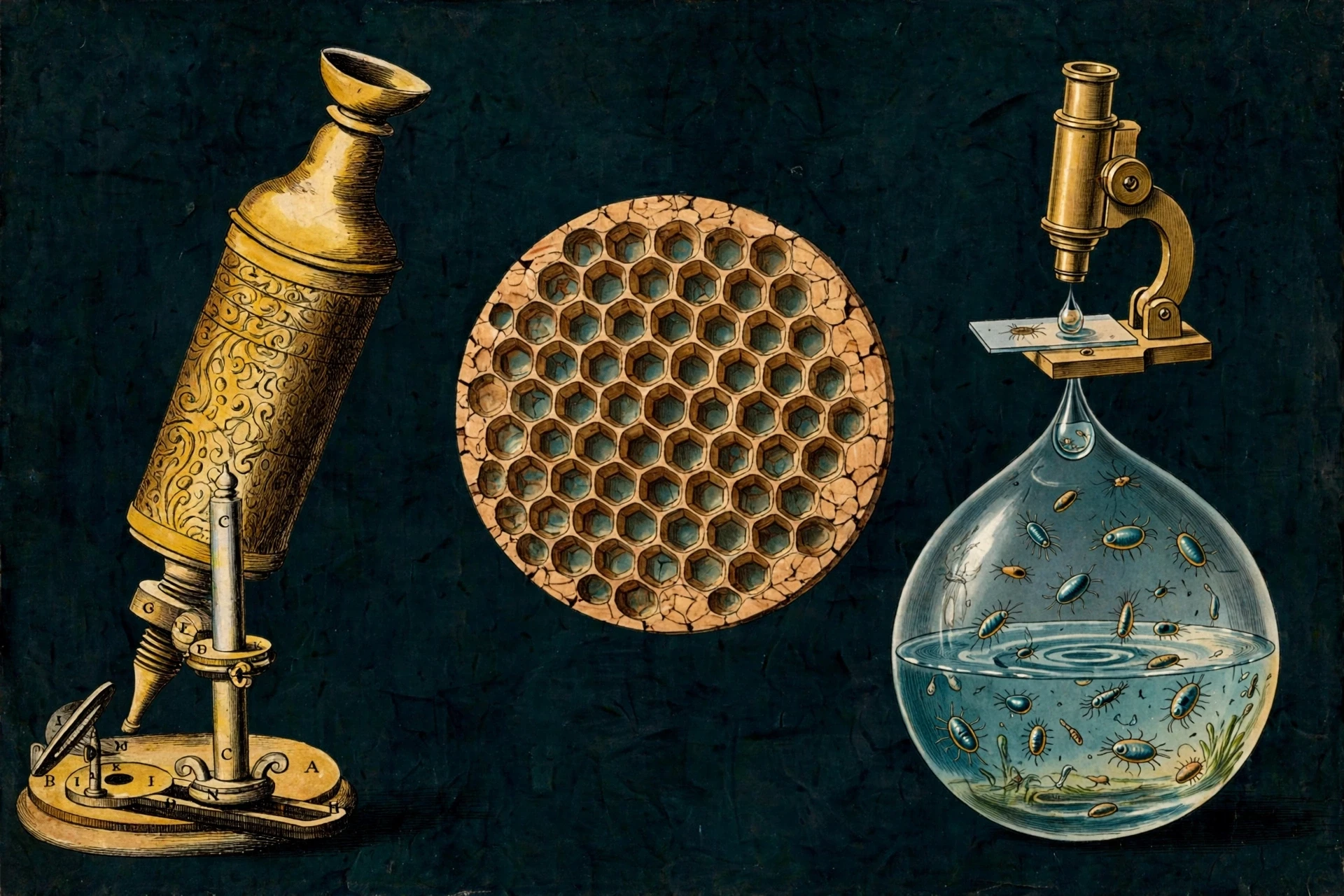
In 1665, Robert Hooke, a curator of experiments at the Royal Society in London, aimed his compound microscope at a thin slice of cork and saw a honeycomb of tiny chambers. He called them "cells," after the small rooms monks lived in. The cork cells were dead — only their walls remained — but the word stuck.
A decade later, Antonie van Leeuwenhoek, a Dutch cloth merchant with no university training, ground lenses so fine that his simple microscopes magnified up to 300 times. He looked at pond water and found it teeming with tiny creatures he called "animalcules." He looked at plaque scraped from his own teeth and saw bacteria — the first human ever to see them. He wrote to the Royal Society, and the Fellows, sceptical, sent a delegation to verify his claims. They confirmed them. Leeuwenhoek had opened a world no one knew existed, using lenses he made in his own workshop.
I think about Leeuwenhoek whenever a student tells me they are not "a science person." He was a draper. He had curiosity, patience, and a willingness to look closely at things other people ignored. That is most of what science is.
2.2 From observation to theory
The microscope improved slowly, and so did the ideas. In the 1830s, better lenses let Schleiden and Schwann see the nucleus in plant and animal tissue and propose that cells were the fundamental unit of life. In 1855, Virchow declared omnis cellula e cellula — every cell from a cell — ruling out the old idea that living things could bubble up spontaneously from non-living matter. In the 1930s, the electron microscope arrived and revealed the organelles in fine detail: the folded inner membrane of mitochondria, the stacked sacs of the Golgi apparatus, the double membrane of the nucleus. Each new instrument rewrote the picture, and each rewrite made the cell look more organised, not less.
2.3 The microscopes we use now
Today's cell biologists have tools Hooke could not have dreamed of. Confocal microscopes build three-dimensional images from glowing tags attached to single proteins. Cryo-electron microscopes freeze cells in a fraction of a second and resolve structures a few atoms across — work that won the 2017 Nobel Prize in Chemistry. Super-resolution microscopy, recognised with the 2014 Nobel Prize in Chemistry, broke what was thought to be a hard physical limit on how small a thing light can resolve, letting researchers watch single molecules move inside a living cell in real time.
The pattern is worth noticing: every time we build a better way to look, the cell turns out to be more intricate than we thought. I would bet heavily that the pattern will hold.
3. Two Kinds of Cell: Prokaryotic and Eukaryotic
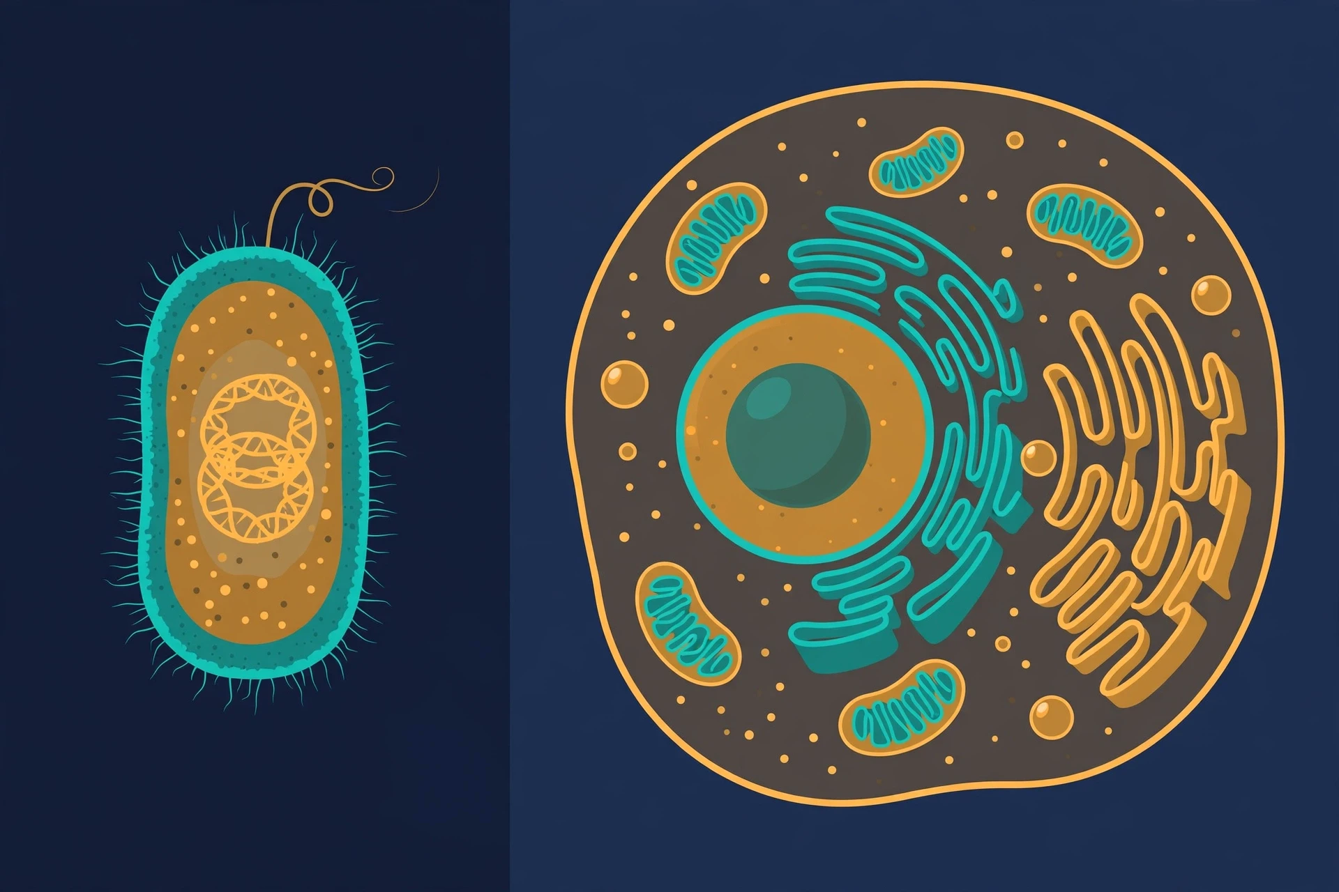
All cells fall into two great designs. Prokaryotic cells — bacteria and archaea — are small and simple, with no nucleus and no membrane-bound organelles. Eukaryotic cells — animals, plants, fungi, and protists — are larger and compartmentalised, with a nucleus and a suite of organelles. You are made of the second kind. The bacteria in your gut, on your skin, and in the soil under your feet are the first.
3.1 Prokaryotic cells: small, tough, and everywhere
A typical bacterium is about 1–2 micrometres long — roughly fifty of them could fit across the width of a human hair. Its DNA floats in a region called the nucleoid, not sealed inside a nucleus. It has ribosomes, a cell membrane, usually a cell wall, and often a flagellum for swimming or pili for sticking to surfaces. That is nearly the whole parts list, and it is enough: bacteria live in boiling springs, in Antarctic ice, in the deepest ocean trenches, and inside your intestines. They were the only form of life on Earth for roughly two billion years, and they still outweigh all other life by some measures of sheer numbers.
Do not mistake simple for primitive. A bacterium can sense chemicals, swim toward food, swap genes with its neighbours, form biofilms that resist antibiotics, and survive conditions that would kill any animal cell in seconds. Simplicity, here, is a strategy that has worked for nearly four billion years.
3.2 Eukaryotic cells: rooms inside rooms
Eukaryotic cells are typically 10–100 micrometres across — ten to a hundred times wider than a bacterium, and a thousand to a million times bigger by volume. Their defining feature is internal membranes that divide the cell into compartments. Each compartment runs different chemistry, at a different pH, with different enzymes. The nucleus guards the DNA. Mitochondria burn fuel. The endoplasmic reticulum builds proteins and lipids. Lysosomes digest waste at a pH so low it would destroy the rest of the cell if it leaked.
OpenStax Biology describes the logic clearly: organelles allow different functions to be compartmentalised in different areas of the cell, so that reactions that would interfere with each other can run side by side (OpenStax Biology 2e, Eukaryotic Cells). A kitchen works the same way: you do not store bleach in the pantry or run the oven inside the fridge. Compartmentalisation is what lets a eukaryotic cell be big and complicated without its chemistry turning into soup.
3.3 How eukaryotes got their power stations
One of the most remarkable stories in cell biology is the origin of mitochondria — and of chloroplasts in plants. About 1.5–2 billion years ago, an ancient archaeal cell engulfed a bacterium, and instead of digesting it, kept it. The bacterium became the mitochondrion, the cell's energy factory. A similar engulfment later gave plants their chloroplasts. This is the endosymbiotic theory, proposed in detail by Lynn Margulis in the 1960s and 70s, when it was widely dismissed. The evidence won out: mitochondria and chloroplasts still carry their own small circular DNA, their own ribosomes, and a double membrane — the fingerprints of a free-living bacterium that moved in and never left.
Margulis spent years being told she was wrong. I keep her story in mind whenever I hear a new idea dismissed too quickly. The history of cell biology is full of correct ideas that sounded absurd at first — and of confident ideas that turned out to be wrong.
Feature | Prokaryotic cell | Eukaryotic cell |
|---|---|---|
Size | Typically 0.5–5 micrometres | Typically 10–100 micrometres |
Nucleus | None; DNA in a nucleoid region | Membrane-bound nucleus |
DNA | Usually one circular chromosome | Multiple linear chromosomes |
Organelles | None membrane-bound | Mitochondria, ER, Golgi, lysosomes, and more |
Ribosomes | Smaller (70S) | Larger (80S in cytoplasm) |
Cell wall | Usually present (peptidoglycan in bacteria) | Plants and fungi only; animals have none |
Division | Binary fission | Mitosis and meiosis |
Examples | Bacteria, archaea | Animals, plants, fungi, protists |
4. The Plasma Membrane: Gatekeeper, Messenger, and Identity Card
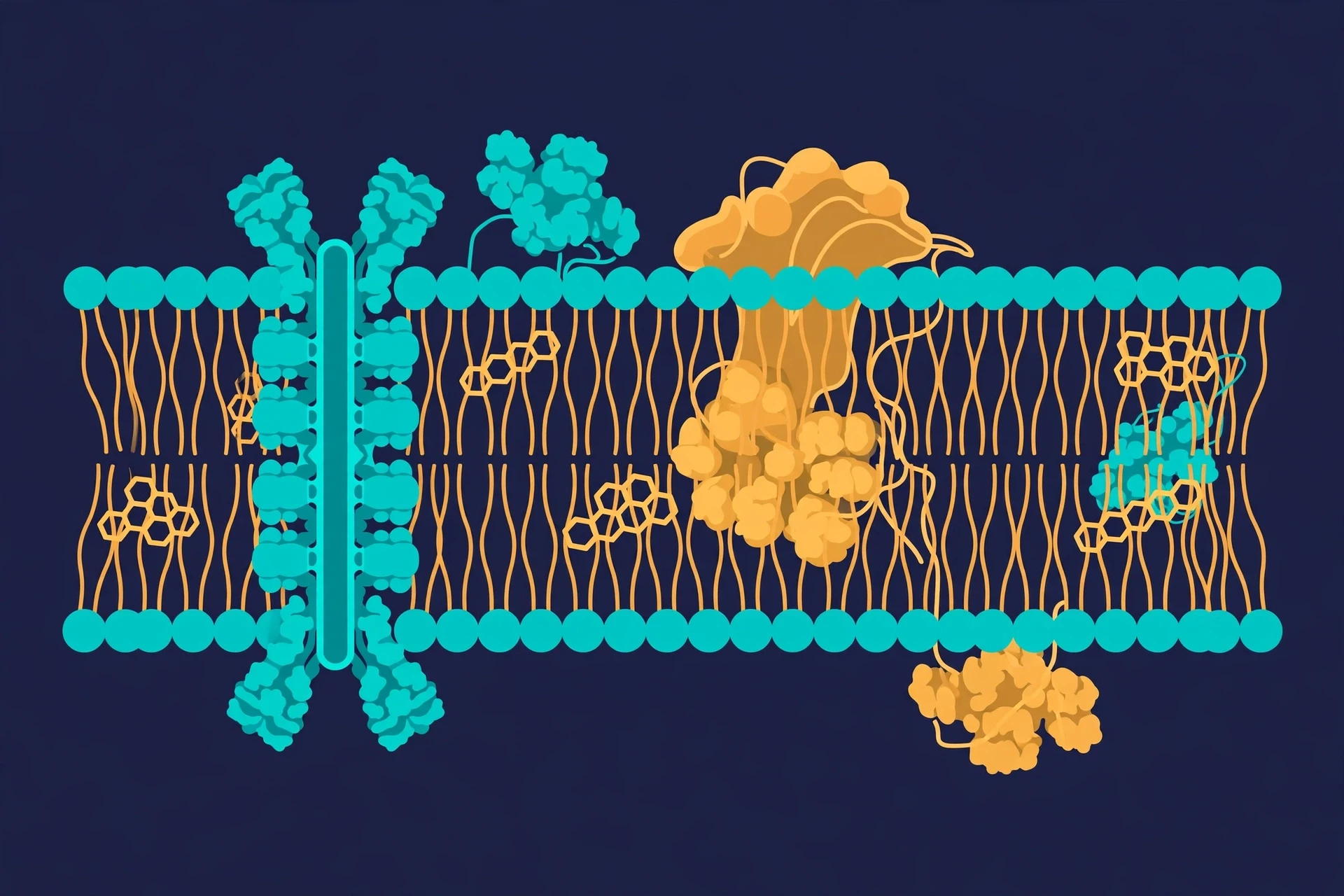
Every cell, prokaryotic or eukaryotic, is wrapped in a plasma membrane. It is only about 5 nanometres thick — roughly one ten-thousandth the width of a hair — yet it does three jobs at once: it holds the cell together, it decides what enters and leaves, and it lets the cell recognise friends, food, and danger.
4.1 The fluid mosaic
The membrane is a phospholipid bilayer: two sheets of fat molecules, tails inward, heads outward, with proteins embedded like boats floating in a sea of fat. That is the "fluid mosaic model," proposed by Singer and Nicolson in 1972. The proteins drift, the lipids flow, and the whole structure repairs itself when torn. It is less like a wall and more like a soap film with machinery stuck in it.
The proteins are the working parts. Some are channels that let specific ions through. Some are pumps that spend energy to move substances against their gradient. Some are receptors that catch chemical signals from outside. Some are identity markers — the molecules that tell your immune system "this cell belongs here." When those markers go wrong, or when immune cells misread them, disease follows. Our guide to How the Human Immune System Works explains how immune cells use these surface signals to tell self from invader.
4.2 How things cross: diffusion, osmosis, and active transport
Small, uncharged molecules such as oxygen and carbon dioxide slip straight through the bilayer by diffusion, moving from where they are concentrated to where they are scarce. Water crosses by osmosis, following salt. Larger or charged molecules need help: channel proteins for facilitated diffusion, or pumps for active transport — moving against the gradient, at the cost of ATP. The sodium-potassium pump, for example, pushes sodium out and potassium in, three ions out for every two in, and your neurons spend a large share of their energy running it. It is the pump that makes nerve signals possible. For the anatomy of the organ those signals serve, see Human Brain Anatomy and Functions: A Complete Guide.
I once watched a student ruin a whole afternoon's experiment by putting red blood cells in pure water instead of saline. Under the microscope, the cells swelled, turned ghostly, and burst — osmosis, in real time, because water rushed in where salt could not get out. She was mortified. I told her she had just performed the clearest demonstration of osmosis I had ever seen, and that the cells had died for science in the most useful possible way. She still remembers it. So do I.
4.3 Endocytosis and exocytosis: the cell eats and ships
Cells also move cargo in bulk. In endocytosis, the membrane folds inward and pinches off a vesicle, swallowing food, fluid, or even whole bacteria — white blood cells eat invaders this way. In exocytosis, vesicles fuse with the membrane from inside and dump their contents outside: hormones, neurotransmitters, digestive enzymes, the proteins that build the extracellular matrix. A pancreatic cell releases insulin by exocytosis. A neuron releases neurotransmitters the same way. The membrane is not a wall with doors. It is a border that constantly remodels itself.
5. The Nucleus and the Genome: The Cell's Library
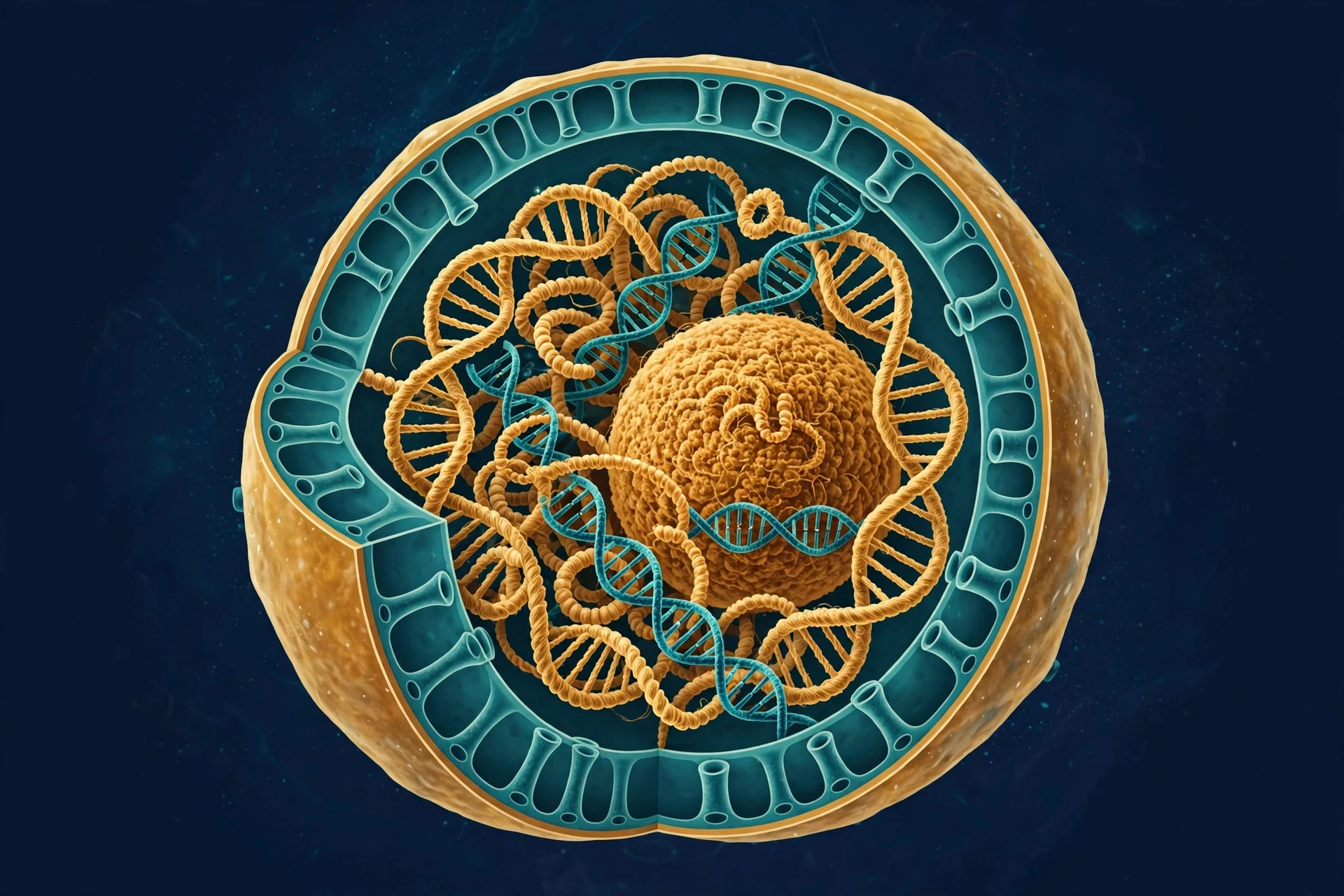
If the cell is a city, the nucleus is its library and its city hall combined. It holds the DNA — the complete instruction set for building and running the cell — and controls which instructions get read, when, and how often.
5.1 DNA, chromatin, and chromosomes
A human cell contains about two metres of DNA, packed into a nucleus roughly six micrometres across. The packing is a marvel in itself. DNA wraps around proteins called histones to form chromatin, which coils and folds into chromosomes when the cell prepares to divide. Humans have 46 chromosomes in most cells — 23 from each parent. When the cell is not dividing, the chromatin loosens, like a library with its books open on the tables, so that the reading machinery can get at the genes it needs.
That DNA is the subject of our guide to DNA: Structure, Replication, and Role in Heredity, which explains the double helix, how DNA copies itself, and how it passes information from one generation to the next. Here, the point that matters is control: the nucleus does not just store DNA, it regulates it. Genes are switched on and off by proteins that bind the DNA, by chemical tags on the histones, and by the three-dimensional folding of the chromosomes themselves. Two cells with identical DNA — a skin cell and a neuron, say — behave completely differently because they read different parts of the same book.
5.2 The nuclear envelope and the nucleolus
The nucleus is wrapped in a double membrane, the nuclear envelope, studded with nuclear pores — gateways that let messenger RNA out and proteins in, under strict control. The envelope is continuous with the endoplasmic reticulum, which we meet in the next section. Inside, the most visible feature is the nucleolus, a dense region where ribosomal RNA is made and ribosome subunits are assembled. Cells that make a lot of protein have large, busy nucleoli; pathologists still use nucleolus size as a clue when examining cells under the microscope.
5.3 From gene to protein: the central dogma in one paragraph
The flow of information runs DNA → RNA → protein. In transcription, a gene's DNA sequence is copied into messenger RNA inside the nucleus. The mRNA travels through a nuclear pore into the cytoplasm, where ribosomes translate it, reading three-letter codons and linking amino acids into a protein chain. That chain folds into a shape, and the shape does the work — an enzyme, a structural fibre, a hormone, an antibody. Every protein in your body was made this way, by ribosomes reading messages that came from the nucleus. When the message is miscopied, or the protein folds wrongly, disease can follow — from cystic fibrosis to many cancers. That is why so much of modern medicine is, at heart, cell biology.
6. The Endomembrane System: Build, Fold, Ship
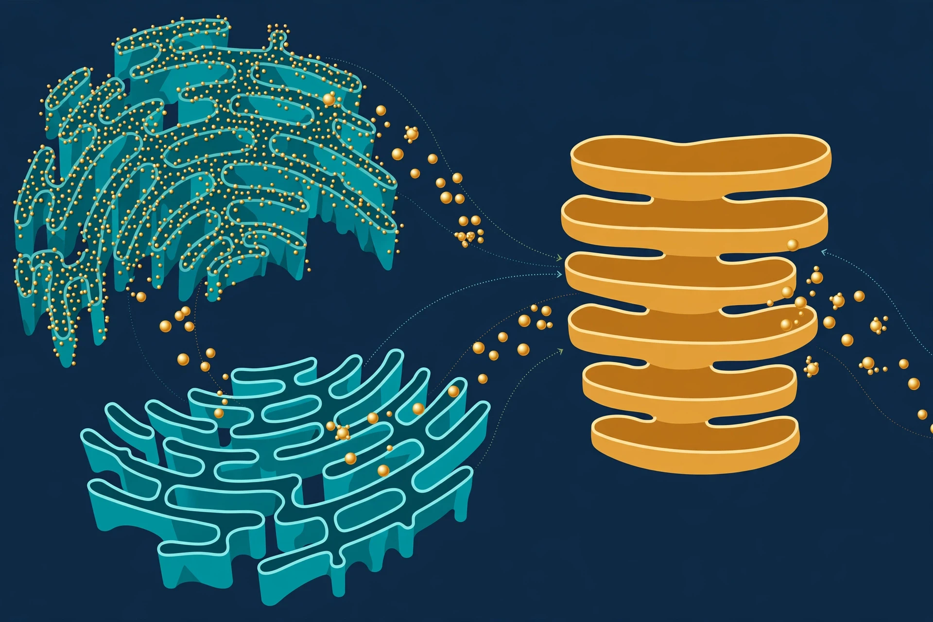
Proteins destined for the membrane, for secretion, or for other organelles travel through a connected network called the endomembrane system: the endoplasmic reticulum, the Golgi apparatus, and the vesicles that shuttle between them. It is the cell's factory floor and its postal service, and it handles a staggering volume — a single cell can secrete thousands of proteins per second.
6.1 Rough and smooth endoplasmic reticulum
The endoplasmic reticulum (ER) is a maze of folded membranes spreading out from the nuclear envelope. The rough ER is studded with ribosomes, giving it its "rough" appearance, and it is where proteins entering the secretory pathway are built and folded. The smooth ER has no ribosomes; it makes lipids, detoxifies drugs and poisons, and stores calcium ions. Liver cells are rich in smooth ER, which is why they handle detoxification — and why they bear the brunt of alcohol. Muscle cells use their specialised smooth ER to release calcium, triggering contraction.
I once spent a long evening in a lab watching a graduate student stain the ER of a living cell with a fluorescent dye. The network glowed like a river delta seen from the air, constantly shifting, branching, and reforming. A still photograph makes it look like plumbing. In life, it moves like weather.
6.2 The Golgi apparatus: the post office
Proteins leave the ER in vesicles and arrive at the Golgi apparatus — a stack of flattened sacs first described by Camillo Golgi in 1898. The Golgi modifies proteins, adding sugar tags that act as address labels, then sorts them into vesicles bound for the membrane, for lysosomes, or for secretion outside the cell. Get the address wrong and the protein ends up in the wrong place, which is how some inherited diseases begin: the protein is made correctly, but it is delivered to the wrong compartment.
6.3 Lysosomes and peroxisomes: clean-up crews
Lysosomes are the cell's recycling centres — sacs of digestive enzymes that break down worn-out organelles, bacteria, and large molecules, working at a pH of around 4.5–5, far more acidic than the surrounding cytoplasm. When lysosomal enzymes are missing or faulty, undigested material piles up inside cells, causing lysosomal storage diseases such as Tay-Sachs. Peroxisomes are smaller sacs that break down fatty acids and detoxify poisons, converting dangerous hydrogen peroxide into water and oxygen. Liver peroxisomes detoxify alcohol. Without these clean-up crews, a cell would choke on its own waste within days. The deliberate dismantling of cell parts — autophagy — is so central to health that it has its own research field; see Fasting and Autophagy: What the Science Says.
7. Mitochondria and Chloroplasts: The Energy Organelles
Every process in this article costs energy, and the currency is a small molecule called ATP — adenosine triphosphate. Break one of its phosphate bonds and energy is released to power pumps, build proteins, and move cargo. The cell's main ATP factories are the mitochondria. In plant and algal cells, chloroplasts do the complementary job: capturing sunlight to build sugars.
7.1 Mitochondria: burning fuel for ATP
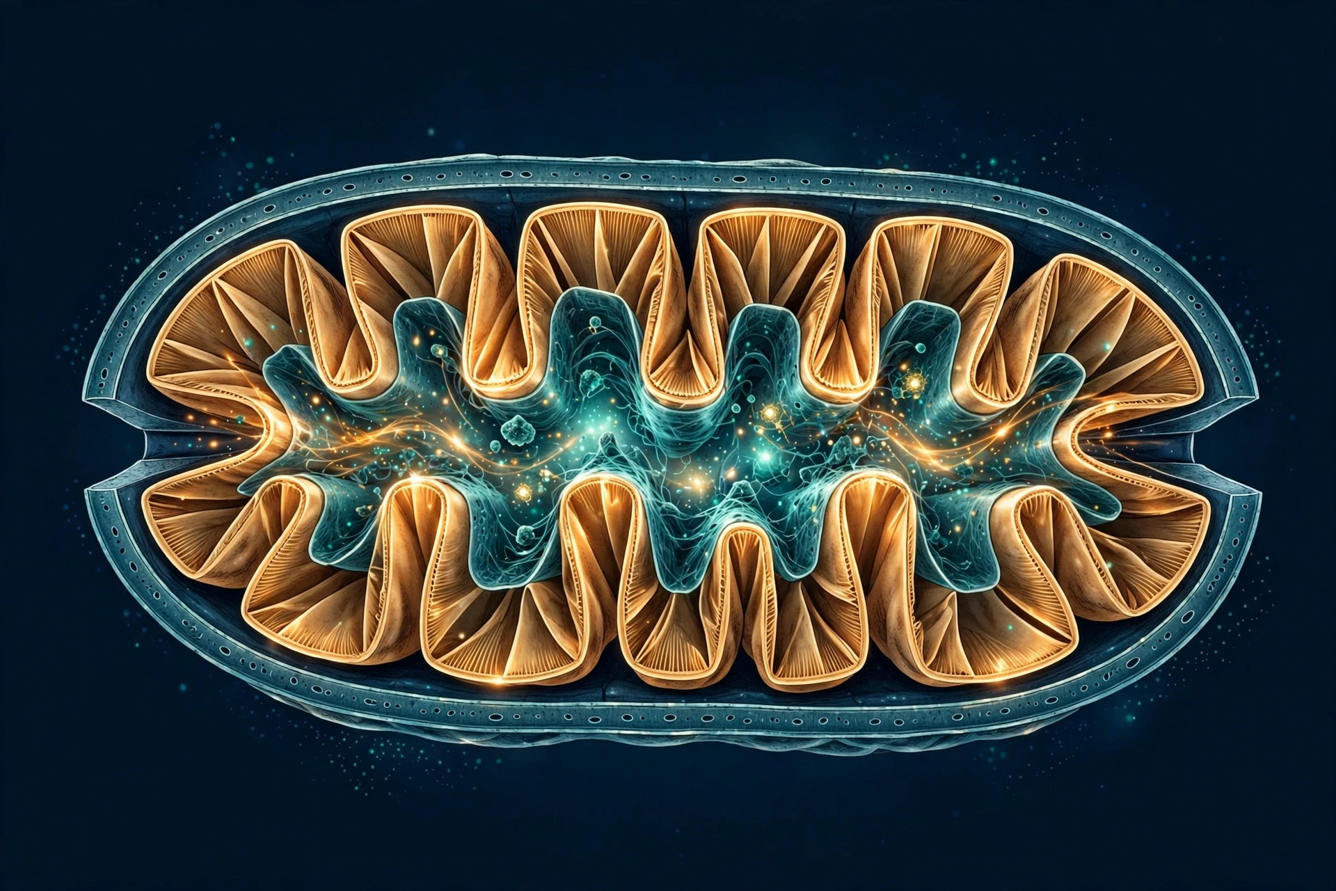
Mitochondria are oval organelles with a double membrane. The inner membrane is folded into cristae, which hugely increase the surface area available for the electron transport chain — the series of protein complexes that pass electrons along, pump protons across the membrane, and use the resulting gradient to make ATP. One glucose molecule can yield roughly 30–32 ATP through cellular respiration. The carbon dioxide you exhale with every breath is the waste product of this process.
OpenStax Biology notes that mitochondria are often called the cell's powerhouses, and that muscle cells — which need a great deal of energy — are packed with them (OpenStax Biology 2e, Eukaryotic Cells). A heart muscle cell can contain thousands. The link between mitochondria and energy explains a whole family of symptoms: when mitochondria fail, the organs that need the most energy — brain, muscle, heart — fail first. Mitochondrial diseases are rare individually but devastating, and they are almost always inherited from the mother, because the egg supplies nearly all the mitochondria in the embryo.
7.2 Chloroplasts: sunlight into sugar
Chloroplasts, found in plant and algal cells, run photosynthesis: capturing light energy with chlorophyll, splitting water, and fixing carbon dioxide into sugars. Their internal stacks of membranes, the thylakoids, are where the light reactions happen; the surrounding stroma is where carbon is fixed. The oxygen in the air you are breathing right now was released by chloroplasts (and by their bacterial cousins, cyanobacteria) splitting water molecules. For the full story of that process, see Photosynthesis Explained: Process & Equation.
The partnership between the two organelles is elegant: chloroplasts build sugars from sunlight, mitochondria burn sugars for ATP, and the waste product of one is the raw material of the other. Life runs on that loop.
7.3 Why these organelles have their own DNA
Both mitochondria and chloroplasts carry a small circular chromosome of their own, and their own ribosomes — the lingering fingerprints of their free-living bacterial ancestors. They still make a handful of their own proteins, although most of their proteins are now coded by the nucleus and imported. That divided control is a source of vulnerability: mutations in mitochondrial DNA accumulate with age and are implicated in some forms of deafness, diabetes, and neurodegeneration. Researchers still disagree on how much of ageing itself is driven by mitochondrial decline — the evidence is real, but the size of the effect is genuinely debated.
8. The Cytoskeleton, Movement, and Cell Division
A cell is not a bag of jelly. It has an internal skeleton — a dynamic network of protein fibres that gives it shape, moves its organelles, and pulls its chromosomes apart when it divides. The cytoskeleton is constantly being built and dismantled, and it is the reason a cell can crawl, change shape, and divide with such precision.
8.1 Three kinds of fibre
Microtubules — hollow tubes of tubulin, the thickest fibres. They act as highways for motor proteins carrying vesicles, form the spindle that separates chromosomes, and build cilia and flagella.
Microfilaments — thin threads of actin, just under the membrane. They let cells crawl, pinch in two during division, and form microvilli that increase surface area in the gut.
Intermediate filaments — tough, rope-like fibres that anchor the nucleus and resist mechanical stress. Hair and nails are made of a related protein, keratin.
When I first saw time-lapse footage of a white blood cell chasing a bacterium, squeezing through gaps half its width and engulfing its prey, I understood the cytoskeleton in a way no diagram had ever managed. The cell was not drifting. It was hunting, and every movement was the cytoskeleton at work.
8.2 The cell cycle and mitosis
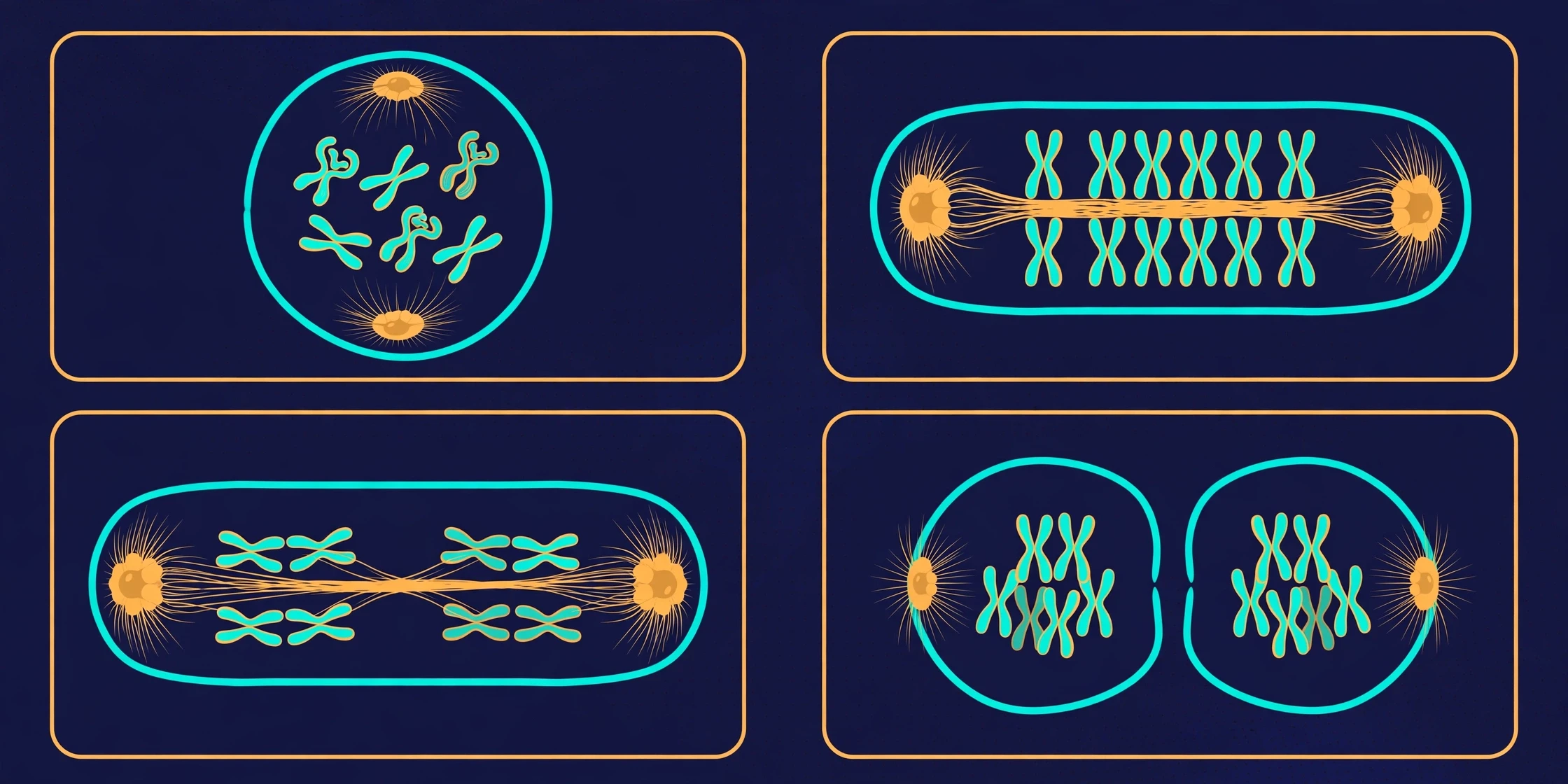
Most cells live by a cycle: grow, copy the DNA, check the copy, divide. The cycle has phases — G1 (growth), S (DNA synthesis), G2 (preparation), and M (mitosis, division) — policed by checkpoints that halt the cycle if the DNA is damaged or the copy is incomplete. Mitosis itself has stages you may remember from school: prophase, metaphase, anaphase, telophase. The chromosomes condense, line up on the spindle, are pulled to opposite ends, and two nuclei form. Then the cytoplasm divides, and there are two cells where there was one.
The checkpoints matter enormously. Cancer is, at its core, a disease of the cell cycle: mutations disable the brakes, and cells divide when they should not. Nearly every cancer drug in use today works by exploiting some feature of cell division — which is also why so many of them harm healthy dividing cells, like those in bone marrow and gut lining, as a side effect. Understanding the cell cycle is what lets researchers design drugs that hit cancer cells harder than healthy ones.
8.3 Meiosis: the division that makes sex cells
Meiosis is a different division, used only to make eggs and sperm. It halves the chromosome number — from 46 to 23 in humans — and shuffles the genetic deck, so that each gamete is unique. When egg and sperm fuse, the full number is restored, but the combination is new. That shuffling is the engine of genetic diversity, and it is why siblings, sharing the same parents, are never identical (except identical twins, who split from a single fertilised egg).
9. How Cells Talk, Specialise, and Die on Schedule
A cell in isolation is interesting. A trillion cells cooperating is a body. That cooperation depends on three things: cells must communicate, they must specialise, and they must know when to die.
9.1 Cell signalling: chemical conversations
Cells talk by sending and receiving chemical signals. A hormone released in one place travels through the blood and is recognised by receptors on distant cells. A neurotransmitter crosses a synapse in milliseconds. A growth factor tells a wound's cells to divide and repair. The signal binds a receptor, the receptor triggers a cascade of proteins inside the cell, and the cascade changes what the cell does — switches a gene on, opens a channel, starts dividing.
The system is precise because receptors are specific: insulin fits its receptor the way a key fits a lock, and a liver cell responds to insulin differently from a muscle cell, even though both carry the same receptor. When signalling goes wrong, the results range from diabetes (cells stop responding to insulin) to cancer (a growth signal stuck in the "on" position). Modern drugs increasingly target signalling pathways rather than the cell as a whole — a more precise intervention, built on cell biology.
9.2 Specialisation: one genome, 200 cell types
Every cell in your body carries the same DNA, yet a neuron, a skin cell, and a red blood cell could hardly look more different. Specialisation — differentiation — happens because each cell type reads a different subset of its genes. A red blood cell makes haemoglobin and ejects its nucleus to make room for it. A neuron grows long branches and fills them with ion channels. A muscle cell packs itself with contractile fibres. A pancreatic beta cell devotes its life to making insulin.
Stem cells are the exception that proves the rule: unspecialised cells that can divide and give rise to specialised ones. Embryonic stem cells can become any cell type; adult stem cells, in bone marrow, gut, and skin, replenish the tissues that wear out fastest. That is why bone marrow transplants work: the donor's stem cells settle in the recipient's marrow and rebuild the blood system from scratch. It is cell biology, applied, saving lives every day.
9.3 Apoptosis: programmed cell death
Cells also need to die — on schedule, cleanly, without spilling their contents. This programmed death is called apoptosis, and it sculpts your body from the start: the webbing between a foetus's fingers dies away by apoptosis, leaving separate digits. Cells with damaged DNA kill themselves rather than risk becoming cancerous. Cells that are no longer needed — the lining of the uterus each month, the extra neurons pruned from a developing brain — are removed the same way.
When apoptosis fails, damaged cells survive and can turn cancerous. When it runs out of control, healthy tissue is lost — as in some neurodegenerative diseases. The balance between division and death is one of the most closely guarded balances in the body, and it is guarded by the cell itself.
A cell that cannot die when it should is as dangerous as a cell that cannot divide when it must. The body stays healthy not by keeping every cell alive, but by keeping the decision to live or die under strict, local control.
9.4 When cell biology goes wrong: disease as cell failure
Almost every disease, looked at closely enough, is a cell biology problem. Cystic fibrosis is a faulty chloride channel in the membrane. Alzheimer's involves proteins misfolding and accumulating inside and around neurons. Type 2 diabetes is cells ignoring insulin's signal. Autoimmune diseases are immune cells attacking the body's own cells. Even ageing, researchers increasingly think, is partly the accumulation of senescent cells — cells that have stopped dividing but refuse to die, leaking inflammatory signals into their neighbourhood. The newest therapies, from CAR-T cells that hunt cancer to gene therapies that fix a faulty channel, work by changing what cells do. Cell biology is no longer just the foundation of medicine. Increasingly, it is the medicine.
10. The Latest Research: What Cell Biologists Are Excited About Now
Cell biology is moving fast, and the last few years have brought tools that are changing what is possible — in the lab, and increasingly in the clinic.
10.1 Seeing and editing single cells
Single-cell sequencing lets researchers read the RNA of one cell at a time, revealing that tissues we thought were uniform are mosaics of cell types, some previously unknown. The Human Cell Atlas project is cataloguing every cell type in the human body, cell by cell — a map as ambitious, in its way, as the Human Genome Project. At the same time, CRISPR gene editing lets researchers change a single letter of DNA inside a living cell and watch what happens. The first CRISPR therapies for sickle cell disease and beta thalassemia were approved in 2023, treating patients by editing their own blood stem cells.
10.2 Organoids: miniature organs in a dish
Grow stem cells in the right conditions and they self-organise into organoids — tiny, simplified versions of a gut, a brain, a kidney, a retina. Organoids let researchers test drugs on a patient's own cells before giving the drug to the patient, and they are replacing some animal testing. Brain organoids have even been used to study how viruses infect developing neural tissue. The ethical questions are real, and they are being debated openly — which is exactly how it should be.
10.3 Senolytics and the biology of ageing
If senescent cells drive part of ageing, can removing them help? Drugs called senolytics, which selectively kill senescent cells, have extended healthspan in mice, and early human trials are underway for conditions including osteoarthritis and lung fibrosis. The results so far are promising but preliminary, and no senolytic is an approved anti-ageing treatment. I mention it here because it shows how cell biology turns into medicine: find the cell, understand the process, intervene precisely, test honestly.
10.4 What we still do not know
For all the progress, the open questions are large. We do not fully understand how a fertilised egg builds a body — how cells know where they are and what to become. We do not know how the first cell arose from non-living chemistry, although laboratory experiments are narrowing the possibilities. We do not know why some cells live for a lifetime (your neurons) while others are replaced every few days (your gut lining). And the origin of the eukaryotic cell — the engulfment that gave us mitochondria — remains one of the great unresolved events in the history of life, with researchers still debating how, and how many times, it happened. A field that admits what it does not know is a field you can trust.
11. Common Misconceptions About Cells — Corrected
"The nucleus is the brain of the cell." The nucleus holds the instructions, but the cell has no single boss. Control is distributed: the membrane, the mitochondria, the signalling pathways, and the cytoskeleton all make decisions. A cell with its nucleus removed can live for hours, and a red blood cell lives for months without one at all.
"Mitochondria are only the powerhouse of the cell." They make ATP, yes — but they also control cell death, make building blocks for other molecules, and signal to the nucleus. Calling them the powerhouse is like calling a city hall "only a post office." It is true, and it misses most of the story.
"All cells look like the textbook diagram." The diagram shows a fried-egg cell with a round nucleus in the middle. Real cells are long, flat, branched, star-shaped, or packed so tightly you can barely see where one ends and the next begins. Shape follows function, and function varies enormously.
"Plant cells and animal cells are basically the same." They share the core machinery, but plant cells have a cell wall, chloroplasts, and a large central vacuole that animal cells lack — and most plant cells have no lysosomes or centrioles. The differences matter for everything from how plants stand upright to how they defend themselves.
"Cells are too small to matter to everyday life." Every cut that heals, every vaccine that works, every meal you digest, and every memory you form is cells doing their jobs. You never see them. You live by them.
"Cancer is one disease." Cancer is hundreds of diseases, all rooted in cell biology gone wrong — different mutations, different cell types, different broken checkpoints. That is why there is no single cure, and why treatments are increasingly tailored to the genetics of each patient's own tumour cells.
Conclusion
We began with an onion skin on a school microscope, a grid of empty-looking boxes. We end with a picture that is almost the opposite: a crowded, humming city of compartments, each running chemistry that would be impossible anywhere else, all of it coordinated by signals, powered by ancient bacterial lodgers, and policed by checkpoints that decide, every hour, whether each cell should grow, divide, or die.
That is the real lesson of cell biology. The cell is not the simple building block we imagined when we first saw it. It is the level at which life actually happens — where DNA becomes protein, where food becomes energy, where a signal becomes an action, where a fertilised egg becomes a person. Everything above the cell — tissues, organs, bodies, ecosystems — is built from what cells do. Everything below it — molecules, atoms, chemistry — only becomes alive when it is organised inside one.
The next time you look at your hand, remember what you are looking at: roughly a hundred billion cells, of dozens of types, cooperating so well that you never notice them. They have been doing it, without a day off, since before you were born. Understanding them is understanding yourself — not as a metaphor, but as a fact.
Frequently Asked Questions
What is cell biology?
Cell biology is the study of cells — their structure, chemistry, and function. It covers how cells are built, how their organelles work together, how they obtain energy, communicate, divide, and die, and how failures in these processes cause disease.
What is a cell?
A cell is the smallest unit of life. Every living thing is made of one or more cells, and every cell has a membrane, cytoplasm, DNA, and ribosomes. Cells carry out all the processes of life: they take in nutrients, make energy, build proteins, respond to signals, and reproduce.
What are the main organelles and their functions?
The main organelles are the nucleus (stores DNA and controls gene expression), mitochondria (make ATP energy), ribosomes (build proteins), endoplasmic reticulum (rough ER builds proteins, smooth ER makes lipids and detoxifies), Golgi apparatus (modifies, sorts, and ships proteins), lysosomes (digest waste), and the cytoskeleton (shape, movement, and division). Plant cells also have chloroplasts (photosynthesis), a cell wall, and a large central vacuole.
What is the difference between prokaryotic and eukaryotic cells?
Prokaryotic cells (bacteria and archaea) are small, have no nucleus, and have no membrane-bound organelles; their DNA floats in a nucleoid region. Eukaryotic cells (animals, plants, fungi, protists) are larger, have a membrane-bound nucleus, and contain many membrane-bound organelles.
What does the mitochondria do?
Mitochondria make ATP, the cell's main energy currency, through cellular respiration — using oxygen to extract energy from glucose and other nutrients. They also help control programmed cell death, produce building blocks for other molecules, and signal to the nucleus.
How do cells divide?
Most cells divide by mitosis: they copy their DNA, line the chromosomes up on the spindle, pull them apart, and split into two genetically identical daughter cells. Sex cells are made by meiosis, which halves the chromosome number and shuffles the genes, producing unique eggs or sperm.
How many cells are in the human body?
The human body contains around 37 trillion cells, in more than 200 different types — including neurons, muscle cells, skin cells, blood cells, and bone cells. Red blood cells are the most numerous, while fat and muscle cells account for most of the body's mass.
What is the cell membrane made of?
The plasma membrane is a phospholipid bilayer with embedded proteins, cholesterol, and carbohydrate chains. The lipids form the barrier; the proteins act as channels, pumps, receptors, and identity markers. This "fluid mosaic" structure is flexible, self-repairing, and selective about what crosses it.
What happens when cells die?
Cells die in two main ways: necrosis, an uncontrolled death from injury that spills the cell's contents and causes inflammation, and apoptosis, a tidy programmed death in which the cell dismantles itself and is cleared away without harming its neighbours. Apoptosis sculpts the body during development and removes damaged cells that could become cancerous.
Why is cell biology important in medicine?
Almost every disease is a cell biology problem at its root — a faulty channel, a broken checkpoint, a misread signal, a protein that folds wrongly. Understanding cells lets doctors design drugs that target the broken process, develop gene and cell therapies, and use a patient's own cells to test treatments before giving them.
Sources / Further Reading
OpenStax Biology 2e — Cell Structure (Chapter 4, Introduction): https://openstax.org/books/biology-2e/pages/4-introduction
OpenStax Biology 2e — Eukaryotic Cells (Section 4.3): https://openstax.org/books/biology-2e/pages/4-3-eukaryotic-cells
National Human Genome Research Institute — Genetics Glossary: Cell: https://www.genome.gov/genetics-glossary/Cell
OpenStax Concepts of Biology — The Cell Cycle (Section 6.2): https://openstax.org/books/concepts-biology/pages/6-2-the-cell-cycle