
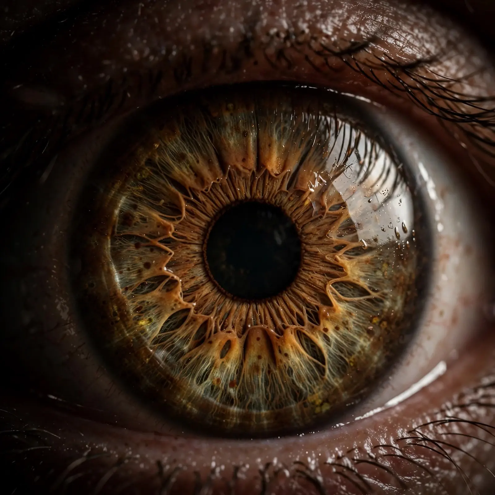
1. The Organ That Gave Darwin a Cold Shudder
The evolution of the eye begins with a confession. In 1860, Charles Darwin wrote to his friend Asa Gray, "The eye to this day gives me a cold shudder, but when I think of the fine known gradations, my reason tells me I ought to conquer the cold shudder." He was not writing as a critic of his own theory. He was writing as a man who knew exactly how strong the objection sounded, and who refused to look away from it.
Picture the scene. Darwin is at his desk at Down House, deep in the long argument over natural selection, and the letters arriving from friends and critics keep circling back to the same organ. A lens that changes shape to focus near and far. An iris that opens and closes like the aperture of a camera. A retina packed with cells that turn light into signals the brain can read. Put the parts together and the whole thing looks planned, as if someone had sat down with a blueprint and built it on purpose. Take one part away and the rest seems useless. That was the puzzle, and Darwin felt its weight in his own body.
I first met that sentence late at night, years ago, in a quiet room with a borrowed copy of Darwin's letters open on my lap and the house asleep around me. I had expected a triumphant passage, the kind of line a great scientist writes when he has solved a problem. Instead I found a man admitting fear. I read the sentence twice, then a third time, because the third time changed how I heard it. Darwin was not surrendering to the shudder. He was naming it so he could answer it. That small act — saying the hardest thing out loud before trying to solve it — has stayed with me ever since, and it is the reason this article starts where it does.
The question Darwin faced still stops people in their tracks today. How could blind natural processes, with no plan and no foresight, build something as precise as an eye? A clock has a maker. A camera has a designer. An eye looks like both at once, and yet it lives inside animals that were never designed at all. If natural selection is true, the eye must have a history written in small, useful steps, each one paying its own way.
That is the claim this article will test, following the evolution of the eye from a patch of light-sensitive cells to the camera eye. We will start with what an eye actually is, stripped down to a single light-sensitive cell. We will follow Darwin's own answer to his critics, word for word. We will climb the ladder from a flat patch of cells to a pit, a pinhole, and a lens, meeting living animals that still occupy every rung. And we will look at a famous calculation from 1994 that put a number on the whole journey: less than 364,000 years from patch to camera eye. The answer turns out to be stranger and more satisfying than either side of the old argument expected. The eye was not built in one leap. It was built the way a path is worn into a hillside — one small step at a time, by creatures that never knew where the path was going.
2. What Is an Eye, Really?
An eye is an organ that detects light and turns it into spatial information. It does more than register brightness: it tells an animal where light comes from, how strong it is, and, in the best cases, what shape cast the shadow. That shift from sensing light to mapping it is what separates an eye from a simple light sensor.
Start small, because the beginning is almost embarrassingly simple. The basic unit of every eye is the photoreceptor cell, a specialised cell that reacts when light strikes it. Inside that cell sit two partners bound together in a membrane: opsin, a light-sensitive protein, and a chromophore, the pigment that actually absorbs the light. When a photon hits the chromophore, the molecule changes shape, the opsin shifts with it, and a chemical chain reaction begins. In animals with a nervous system, that reaction ends as an electrical signal travelling toward the brain. Light in, signal out. That is the whole trick, and everything else in the eye exists to make that trick more precise.
A single photoreceptor is not an eye. A group of them, gathered into a patch, is the humblest version of one. Biologists call such a patch an eyespot. An eyespot can tell light from dark, and that alone is worth having. A tiny animal drifting in shallow water can use it to tell day from night, to stay near the sunlit surface where food grows, or to sink away from the glare when a shadow passes overhead. It cannot tell the shape of the shadow, or the direction it came from, or whether the thing that cast it is food or a predator. It can only say: brighter now, darker now. For many creatures, through much of the history of life, that was enough.
Photoreception answers one question: how much light is there? Vision answers a harder one: where is the light coming from, and what does its pattern mean? A flat eyespot fails the second question because light from every direction strikes the same cells in the same way. To answer it, the eye needs structure — shading on one side, a cup to block stray light, an opening small enough to sort rays by angle.
The deep history here is older than animals with proper eyes. More than 600 million years ago, early organisms had already evolved photoreceptors that could signal the presence of light, probably helping them move toward light, dodge predators' shadows, and keep their daily rhythms in step with the sun. The machinery came first. The organ came later, built on machinery that had been doing a simpler job for a long time. That pattern — old parts, new uses — runs through the whole story of the evolution of the eye.
How many times did eyes themselves appear? The honest answer is: many. Eyes — photoreceptive organs, not just scattered light-sensitive cells — have evolved independently at least 40 times, and possibly as many as 65 times, according to the estimate by Salvini-Plawen and Mayr in 1977. That number surprises people who imagine the eye as a single invention, passed down from one ancestor to all the rest. The truth is closer to the invention of the wheel, or the wing, or the habit of living in burrows. When a trick is useful and the raw materials are lying around, life stumbles on it again and again. Opsins were already there. Light was already there. All that was needed was for a patch of cells to gather in one place and start reporting.
The chemistry of a photoreceptor is the same kind of chemistry that runs the rest of a cell: proteins changing shape, signals passing from one molecule to the next. The cell biology article walks through how cells organise that work. What matters here is that the eye did not begin as an eye. It began as a patch of ordinary cells doing an ordinary chemical job, and natural selection slowly turned that job into sight.
3. Darwin's "Absurd" Problem — and His Answer
Darwin admitted that the evolution of the eye seemed absurd in the highest possible degree, then answered his own objection in the same passage of On the Origin of Species. His reply rested on three conditions: useful gradations must exist, variation must be inherited, and some variations must sometimes be useful to their owner. If those held, the difficulty was apparent, not real.
Darwin put the objection in the strongest words he could find. He did not soften it, hide it in a footnote, or hand it to a critic to state for him. In Chapter 6 of On the Origin of Species, published in 1859, he wrote it out himself, in full:
"To suppose that the eye, with all its inimitable contrivances for adjusting the focus to different distances, for admitting different amounts of light, and for the correction of spherical and chromatic aberration, could have been formed by natural selection, seems, I freely confess, absurd in the highest possible degree."
Read that sentence slowly, because every word is doing work. "Inimitable contrivances" — devices so fine that no human craft could copy them. Adjusting focus to different distances: the lens changing shape as you look from this page to the far wall. Admitting different amounts of light: the iris narrowing in bright sun and widening in a dark room. Correcting spherical and chromatic aberration: the eye fixing two optical flaws that lens-makers struggle with, bending different colours of light to the same point without smearing the image. Darwin knew the optics. He had read the anatomists. He was not pretending the eye was simple. He was admitting, in public, that it looked impossible.
Then, in the same breath, he answered himself. The passage continues:
"Yet reason tells me, that if numerous gradations from a perfect and complex eye to one very imperfect and simple, each grade being useful to its possessor, can be shown to exist; if further, the eye does vary ever so slightly, and the variations be inherited, which is certainly the case; and if any variation or modification in the organ be ever useful to an animal under changing conditions of life, then the difficulty of believing that a perfect and complex eye could be formed by natural selection, though insuperable by our imagination, can hardly be considered real."
That is the whole argument, and it is still the argument used today. Darwin set three conditions, each checkable against the living world.
First, gradations must exist. There must be a series of eyes running from very simple to very complex, with each grade useful to the animal that has it. Darwin was not asking his readers to imagine these steps. He was pointing at them. Flatworms with eyespots. Snails with pit eyes. The nautilus with a pinhole eye. Each one alive, each one getting by with the eye it has. If the ladder exists in nature, then the eye does not need to appear in one jump. It can climb.
Second, the eye must vary, and the variation must be inherited. This was the easy condition for Darwin, and he says so plainly: "which is certainly the case." Eyes differ between individuals, just as noses and wings differ. Some eyes focus a little better. Some gather a little more light. And those differences pass from parent to offspring, the way any inherited trait does. Without variation there is nothing for selection to act on. With it, selection has raw material in every generation.
Third, each variation must sometimes be useful. A slightly deeper pit, a slightly clearer covering, a slightly better lens — if any of those helps an animal find food, dodge a predator, or find a mate under changing conditions, then animals carrying it will leave more descendants, and the trait will spread. Note the word "ever" in Darwin's sentence: "if any variation or modification in the organ be ever useful." He did not require every change to help. He required only that useful changes happen sometimes. That is a low bar, and life clears it constantly.
The most important word in the whole passage is the one Darwin put in the first sentence: "seems." The eye seems absurd. The difficulty is "insuperable by our imagination" — our imagination cannot picture the climb, so it feels impossible. But feeling impossible and being impossible are different things. Darwin's point was that imagination is a poor judge of what small steps can do over long stretches of time.
Critics in Darwin's day heard the first sentence and stopped reading. Some still do, quoting the passage as though Darwin were confessing defeat, with the second half left out. The answer sits in the same paragraph, in the same book. Darwin raised the objection himself so he could show what would settle it: real gradations, real variation, real usefulness. The rest of this article is the evidence he asked for about the evolution of the eye. The mechanism behind it, natural selection, and the evidence for Darwin's theory, are covered in their own articles.
4. The Ladder of Light: From Patch to Pit to Pinhole
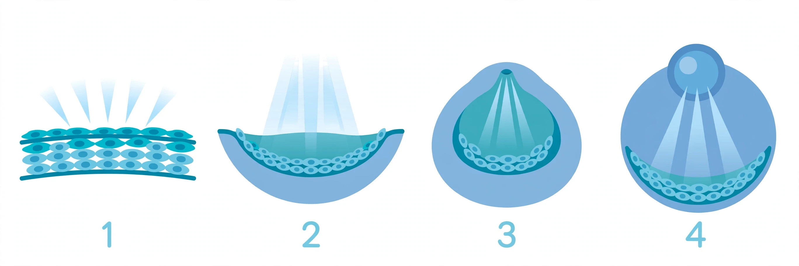
The evolution of the eye climbs a ladder of four stages: a flat patch sensing light, a pit sensing direction, a pinhole forming a crude image, and a lens sharpening it. Every rung exists in a living animal, so the ladder is not a guess about vanished forms. Each step works on its own, which is the whole point.
The clearest map of that ladder comes from the work of Dan-Eric Nilsson, a biologist at Lund University in Sweden, who spent years asking a simple question: what is the smallest useful improvement you can make to an eye? His answer was a sequence of four stages, each one a small change to the shape of a light-sensitive patch, and each one paying for itself the moment it appears. The stages are not a story about one species turning into another. They are a map of what is physically possible, and living animals happen to occupy every stop along the way.
Stage one is non-directional photoreception: a flat patch of light-sensitive cells, lying open to the sky. It can tell light from dark, and nothing else. The humblest eyespots are flat patches of this kind, with no cup to shade them: they can tell light from dark, and nothing else. That is a modest talent, and it is enough. A flatworm that can tell light from dark can hide under a stone when the sun is high, and come out to feed when the light fades. It does not need to know what cast the shadow. It only needs to know that the light changed, and to move.
Stage two is directional photoreception: the patch sinks into a pit, or a cup. Now the walls of the cup shade the cells on one side. Light coming from the left strikes a different set of cells than light coming from the right, because the rim of the cup blocks the rest. The animal has gained something new: direction. It can tell not just that there is light, but roughly where it is coming from. The slit-shell mollusc Pleurotomaria carries a pigment-cup eye of this kind, its cup shading the cells on one side so the animal can tell roughly where light is coming from. Pleurotomaria does not need a sharp image. It needs to know when a shadow falls across it from above, because that shadow might be a crab or a bird. Direction is the difference between freezing and fleeing the right way, and a pit gives it that.
No new chemistry was needed for that step. The patch only had to sink a little deeper, and the geometry did the rest.
Stage three is low-resolution vision, and it arrives when the cup deepens and its opening narrows. At some point, shrinking the opening becomes a better way to sharpen the picture than deepening the cup any further. Light can now enter only through a small hole, and each point on the retina receives light from only one direction. The result is a real image: dim, blurry, and upside down, but an image all the same. The animal can now make out shapes, not just directions. It can tell a large dark blob from a small one, a moving edge from a still one.
The living example is the nautilus, a shelled cephalopod that swims along deep reef slopes in the Indo-Pacific. The nautilus has no lens at all. Its eye is a pinhole camera, a dark chamber with a tiny opening, working on the same principle as a camera obscura, the darkened room with a small hole that painters used for centuries to project an image onto a wall. The image is dim, because the eye lets in so little light. But it works. A nautilus can find its way and sense the shapes around it with an eye that has no lens, no cornea, and no focusing mechanism of any kind.
That matters because it answers the most common objection to the ladder. People ask: what good is half an eye? The nautilus answers: a pinhole eye is not half an eye. It is a complete eye for the life the nautilus lives. It does not need to read fine print. It needs to navigate open water and spot large shapes, and a dim image does that job. The same is true of every rung. A flat patch is not a failed camera eye. It is a working light sensor. A pit is not a broken pinhole. It is a working direction finder. Each stage is a finished product for the animal that carries it, and also a starting point for the next stage.
Stage four in the evolution of the eye is high-resolution vision: a lens. A layer of transparent cells grows across the opening of the pinhole, and that layer begins to bend light, focusing it sharply onto the retina. At first the lens is crude, and the image it makes is only a little better than the pinhole's. But a little better is enough. An animal that can make out the fuzzy outline of prey a moment sooner, or spot the edge of a predator a moment earlier, eats more often and gets eaten less often. Over generations, small improvements to the lens — a clearer material, a better curve, a shape that can change — are favoured, and the image sharpens. The eye becomes a camera.
The lens itself is a story of borrowed parts. The proteins that make a lens transparent, the crystallins, did not evolve for vision. Many began as ordinary proteins elsewhere in the body, including heat-shock proteins that help other proteins keep their shape when a cell gets hot. At some point, one began to be made in the cells covering the eye, and it happened to have the right optical properties: clear, stable, able to bend light. The eye did not invent a lens from nothing. It recruited a protein that was already good at the job, and put it to new work.
Stand back and look at the ladder whole, and Darwin's three conditions for the evolution of the eye fall into place. Gradations exist, and they are swimming, crawling, and clinging to rocks right now. Variation is inherited, as it is for every trait. And each step is useful on its own, because each step answers a question the animal needs answered: brighter or darker, this way or that way, big shape or small shape, sharp or blurred. No step is a useless bridge to a future eye. The ladder is not a guess about the past. It is a description of the present.
5. The Nilsson–Pelger Model: A Camera Eye in Under 400,000 Years
In 1994 Dan-Eric Nilsson and Susanne Pelger calculated that, in the evolution of the eye, a camera eye could evolve from a light-sensitive patch in under 364,000 years. Their model needed 1,829 tiny steps, each improving the optics a little. The assumptions were deliberately pessimistic, which makes the short timescale the striking part.
The question Nilsson and Pelger asked was a simple one, and it was the same question Darwin had left open. Grant the ladder. Grant that each step is useful. How long would the climb actually take? If the answer were hundreds of millions of years, the eye would still be a problem, because the fossil record shows complex eyes appearing relatively quickly in the Cambrian period, when animal body plans were diversifying fast. If the answer were short, the problem would dissolve. Eyes would be not just possible, but easy, on geological timescales.
Their paper, published in 1994 in the Proceedings of the Royal Society B under the title "A Pessimistic Estimate of the Time Required for an Eye to Evolve", set out to answer the question in the most cautious way they could. The word "pessimistic" in the title is doing important work. They did not try to make the eye evolve as fast as possible. They tried to make it evolve as slowly as they reasonably could, and then asked whether even that slow pace was fast enough.
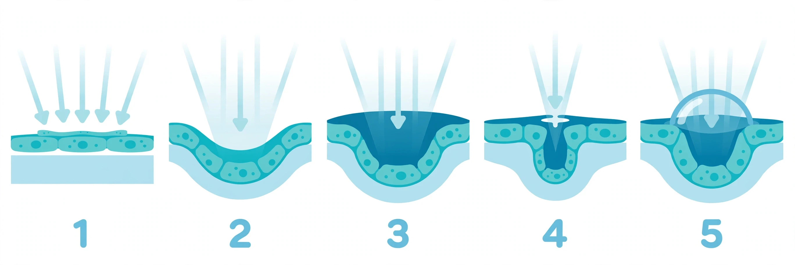
Here is how the model works. Start with a flat patch of light-sensitive cells. Allow its shape to change, but only in tiny steps: no single step may change any dimension by more than 1 percent. At each step, keep only the changes that improve the optics, judged by one measure — spatial resolution, how finely the eye can separate two points of light. Then repeat, letting the patch deepen into a cup, the cup narrow into a pinhole, and a lens grow across the opening.
Count the steps. The model needed 1,829 of them. That sounds like a lot, until you translate steps into time. Nilsson and Pelger estimated how many generations each step would take, based on the amount of variation available in a population and the strength of selection favouring a better eye. They used deliberately small values for both: little phenotypic variation (1%), and only weak selection (a selection intensity of 0.01). Even with those cautious numbers, the whole journey added up to 363,992 generations. Assume roughly one generation per year, which is common in small animals, and the total comes to under 364,000 years. Less than half a million years, from a flat patch to a camera eye.
Set that number against the timescale of life. The Cambrian period, when complex eyes first appear in the fossil record, began around 540 million years ago. If a camera eye takes under 364,000 years to evolve, then eyes could have evolved roughly 1,500 times over in the time since the Cambrian. That is not a claim that eyes did evolve 1,500 times. It is a way of saying that time was never the limiting factor.
The honesty of the model matters as much as its result, so here are its limits, stated plainly. The Nilsson–Pelger model covers optics and geometry only. It tracks how the shape of the eye changes, and how sharply that shape can focus light. It does not model the brain, or the neural processing that turns a pattern of light on the retina into a picture the animal can use. It does not model the genetics, the specific mutations that would have to occur, or the developmental rewiring that builds an eye from an embryo. The authors themselves were clear about this: the estimate applies where "selection for eye geometry and optical structures imposed the only limit." In other words, the model answers one question — how long would the optics take, if optics were the only constraint? — and it leaves the rest of the eye, the nerves and the brain and the genes, for other work.
That limit does not make the model useless. It makes it precise. Nilsson and Pelger isolated one part of the problem, made cautious assumptions about it, and showed that this part, at least, is fast. The brain and the wiring are real challenges, and later research, including the 2007 review of the evolution of the vertebrate eye by Lamb, Collin, and Pugh, has traced how photoreceptors, the retina, and the eye cup developed together. But the optical problem — the part that looks most like design — turns out to be the easy part. Shape changes a little, the image gets a little sharper, and selection keeps the improvement.
The model is also a theoretical calculation, not a simulation of random mutation: it asks what the best small improvement would be at each step, not the exact genetic route life took.
Even with those limits, the result changed the argument. Before 1994, the eye's complexity could be waved around as a vague problem: surely something this intricate must take an impossibly long time. After 1994, there was a number on the table, built from cautious assumptions, and the number was small. Critics could argue with the assumptions, and some did, but they could no longer argue from a feeling that the time must be vast. Anyone who wanted to claim the eye could not evolve had to explain why 1,829 small steps could not happen in under 364,000 years, when the fossil record gives life more than a thousand times that long.
That is the point to carry forward. The evolution of the eye did not need a miracle, and it did not need forever. It needed a patch of cells that could sense light, a series of small changes that each helped a little, and a stretch of time that, by the standards of geology, counts as a blink. The ladder exists. The climb is short.
6. The Master Switch: Pax6 and the Deep Unity of Eyes
One gene, Pax6, switches on eye development in animals from fruit flies to humans, and switching it on in the wrong place grows an extra eye. That single fact rewrote the story of the evolution of the eye, turning a puzzle about many separate inventions into a story about one ancient toolkit used again and again.
The story starts in a fly room, more than a century ago. In 1915, Mildred Hoge, a young geneticist working with fruit flies, found a mutant fly born with no eyes at all. She named the mutation eyeless.
The flies were fragile and short-lived, and the effect varied from fly to fly. Some had tiny, shrunken eyes. Others had none. Hoge bred them carefully, traced how the trait was inherited, and mapped the gene to the fourth chromosome of the fly.
For decades, eyeless sat in the textbooks as a curiosity: a fly gene that broke a fly eye. Nobody guessed it would turn out to be one of the most important genes in all of animal biology.
In 1994, Quiring and colleagues in Walter Gehring's laboratory in Basel cloned the eyeless gene. The sequence was a shock. The fly gene matched a mouse gene called Small eye, and both matched the human gene behind aniridia, a condition in which a person is born with little or no iris.
Three animals that could hardly look more different — a fly, a mouse, a human — carried the same gene, doing the same job. The gene was Pax6.
Pax6 is not a building material. It does not make lens protein or pigment. It is a switch. It codes for a transcription factor, a protein that binds DNA and turns other genes on and off.
Pax6 carries two DNA-binding regions, a paired domain and a homeodomain. From its position near the top of a long chain of command, it can set a whole eye-building programme in motion. Flip the switch, and a programme of roughly two thousand downstream genes follows.
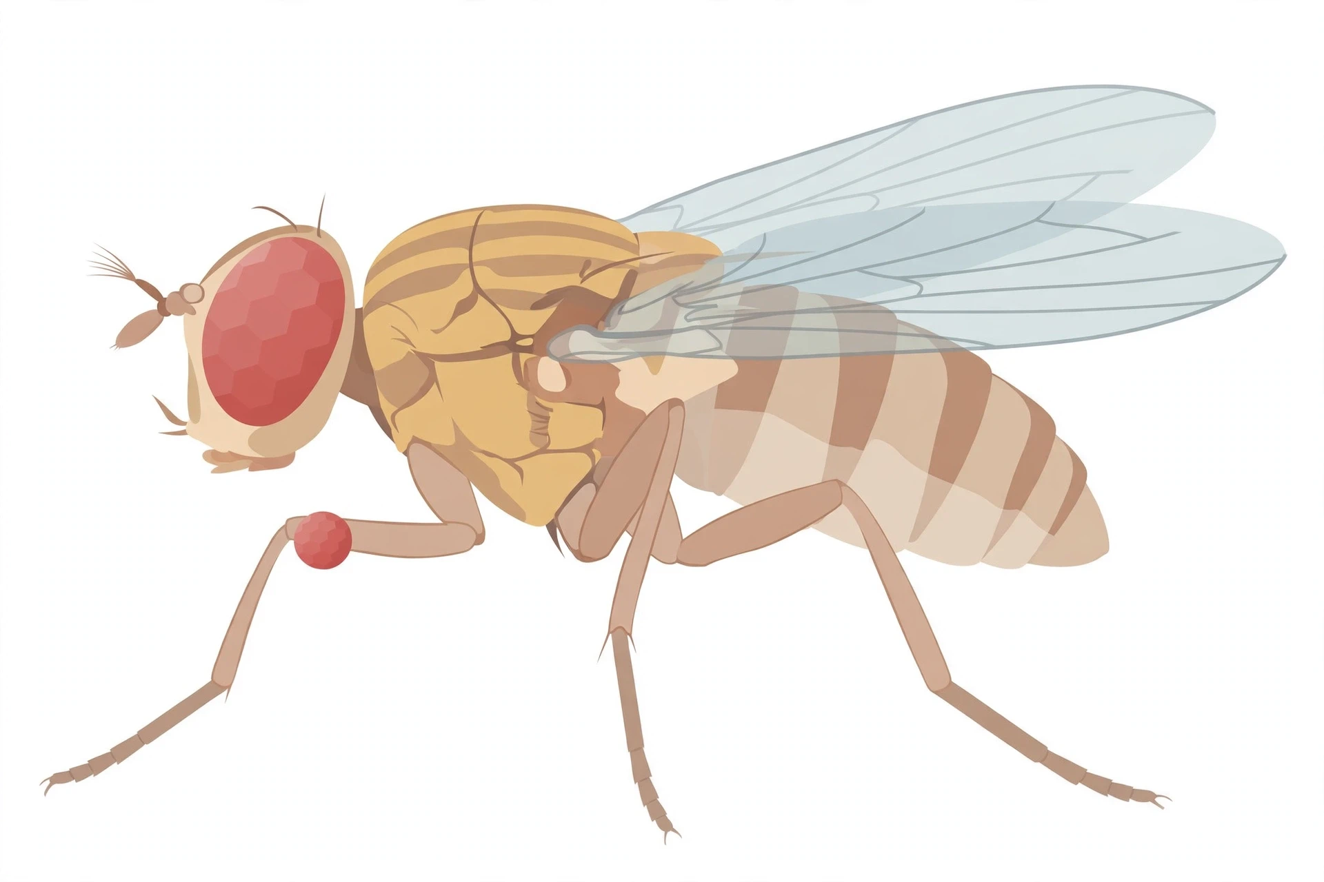
The proof came a year later, in an experiment that still sounds like science fiction. In 1995, Halder and colleagues took the fly version of the gene and switched it on in tissues where it had no business being active: the discs of tissue that would become a leg, a wing, or an antenna.
The result was ectopic eyes — real, extra eyes growing on legs, wings, and antennae. Under the microscope they looked like proper fly eyes, built from fully formed ommatidia with a complete set of photoreceptor cells. Later recordings showed that some of the eyes on the antennae could even respond to light.
Then came the experiment that made the point impossible to ignore. The team took the mouse version of Pax6, put it into a fly, and switched it on in the same wrong places. The mouse gene grew fly eyes.
Not mouse eyes — the fly provided all the local building instructions — but the command itself crossed a gap of hundreds of millions of years of separate evolution and still worked. A mammalian switch could run an insect eye programme. Gehring later recalled that after several months of failure, the success made the front page of the New York Times — in his 2005 account he gives the headline as "Scientists Out Do Hollywood."
When the switch breaks
The same gene that builds eyes can also break them, and the breakage looks strikingly similar across species. In humans, mutations in Pax6 cause aniridia, the absence or severe reduction of the iris. In mice, the matching mutation is called Small eye, because animals carrying one faulty copy grow up with small, reduced eyes. Animals with two faulty copies die at or shortly after birth, lacking eyes and a nose, and missing a large part of the forebrain.
In flies, the classic eye-disc eyeless mutants shrink or erase the compound eyes while leaving the simple eyes on top of the head untouched. Lose the switch, and the eye programme never starts. The organs differ wildly, yet the command that starts them is shared.
One toolkit, many eyes
For most of the twentieth century, the standard view was that eyes had evolved independently 40 to 65 times. The reasoning was straightforward. A camera eye, a compound eye, and a mirror eye look nothing alike.
They develop in different ways, from different tissues, in animals whose last common ancestor lived long before any of them had eyes. How could such different organs share a single origin? They must have been invented again and again.
Pax6 seemed to break that logic. If eyes were invented separately dozens of times, why would they all be switched on by the same gene? Gehring argued in his 1996 review that Pax6 was "the master control gene for morphogenesis and evolution of the eye," and that the many kinds of eyes might have grown from a single prototype.
The puzzle needed a careful answer, and Russell Fernald gave it in 2004. The genetic toolkit is ancient and shared, while the organs themselves were built many times by recruiting that same toolkit.
Think of it like a set of Lego bricks handed down through a family. The bricks are old, and every cousin has the same set. But each cousin builds something different with them — a house, a bridge, a tower.
The shared bricks do not mean the buildings are the same building. They mean the builders started from the same box.
In the case of eyes, the ancient toolkit included Pax6 and its partners, such as the Six genes, which help direct eye development in animals from planarians to humans.
Long before there was a camera eye or a compound eye, the evolution of the eye began with some early animal that probably had a simple patch of light-sensitive cells, switched on by Pax6. Each lineage then recruited that old programme and built onto it. The toolkit stayed. The organs diverged.
This is why the story of the evolution of the eye is not a simple choice between "evolved once" and "evolved many times." The genes are monophyletic, descended from a common ancestral programme. The organs are polyphyletic, built repeatedly. Both statements are true at once, once you separate the switch from the machine it controls. It also explains a pattern that runs through the whole history of animal classification: animals that look nothing alike on the outside can share deep genetic machinery on the inside, because the machinery predates the differences.
7. Two Kinds of Light Catchers
The evolution of the eye produced two different photoreceptor designs, ciliary cells in vertebrates and rhabdomeric cells in most invertebrates, yet both catch light with the same opsin molecules. The split is old, deep, and visible in your retina, where a leftover rhabdomeric sensor keeps time instead of forming images — a living record of the evolution of the eye.
Every photoreceptor faces the same engineering problem. Light is faint, and a single opsin molecule can only catch one photon at a time. To catch enough light to be useful, a cell needs to pack as many opsin molecules as possible into its membrane.
The trick is surface area. A flat cell has very little. A folded cell has far more. The two kinds of photoreceptors solve this folding problem in opposite ways.
A ciliary photoreceptor folds its membrane the way you might fold a stack of paper. The light-catching surface grows out of a cilium, the tiny hair-like structure that cells use for sensing and movement, and stacks itself into discs or folded sheets.
Your rods and cones are ciliary photoreceptors. Each rod carries a tall stack of discs, packed with the opsin called rhodopsin, like a roll of coins in a tube.
A rhabdomeric photoreceptor folds its membrane the way a brush bristles. The surface grows into a dense forest of tiny finger-like projections called microvilli. Together they form a structure called a rhabdom, and the opsins sit in the walls of those fingers.
This is the design used by insects, spiders, squid, and most other invertebrates. A fly's photoreceptor is a brush. Yours is a stack of plates.
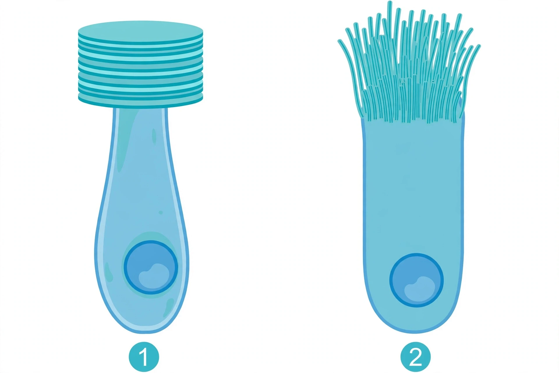
Two opsins, two signalling chains
The difference runs deeper than shape. Each cell type uses its own family of opsin, and its own chemical chain of events after light arrives. Vertebrate ciliary cells use c-opsins. Invertebrate rhabdomeric cells use r-opsins.
When light hits a c-opsin, the signal travels through a G protein called transducin and an enzyme that lowers the level of a messenger called cyclic GMP inside the cell. When light hits an r-opsin, the signal travels through a different G protein and an enzyme called phospholipase C. Same starting point — an opsin wrapped around a light-absorbing retinal chromophore — and a different route to the same destination, a nerve signal.
For a long time, biologists assumed the split was neat: vertebrates had ciliary cells, invertebrates had rhabdomeric cells, and the two lines had diverged long ago. Detlev Arendt's work showed that the truth is older and stranger.
The two cell types descend from two distinct ancestral photoreceptor cells that already existed in the common ancestor of bilaterians, the animals with left and right sides, more than 600 million years ago.
The evidence came from a small marine worm, Platynereis dumerilii, that still carries both designs. Its eyes use rhabdomeric cells with r-opsin, as expected for an invertebrate. But deep in its brain sit ciliary photoreceptors that use c-opsin, the vertebrate kind.
One animal, both cell types. The simplest reading is that the common ancestor of bilaterians already owned both kinds of light catcher.
That reading reshapes the family tree of your own retina. Evidence suggests that your rods and your cones descend from that ancient ciliary photoreceptor, while the bipolar cells that carry their signals appear to have a mixed origin, linking the ciliary and rhabdomeric lines.
Your ganglion cells, amacrine cells, and horizontal cells, by contrast, appear to descend from the rhabdomeric line. Your retina is not the product of one cell type. It is a layered collaboration between two ancient lines, folded together into one sheet of tissue.
The sensor that tells time
The most surprising leftover sits in plain sight. Vertebrates kept an r-opsin-like molecule too, even though their image-forming photoreceptors are all ciliary. It is called melanopsin, and it lives in a small set of retinal ganglion cells, the very cells that carry visual signals out of the eye toward the brain.
These melanopsin cells are intrinsically photosensitive: they can sense light on their own, even when every rod and cone is silenced. They make up only a tiny fraction of retinal ganglion cells — about 0.2% in primates, roughly 3,000 cells per human eye — yet they carry a message the rest of the retina does not.
They report how bright the world is, not what it looks like. Their axons run to the suprachiasmatic nucleus, the brain's master clock, and to the olivary pretectal nucleus, which controls pupil size; through the clock, they also govern the nightly release of melatonin. When morning light strikes melanopsin, which responds most strongly to blue light near 480 nanometres, the clock resets, melatonin falls, and the body learns that the day has begun.
This is non-image-forming vision. Some people who have lost all rods and cones cannot see at all, yet their sleep rhythms can still follow the sun, because melanopsin still reports brightness to the brain's clock. Their image vision is gone, but their clock vision remains.
Counting the sensors
Your retina is a dense census of ciliary cells. As approximate textbook figures, a human retina holds roughly 120 million rods and 6 million cones. The rods spread across the periphery, where they catch faint light and movement in the dark.
The cones crowd into the centre, especially in the fovea, the tiny pit that gives you sharp central vision. The fovea has no rods at all, only cones, packed tighter than anywhere else in the eye. That is why you see fine detail best when you look straight at it, and why a faint star vanishes when you stare directly at it but reappears when you glance slightly to the side.
The numbers also explain the trade-off built into your own vision. Rods are sensitive but colour-blind and blurry. Cones are sharp and colourful but need bright light.
You carry both because your ancestors needed both: the night watch and the day shift, in the same eye. Signals from both travel through the nervous system to the brain, where the picture is assembled.
Feature | Ciliary photoreceptors | Rhabdomeric photoreceptors |
|---|---|---|
Animals | Vertebrates, including humans | Most invertebrates, including insects and molluscs |
Opsin type | c-opsins | r-opsins |
Membrane shape | Folded into discs or stacked sheets from a cilium | Folded into microvilli forming a rhabdom |
Example | Human rods and cones | Photoreceptors in a fly compound eye |
8. Camera Eyes vs Compound Eyes
The evolution of the eye produced two different engineering solutions to the same problem: vertebrates focus light through one lens onto one retina, while insects build thousands of tiny lenses into a compound eye. Each wins in a different kind of life, one prizing sharp detail and the other prizing a wide, watchful view.
Your eye works like a camera, because in a deep sense it is one. Light enters through the cornea, passes through the pupil, and is bent by the lens onto the retina at the back. The lens changes shape to focus on near or far objects.
The iris opens and closes to control how much light gets in. The image lands upside down on the retina, and the brain flips it the right way up. It is a compact, elegant design, and it puts a huge number of photoreceptors behind a single optical path.
An insect eye works nothing like that. A fly or a dragonfly does not have one lens. It has thousands. Each unit, called an ommatidium, is a tiny eye in its own right: a small lens on the surface, a cone-shaped light guide beneath it, and a cluster of photoreceptors at the bottom.
Each ommatidium looks in a slightly different direction and reports the brightness in that one direction. The brain stitches those thousands of separate reports into a single mosaic picture.
A dragonfly carries this design to an extreme. An adult dragonfly's compound eyes have nearly 24,000 ommatidia each — the largest species reach up to about 30,000 — and the eyes wrap around most of its head like a helmet. The animal lives by sight, hunting other insects in mid-air, and its eyes are built for that chase.
Sharpness against watchfulness
No design wins everywhere, because physics forces a trade. A camera eye wins on resolution. All of its light passes through one lens onto a dense sheet of receptors, and the centre of the retina — the fovea — packs cones so tightly that you can read small print or recognise a friend's face across a street.
A compound eye cannot match that. Each ommatidium is one pixel, and the pixel is the size of the lens. To see finer detail, an insect would need smaller lenses, but smaller lenses catch less light and blur the image through diffraction. There is a hard physical limit to how sharp a compound eye can be at a given size.
A compound eye wins on the things a camera eye cannot do. Its field of view is enormous. Because the lenses curve around the head, an insect can see in nearly all directions at once, with only a small blind spot behind it.
A dragonfly sees almost the whole sky without turning its head. A camera eye, by contrast, sees a narrow cone of the world clearly, and must swivel in its socket to look elsewhere.
Compound eyes also win on motion. Each ommatidium samples the world quickly, and the eye as a whole is exquisitely sensitive to anything that moves across its field. A fly notices your hand coming long before you reach it, not because it sees your hand clearly, but because a ripple of brightness sweeps across hundreds of ommatidia in a fraction of a second.
That is why swatting a fly is so hard. You are aiming at an animal whose entire visual system is built to detect your approach.
Night brings a third trade-off. Day-flying insects such as bees and butterflies use apposition compound eyes, in which each ommatidium is screened off from its neighbours, so each one sees only its own narrow slice of light. The image is crisp, but each receptor gets very little light.
Nocturnal moths use a different design, the superposition compound eye. A clear zone sits between the lenses and the retina, and light from many facets is pooled onto a single receptor. The moth gains a far brighter image at the cost of fine detail. It is the same bargain a photographer makes when opening the aperture wide in dim light: more light, less sharpness.
Convergence, written by physics
Camera eyes and compound eyes each evolved multiple times independently. Vertebrates built a camera eye. Cephalopods — squid and octopuses — built another, so similar in outline that early anatomists were startled, yet wired differently inside.
Box jellyfish, animals without a brain at all, carry camera-like eyes too. Compound eyes arose repeatedly among arthropods, and in different forms in different groups.
This is convergence driven by physics. The laws of optics allow only a small number of workable designs — Fernald counted eight known optical systems in animal eyes — and evolution keeps rediscovering them. Light can be focused by a lens, reflected by a mirror, or channelled through many small tubes. There are only so many ways to bend light onto a sensor.
The variety is part of what makes the biodiversity of animal vision so rich. A scallop lines the rim of its shell with dozens of tiny mirror eyes. A deep-sea fish grows a tubular eye that stares upward through the dark.
Each is a local answer to a local life, built from the same ancient toolkit, shaped by the same laws of light.
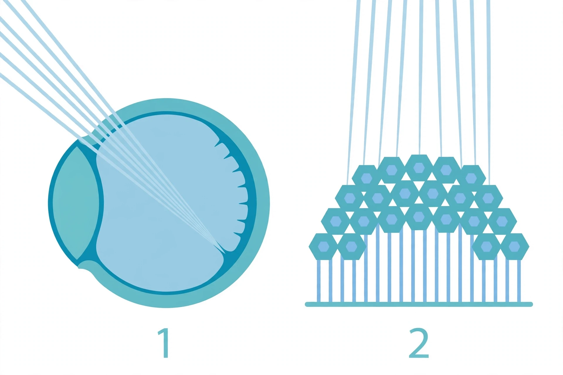
Feature | Camera eye | Compound eye |
|---|---|---|
Structure | One lens focusing light onto one retina | Thousands of ommatidia, each with its own tiny lens |
Resolution | High, especially at the fovea | Low to moderate, limited by ommatidium size |
Field of view | Narrow, must turn the eye or head to look around | Very wide, near-panoramic |
Motion detection | Good, but best in the centre of the field | Excellent across the whole field |
Animals | Vertebrates, cephalopods, box jellyfish | Insects, crustaceans, and other arthropods |
Neither design is the "best" eye in any absolute sense. In the evolution of the eye, the best eye is the one that fits the life its owner leads.
A dragonfly needs to catch a mosquito in mid-air without being eaten itself. A moth needs to find a flower in the dark. The evolution of the eye is not a ladder climbing toward the human eye. It is a branching tree of bargains, each branch trading sharpness for watchfulness, or brightness for detail, in the currency of survival.
9. The Backward Retina and the Blind Spot
Your retina is wired backwards — one of the strangest quirks in the evolution of the eye. The light-sensing cells point away from incoming light, and the optic nerve punches a hole through the retina, leaving a blind spot your brain quietly paints over. The flaw is real, and it survives because evolution works with what already exists.
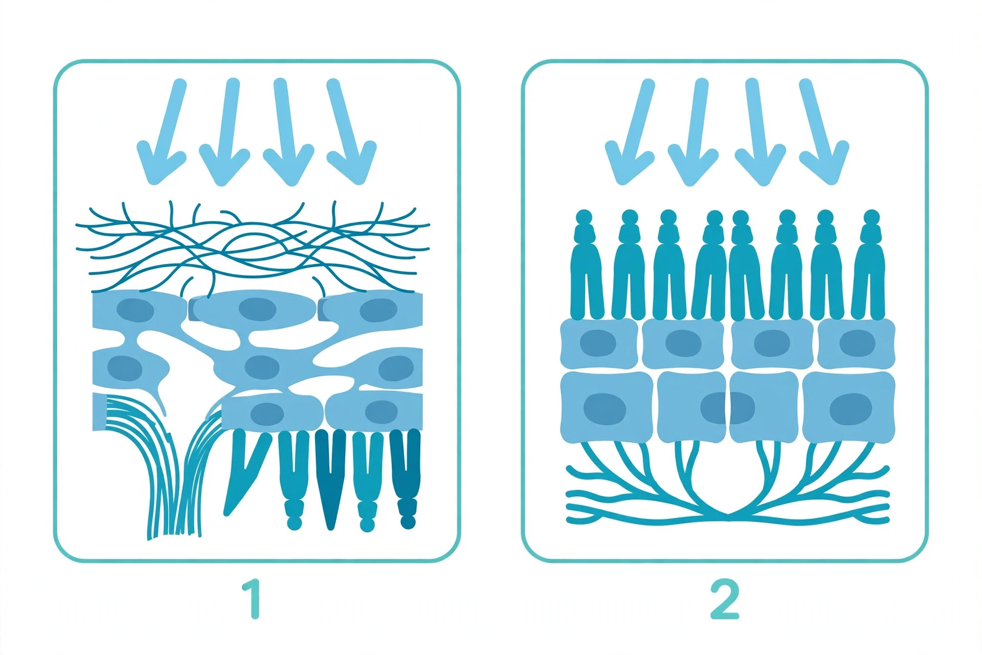
Picture the retina as a sheet of film at the back of the eye. In a well-designed camera, the film faces the lens, and the wiring runs out the back. In your eye, the film faces away from the lens.
The rods and cones sit at the very back of the retina, pointing away from the incoming light, while the nerve fibres and blood vessels that serve them sit in front, between the light and the sensors. Every photon that reaches a photoreceptor must first pass through a tangle of wiring.
It is a strange arrangement, and it has a visible consequence. All the nerve fibres from the retina gather into a single bundle, the optic nerve, which must leave the eye to reach the brain. To get out, it punches straight through the retina.
Where it exits, there is a patch with no photoreceptors at all. That patch is the blind spot, about 15 degrees out from the centre of your visual field, on the outer (temporal) side, away from your nose. Anything whose image falls entirely on that patch simply is not seen.
Try it yourself
You can find your blind spot in less than a minute. Close your left eye. Fix your right eye on a spot straight ahead of you, perhaps a mark on the wall. Hold your right thumb out to your right at arm's length, so it sits well out in your peripheral vision.
Now move it slowly inward, toward the centre of your view, without moving your gaze. At some point the thumb will vanish. Keep moving it, and it will pop back into view on the other side.
I first tried this test as a teenager, sitting at a kitchen table, certain my eyes could not possibly have a hole in them. I moved my thumb back and forth, waiting for a trick, and then it was simply gone — not black, not blank, just absent, as if that piece of the room had never been there.
What startled me was not the gap itself. It was how smooth the wall behind it looked. My brain had filled the hole with more wall, and I would never have known unless someone had told me where to look.
That filling-in is the second half of the story. The blind spot does not look like a hole, because the brain refuses to show you one. It takes the colour and pattern from the surrounding retina and paints across the gap.
With both eyes open, each eye covers the other's blind spot anyway, so the gap almost never matters. The system is flawed and patched at the same time, and the patch is so good that most people go their whole lives without noticing the flaw. It is the same kind of quiet editing the brain performs in optical illusions, where what you see is the brain's best guess about the world, not a raw photograph of it.
Why the wiring ended up backwards
The backward retina is not a mistake a designer made once and never fixed. It is a consequence of how the eye is built in the embryo. The vertebrate eye does not grow inward from the skin. It grows outward from the brain.
Early in development, a pouch called the optic vesicle bulges out from the developing forebrain, then folds in on itself to form a cup. The inner wall of that cup becomes the retina. Because the retina is brain tissue folded inside out, its wiring ends up on the wrong side, facing the light, and the photoreceptors end up at the back, facing away.
Once that arrangement exists, evolution cannot simply flip it. Rewiring the retina would mean rebuilding the eye from scratch, and every intermediate step would have to work at least as well as the eye it replaced.
A half-rewired eye would be worse than the one it replaced, not better, and its owner would be outcompeted long before the new design was finished. Natural selection has no foresight. It cannot accept a worse eye now in exchange for a better eye a million years later. So the backward retina persists, patched by the brain, because it works well enough.
Cephalopods show that a different history was possible. A squid or an octopus builds a camera eye that looks, from the outside, much like yours: a lens, an iris, a retina. But its retina is wired the "right" way round.
The photoreceptors face the light, and the nerve fibres run out behind them. There is no blind spot, because no nerve bundle needs to punch through the light-sensitive sheet. The difference is not that cephalopods are better designed. It is that their eye grew from the skin, not from the brain, so the wiring never ended up on the wrong side.
Two animals, two camera eyes, two developmental histories — and only one of them carries the flaw.
That contrast is the point of the backward retina — and of the evolution of the eye as a whole. In the evolution of the eye, nature is a tinkerer, not an engineer with a blank sheet.
It builds on what came before, keeps what works, and patches what does not. The eye you see with is very good. It is also, in one small and measurable way, wired backwards. And the fact that you can read this sentence at all, through a retina that faces the wrong way, past a blind spot your brain has quietly painted over, is a measure of how well the patch works.
10. The Octopus Eye: Evolution's Second Draft
The octopus built a camera eye almost identical to ours — lens, iris, retina — but it evolved independently, and in one respect it is built better than ours. Its photoreceptors face the light, so it has no blind spot. That second draft of the design is one of the clearest proofs that evolution can reach the same answer twice.
Hold an octopus eye beside a human eye and the resemblance is startling. Both have a clear cornea at the front, an iris that opens and closes, a lens that focuses light, and a retina at the back that turns light into nerve signals. A doctor who had never seen either animal before might guess they were close cousins. They are not.
The last common ancestor of an octopus and a human lived more than 500 million years ago, long before either lineage had anything you could call a camera eye. Each line invented the design on its own.
That is what biologists mean by convergent evolution — and the evolution of the eye is full of it. Two separate histories, facing the same problem — how to form a sharp image in water or air — arrived at the same solution. The story of the evolution of the eye is full of such repeats, but the octopus is the most famous. Squid and cuttlefish, its fellow cephalopods, carry the same design.
Their eyes are not copies of ours. They are parallel inventions, built from different starting materials, and the differences between the two versions tell us as much as the similarities do.
The most important difference sits in the retina. In your eye, the light-sensitive cells face away from the incoming light. Nerve fibres and blood vessels lie in front of them, and the fibres must bundle together and punch through the retina to leave the eye. Where they punch through, there are no light-sensitive cells at all.
That gap is your blind spot. Your brain papers over it so neatly that you never notice, but it is there in every human eye, a small hole in the middle of the picture.
The octopus has no such hole. Its photoreceptors face forward, toward the light, with the nerve fibres running behind the retina. The fibres can leave through the back of the eye without disturbing a single light-sensitive cell.
Light reaches the receptors directly, with nothing in the way. In this one respect, the cephalopod eye is wired the right way round, and ours is wired backwards.
The two eyes also start life differently. Your eye grows as an outgrowth of the brain, a pocket of brain tissue that pushes outward toward the skin. The octopus eye grows the other way. It forms as an invagination of the skin, a dimple in the surface that sinks inward and closes into a ball.
Same finished shape, opposite construction. It is as if two builders were asked to make the same house, one starting from the basement and the other from the roof, and both ended up with the same rooms in the same places.
Look into an octopus eye and you will see another small difference. The pupil is not a round hole. In octopuses and cuttlefish it is a horizontal slit, held level even when the animal turns.
The shape may help the animal judge colour by the way different wavelengths blur, a trick that matters for an animal with only one type of photoreceptor, where humans have three. Yet an octopus can match the colour and texture of a rock or a reef in seconds, changing its skin like a living screen, and vanish in front of your eyes.
I remember the first time I stood in front of an octopus at an aquarium and felt watched in a way I had never felt watched before. The animal was curled in a corner of its tank, the colour of wet stone, and one eye was fixed on me. It was a large eye, dark and gold-ringed, and it moved as I moved. I shifted a step to the left, and the eye followed.
I leaned closer, and the pupil narrowed to a thin dark line. I had looked into the eyes of dogs and cats and horses, and there was always a sense of shared history in the look, a distant kinship. This was different. This eye had been built by a separate history, along a separate road, and it was looking back at me anyway.
I stood there longer than I meant to, caught between feeling like an observer and feeling like prey, and I left thinking that the strangeness was the point. Two kinds of mind, two kinds of eye, meeting across half a billion years of separate evolution.
Yet beneath that strangeness, the two eyes share an old toolkit. The gene Pax6 acts as a master switch for eye development in animals from fruit flies to mice, and versions of the same gene family guide eye formation in cephalopods too. In squid, a close cousin of the octopus, Pax6 is switched on as the eye forms. The shared toolkit does not mean the two camera eyes were inherited from one ancestral eye.
It means the ancestor of both animals already carried the genetic switches for sensing light, and each lineage recruited those switches again, independently. Deep homology at the level of genes, convergence at the level of the organ — a pattern that runs through the whole evolution of the eye.
The nautilus is the living counterpoint to all of this, a reminder that the camera eye was not the only answer. The nautilus is a cephalopod too, a distant cousin of the octopus that still swims in a coiled shell, much as its ancestors did hundreds of millions of years ago. Its eye has no lens at all. It is a pinhole eye, a dark chamber with a small opening, working on the same principle as a pinhole camera.
Light passes through the tiny hole and paints a dim, upside-down image on the retina. The picture is faint and fuzzy, and the animal cannot focus it, but it is a real image, and it is enough to tell light from dark and a shadow passing overhead from empty water.
The nautilus eye matters in the evolution of the eye because it is a surviving intermediate, a design that works well enough to persist. It shows that evolution did not need to leap straight to a lens. A cup of light-sensitive cells, deepened into a pit, narrowed at the mouth, could already see. Add a lens later and the image sharpens.
The octopus took that road all the way to a camera eye. The nautilus stopped at the pinhole and stayed there, because for its slow, deep-water life, a dim picture was enough. Both are honest answers to the same question, and both are still swimming.
11. Eyes That Break the Rules
In the evolution of the eye, nature's strangest eyes break every rule of thumb — a shrimp with a dozen colour channels, a jellyfish with 24 eyes, and a scallop whose eyes are tiny mirrors. Each one forces us to rethink what an eye is for.
The Mantis Shrimp: Twelve Channels and a Surprise
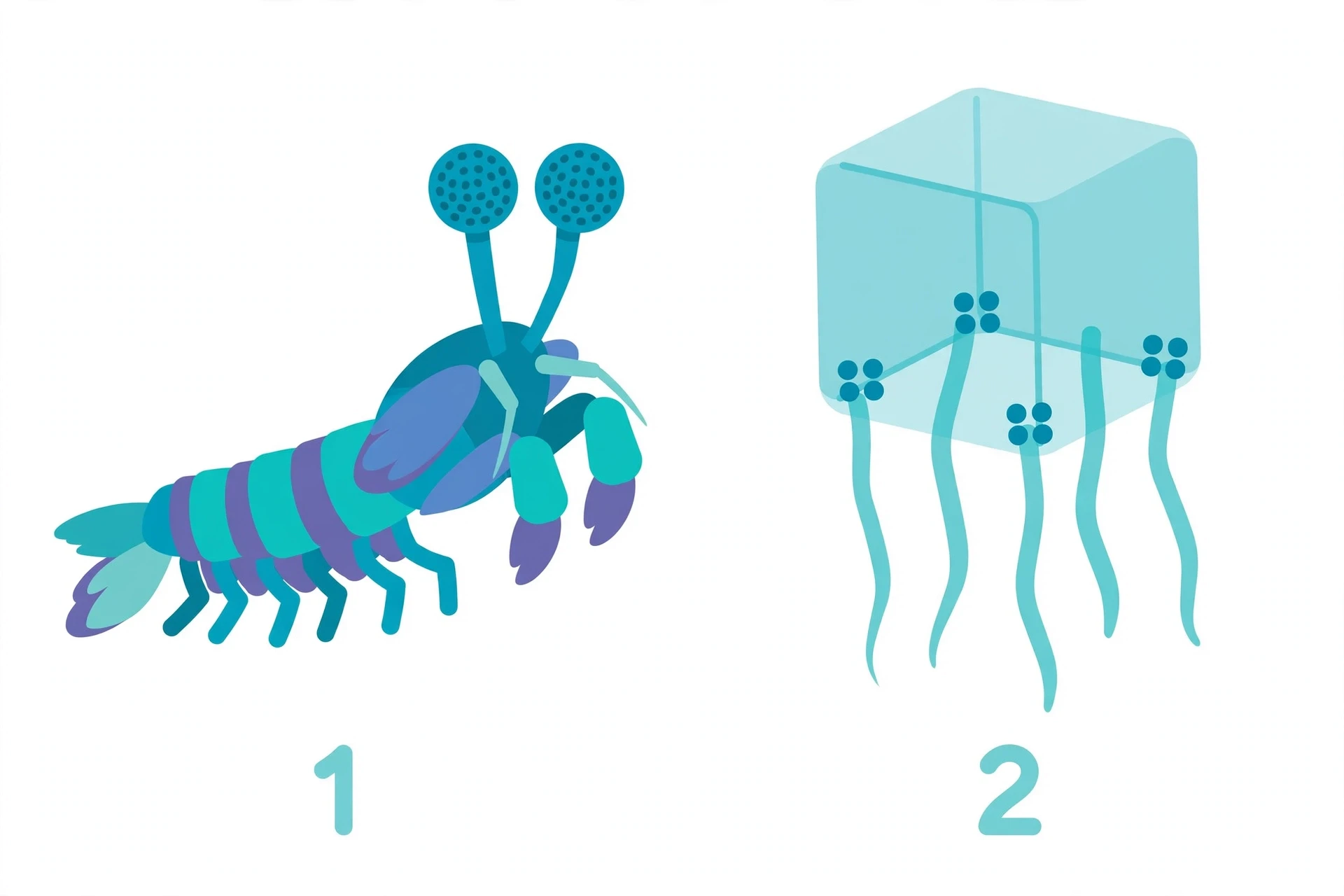
The mantis shrimp is a small, brightly coloured crustacean that lives on tropical coral reefs, and it carries what Guinness World Records calls the most complex eyes in the animal kingdom. Each eye is a compound eye built from thousands of tiny units, and it can contain 12 to 16 different photoreceptor types.
In the most complex species, twelve of those are devoted to colour, covering wavelengths from 300 to 720 nanometres, from deep ultraviolet through the whole visible spectrum to far red. The remaining four detect polarised light. Humans, by comparison, have three colour receptors.
Each mantis shrimp eye is also divided into three regions that can focus independently, so a single eye has what researchers call trinocular vision — three views from one eye, where we need two eyes for depth. And the shrimp can see circularly polarised light, a twisting form of light that no other animal is known to detect. Parts of the shrimp's own shell reflect it, which suggests the animals may be sending each other secret signals, messages written in a kind of light that their predators and prey cannot read.
With all that hardware, you would expect the mantis shrimp to be the finest colour judge on the reef. It is not. That is the twist, and it came from a study led by Hanne Thoen at the University of Queensland, published in Science in 2014. Thoen trained mantis shrimp of the species Haptosquilla trispinosa to associate food with a particular colour, then offered them colours closer and closer to the one they had learned.
If more colour channels meant finer colour vision, the shrimp should have beaten humans easily. Instead, they were worse than humans at telling similar colours apart, and worse than bees, fish, and butterflies too. When two colours differed by only a small amount, the shrimp guessed at chance level, while a human could still tell them apart without effort.
The explanation seems to be speed, not precision. Human colour vision works by comparison. The brain takes the signals from three receptors and compares them, a slow calculation that yields fine distinctions. The mantis shrimp seems to skip the comparison.
Each of its many receptors appears wired to recognise its own narrow slice of the spectrum directly, like a set of labelled bins. The animal does not work out exactly which shade it is seeing. It recognises the bin, and moves on. That is a fast system, and for a reef animal that must spot prey and predators in a flickering, multicoloured world, fast may matter more than fine.
The Box Jellyfish: Twenty-Four Eyes, No Brain
The box jellyfish sounds like a creature that should not be able to see at all. It has no brain, no heart, and a body that is mostly water. Yet it is an active hunter, and it hunts with 24 eyes.
The eyes are arranged in four clusters, called rhopalia, one hanging from each side of the bell-shaped body. Each cluster holds six eyes. Four of them are simple pits and slits that sense the presence and direction of light. The other two are genuine image-forming eyes, with a lens, a retina, and a cornea, built with very similar genes to the ones that build our eyes.
The jellyfish has no brain to process the pictures, so the work is done locally, in the nerve net around each cluster. The eyes send signals straight to the muscles, and the animal steers. The clusters are weighted with heavy crystals, so they always hang upright, even when the jellyfish swims upside down, giving it a steady view of the sky to navigate by near the mangrove forests where its prey gathers. Eyes came before brains in the evolution of the eye, and the box jellyfish kept that ancient arrangement: a light-sensitive patch needs only to be wired to something that can move.
The Scallop: A Telescope in a Shell
Scallops are easy to underestimate. They sit on the sea floor, filtering food from the water, and they look like nothing so much as a pair of ridged shells. Lift the shell, and you will find up to 200 tiny eyes along the edge of the soft mantle, each one no wider than a grain of sand, each one shining blue.
Each scallop eye works like a reflecting telescope. Light enters through the pupil, passes a lens and two retinas, and strikes a curved mirror made of guanine crystals at the back of the eye. The mirror reflects the light back onto the retinas, where the image forms. It is the same principle as the great telescopes that astronomers point at the sky, shrunk to the size of a pinhead and repeated two hundred times around a shell.
For years, the optics puzzled researchers. The main retina sits so close to the mirror that the image falling on it should be badly out of focus, a blur rather than a picture. A study published in Current Biology in 2019, led by researchers at the University of South Carolina, found part of the answer. Scallop eyes have pupils that dilate and contract in response to light.
The pupil can shrink to about 50 to 60 percent of its fully open area, though the change takes several minutes, far slower than the blink of a human iris. The scallop eye has no iris like ours. Instead, muscle-like fibres in the cornea may change its shape, going from thin and flat to tall and long, and that may reshape the eye itself.
Narrowing the pupil sharpens the image, trading a little sensitivity for a clearer picture, just as squinting sharpens the view for us. Two hundred dim eyes add up to a full circle of vigilance, telling the animal when to snap shut or swim away.
12. The Fossil Record of Vision
The oldest certain fossil eye is about 530 million years old — and it belonged to a trilobite. Stone can preserve what soft flesh cannot, and the trilobite's eyes are the earliest hard evidence that animals had learned to see.
The fossil comes from Saviranna in northern Estonia, and it belongs to a trilobite called Schmidtiellus reetae. It lived about 530 million years ago, in the early Cambrian, when the seas were filling with the first complex animals. Its right eye was partly worn away, and that accident of preservation gave researchers a view inside the organ.
They counted about 100 ommatidia, the tiny visual units of a compound eye, spaced further apart than in a modern insect eye. The structure was similar to the compound eyes of present-day dragonflies and bees, but simpler, a first draft of a design that would later become one of the most successful in the animal kingdom.
There may be older eyes still. Animals were already sensing light before the Cambrian began, but no eye fossil is known from the Ediacaran period, about 555 million years ago — those early eyes were soft tissue, and soft tissue almost never fossilises. The trilobite from Estonia is the oldest eye we can be sure about, and it is a good place to start the story in stone.
Trilobites are the great eyewitnesses of the early fossil record. They were marine arthropods, distant relatives of spiders and crabs, and they dominated the seas of the Palaeozoic era for nearly 300 million years. Many of them carried elaborate compound eyes, and later trilobites built those eyes in a way no other animal ever has. Their lenses were made of calcite, a crystal form of calcium carbonate, the same mineral as limestone and marble.
Trilobites are the best-known animals to have made lenses from calcite. Almost every other lens in the animal kingdom is made of protein, soft and transparent — a rare living exception is the chiton, whose lenses are made of aragonite, another mineral form of calcium carbonate. Trilobite lenses were stone, and stone survives. Some trilobites carried hundreds of lenses in each eye, the calcite crystals lined up in a single direction so light was not split into double images.
The Burgess Shale, in the Canadian Rockies, shows how far vision had already diversified by about 508 million years ago. The shale was laid down in the middle Cambrian, in fine mud that buried animals quickly and preserved soft parts that almost never fossilise. When the rock is split open, it reveals a crowded sea, and many of its animals had eyes. Anomalocaris, a predator about a third of a metre long, carried stalked compound eyes that could scan the water for prey.
Opabinia, a smaller animal with a long snout like a vacuum hose, had five eyes. Within a few tens of millions of years of the first certain fossil eye, the seas already held many different designs, for many different lives.
The story the fossils tell about the evolution of the eye is real, but it is also incomplete, and honesty requires saying so. Fossils are biased toward hard parts. Shells, bones, and calcite lenses survive. Retinas, optic nerves, and brains almost never do.
A fossil eye can show us the shape of a lens and the number of facets, but it cannot show us what the animal saw. We cannot look at a trilobite and learn whether it saw in colour, or how sharp its vision was, or how its brain turned patterns of light into behaviour. The soft machinery of sight rots away, and the rock keeps only the housing.
That bias shapes the whole record of the evolution of the eye. The earliest eyes were almost certainly soft, simple patches of light-sensitive cells on animals with no shells and no skeletons, and those animals left no trace at all. The fossil record of vision begins, in effect, at the moment eyes became hard enough to fossilise, not at the moment eyes first evolved. There is a long, dark stretch of history before Schmidtiellus reetae in which eyes were already evolving, and we will never see it directly.
We can only infer the early evolution of the eye from the genes that living animals still carry, and from the designs that survive in creatures like the nautilus and the box jellyfish. To read more about how palaeontologists piece together deep time from such fragments, see the fossil record article, and for the great dyings that repeatedly reset the seas those eyes swam in, see mass extinctions.
13. The Cambrian Explosion: When Seeing Changed Everything
About 541 million years ago animal life diversified explosively — and one popular idea says the trigger was vision itself: once animals could see, predators and prey entered an arms race. If that idea is right, then the evolution of the eye did not just change how animals sensed the world. It changed the world.
The Cambrian explosion is the name for a burst of diversification at the start of the Cambrian period, about 541 million years ago. Before it, the seas held mostly soft-bodied creatures, many of them flat, slow, and simple, drifting or creeping across the sea floor. Within about 20 to 25 million years — a blink in geological time — most of the major animal body plans appeared in the fossil record: arthropods, molluscs, annelid worms, echinoderms, and the chordates, the group that includes all vertebrates, ourselves among them.
Hard parts appeared too: shells, exoskeletons, spines, and teeth. The seas went from quiet gardens to crowded, competitive cities, and they have stayed that way ever since.
Why did it happen so fast? In 2003, the zoologist Andrew Parker offered a bold answer in his book In the Blink of an Eye, which he called the light switch hypothesis.
Once one animal evolved a working image-forming eye, it could hunt by sight, spotting prey from a distance and chasing it down. Prey that could not see the hunter coming were easy meals. The only prey that survived were the ones that evolved defences: armour, spines, shells, camouflage, burrows, speed, and eyes of their own.
Those defences made hunting harder, which favoured better hunters, which favoured better defences, and so on, round and round. Vision flipped a switch, and an arms race began, and the arms race drove the diversification we call the Cambrian explosion.
It is a beautiful idea, and it fits some of the evidence well. Eyes appear in the fossil record early in the Cambrian, in trilobites and other early arthropods. Armour and spines appear at about the same time. Fossil shells from the period sometimes carry scars that look like the marks of failed attacks, as if predators were already testing the defences of their prey.
And the logic of the arms race is sound. Once a predator can see, hiding becomes harder, and every defence becomes worth its cost.
But a beautiful idea is not a proven one, and the light switch hypothesis is still debated. Other forces were at work at the same time, and any one of them could have helped drive the diversification. Oxygen levels in the oceans were rising, giving animals more energy to build larger bodies and more active lives. New developmental toolkits were coming into play, above all the Hox genes, which control the layout of the body — where the head goes, where limbs form, how segments are arranged — and which gave evolution a flexible way to build new body plans from old parts.
Ecological feedbacks were multiplying too: as animals began to burrow, they stirred the sea floor and changed the chemistry of the mud; as they began to eat each other, they created new niches for scavengers and parasites. Correlation is not proof. Eyes and armour appearing together does not show that one caused the other. Both could have been driven by a third factor, or by several at once.
What is solid is the timing, and the scale of what followed. Image-forming eyes show up in the fossil record close to the start of the Cambrian, and from then on, vision becomes central to animal life. Predators with stalked eyes patrol the Burgess Shale seas. Prey animals grow shells and spines.
The dance between seeing and being seen, between hunter and hunted, becomes one of the great engines of evolution, and it has never stopped. The genetic toolkit that let a trilobite spot a shadow above it is, in a direct line of descent, part of the toolkit that built the eye that lets you read these words.
The Cambrian explosion was not a single event with a single cause, and it would be wrong to present it that way. It was a convergence of chemistry, genetics, ecology, and chance, and vision may have been one thread among many, or the thread that pulled the others tight. Either way, the world after the Cambrian was a seen world, and the evolution of the eye had helped make it so. Life before had sensed light and dark.
Life after could look, and be looked at, and that changed everything.
For the longer story of how life itself began, see the origin of life article, and for how the fossil evidence is read, the fossil record.
14. How Your Eye Is Built: The Human Camera in Detail
The evolution of the eye built a camera about two-thirds the size of a ping-pong ball — a cornea and lens focus light onto a retina packed with roughly 120 million rods and 6 million cones, and your brain turns that signal into sight. Light travels through each part in turn, and every step shapes the picture you finally see.
Hold a ball about two-thirds the size of a ping-pong ball — roughly the size of a large marble — in your palm and you are holding a fair model of the eyeball. The white outer coat, the sclera, gives the ball its shape and protects what sits inside. At the front, that coat turns clear, forming the cornea.
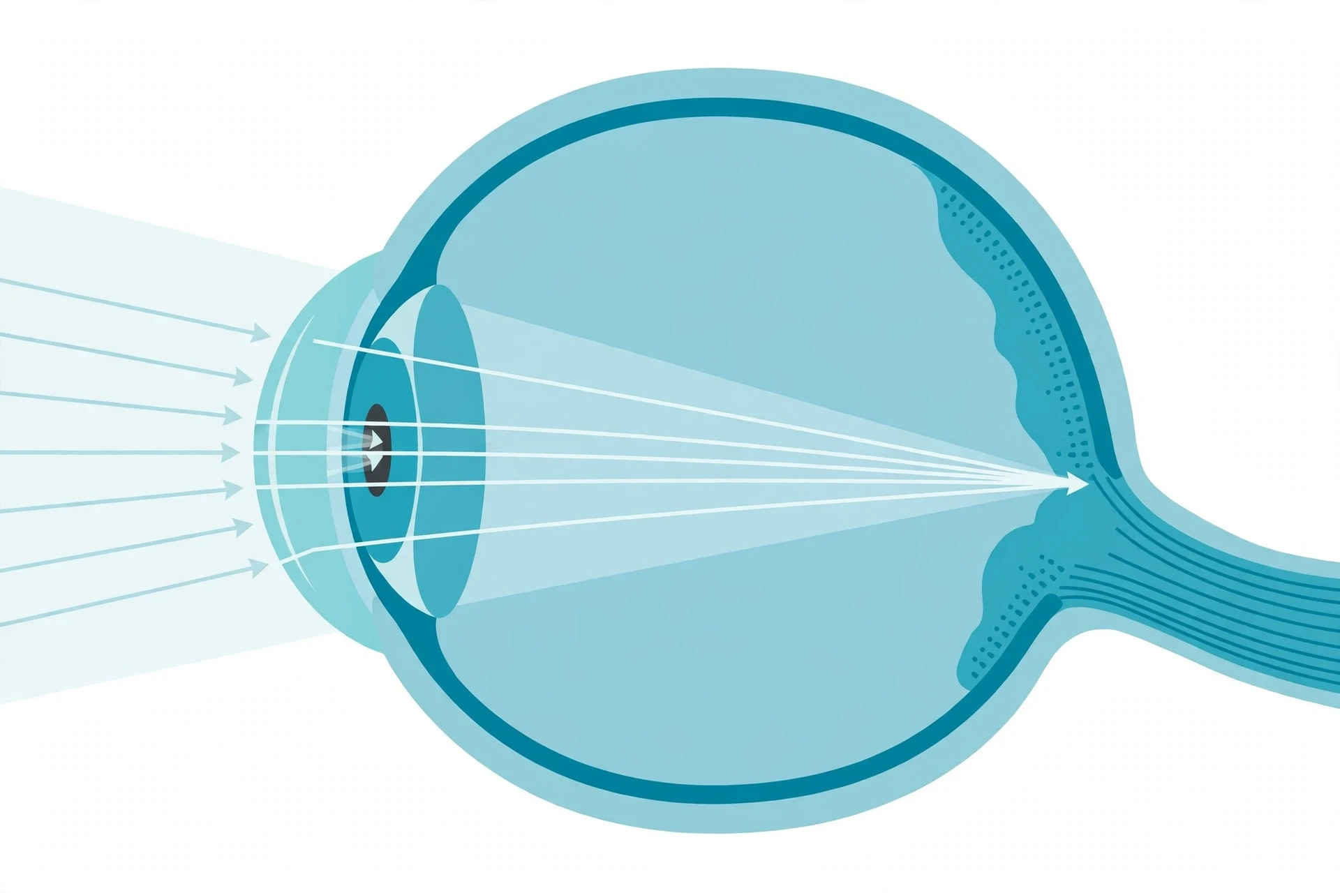
Light meets the cornea first. Its curved surface bends incoming rays sharply, supplying about two-thirds of the eye's focusing power. The lens gets most of the credit in everyday talk, but the cornea does the heavy lifting before light ever reaches it.
Behind the cornea sits a small chamber filled with clear fluid. Then comes the iris, the colored ring that gives eyes their brown, blue, or green. The iris holds two small muscles, and the hole at its center is the pupil. In bright light it tightens and the pupil shrinks. In a dark room it opens wide, like a camera aperture gathering every stray ray.
I still remember my first eye test, sitting in a dim room with a metal frame perched on my nose while the optician flipped lenses in front of me and asked, again and again, which view looked clearer. I kept waiting for a dramatic moment, but the change came in small steps, one lens at a time. When the right lens finally slid into place, the letters on the far wall stopped swimming and stood still. That quiet click of clarity is what your own lens does all day, without you noticing.
The lens sits just behind the pupil, held in place by fine fibers and a ring of muscle called the ciliary body. Unlike a glass camera lens, it changes shape. When the muscle relaxes, the lens flattens for distant objects. When it tightens, the lens grows rounder and stronger for close work. This change of shape is called accommodation.
From lens to retina
Past the lens lies the largest space in the eye, filled with vitreous gel. The gel is mostly water, clear as glass, and it holds the eyeball round while letting light pass through. Floaters drift in this gel. They are tiny clumps that cast shadows on the retina, one more sign that the eye is a living structure, not a polished machine part.
At the back of the ball lies the retina, a thin layer of nerve tissue, at most about half a millimetre thick. It holds the photoreceptors that catch light and turn it into electrical signals. Rods, about 120 million in each eye, work in dim light and see in shades of gray. Cones, about 6 million, work in bright light and give us color and fine detail.
The cones are not spread evenly. Most of them crowd into the fovea, a small pit at the center of the retina, about the size of a pinhead. When you look straight at a word, its image falls on the fovea, and that is why it looks sharp. Words at the edge of your view fall on parts of the retina with fewer cones, and they look softer.
Where the optic nerve leaves the eye, there is a patch with no rods and no cones at all. That patch is the blind spot. Light that falls there sends no signal, and your brain fills in the gap with a best guess based on the scene around it. You never notice the hole, because the brain is a skilled editor.
The image that lands on the retina is upside down. Light from the top of a tree passes through the lens and strikes the lower retina, while light from the ground strikes the upper retina. The brain sorts this out so completely that the flip never reaches your awareness. Signals travel along the optic nerve to the visual cortex, where edges, movement, color, and depth are pulled apart and rebuilt into the scene you experience. To see how that processing fits into the wider system, read the guide to human brain anatomy and the guide to how the nervous system carries signals.
Why the wiring runs backwards
The retina has one more odd feature, and it makes sense only when you know how the eye grows. In the embryo, the eye does not bud from the skin. It grows out from the brain as a hollow pouch called the optic vesicle, which folds in on itself to form the eye cup. The light-sensitive cells end up facing away from the incoming light, with their wiring laid across the front of the retina.
That is the deep reason for the blind spot, and for the inverted retina described earlier in this article. The eye is built from brain tissue that pushed outward, not from skin that pushed inward. The design works well, but it carries the marks of its history in every eye that reads these words.
15. The One-Eyed Ancestor: New Research from 2026
A 2026 study in Current Biology proposes a new chapter in the evolution of the eye: our paired eyes descend from an eye on top of a worm-like ancestor's head — an ancestor that lost its paired eyes, then rebuilt them. No fossil of that ancestor exists, so the case rests on living animals and the wiring they still share.
The study is by George Kafetzis, Michael Bok, Thomas Baden, and Dan-Eric Nilsson, published as "Evolution of the vertebrate retina by repurposing of a composite ancestral median eye" in Current Biology, volume 36, issue 4, pages R153 to R170. The team did not dig up a new fossil. They compared living animals, looking for a pattern that could explain why vertebrate eyes look so different from the eyes of insects, snails, and octopuses.
They surveyed 36 major groups of living animals, covering nearly all bilateral animals, the animals whose bodies split into rough mirror-image left and right halves. In group after group, eyes and light-sensing cells turned up in the same two places. Some sat in pairs on the sides of the head. Others sat on the midline, on top of the brain.
Two jobs, two places
The two positions matched two jobs. Paired eyes on the sides of the head were used to steer movement, to guide swimming, crawling, and hunting. Light-sensing cells on the midline did quieter work. They told the animal whether it was day or night, and which way was up.
Picture a small worm-like animal living in the sea about 600 million years ago. At first it likely had paired light-sensing cells on the sides of its head, as many animals still do. Then its way of life changed. It settled down, burrowed into the seabed, and began to filter food from the water around it.
A settled filter feeder has little use for steering eyes. Eyes are costly to build and costly to run, and an animal that stays in one place gains little from sharp vision. Under those conditions, the paired eyes faded away. The midline cells stayed, because the animal still needed to know day from night and up from down. Over time, those cells formed a small median eye, a single light-sensitive organ on top of the head.
Then the story turned again. Between 600 and 540 million years ago, descendants of that burrowing animal returned to active swimming. They needed to steer again, to find food, to judge their own motion, and to avoid predators. The need for paired eyes came back, but the old paired eyes were gone.
Rebuilding from spare parts
Evolution did not start from scratch. The new paired eyes were built by repurposing parts of the median eye. Small eye cups formed on each side of the midline organ, then separated from it, moved out to the sides of the head, and grew into the paired image-forming eyes that vertebrates carry today.
That reuse explains a long-standing puzzle. The vertebrate retina develops from brain tissue, while the eyes of insects and squid develop from skin on the sides of the head. If our eyes came from a median eye that was already part of the brain, the brain origin stops looking strange.
The median eye did not vanish completely. Parts of it remained in place and became the pineal organ in the brain. In many vertebrates, the pineal organ still senses light directly. In mammals, including us, it has lost that direct light sense, but it still makes melatonin, the hormone that helps set the sleep-wake cycle.
The model also helps explain why the vertebrate retina is so complex. Our retina holds more than 100 types of neurons, while octopus and snail retinas hold only a handful. The 2026 team argues that much of that complexity was already present in the median eye, in cells from two different photoreceptor lineages living side by side. The retina inherited that mixture.
State the caveat plainly: no fossil of this one-eyed ancestor exists. This is a reconstruction from living animals, a model, not a fossil discovery. Fossils of soft, worm-like animals from 600 million years ago are rare, and a tiny median eye would leave almost no trace in rock. The model could be wrong in its details, and later work may revise the timing or the steps. What makes it worth attention in the evolution of the eye is how many separate facts it ties together: the two positions of light-sensing cells, the brain origin of the retina, the pineal organ, and the mixed cell types in the retina itself.
16. What the Eye Cannot Do — and What It Gets Wrong
In the evolution of the eye, the result is a magnificent compromise, not a perfect design — it has a blind spot, backwards wiring, and quirks that mislead it, and the popular claims about it are often wrong. Some limits come from its history, some from physics, and some from the shortcuts your brain takes to finish the picture.
Start with the idea of perfection. A well-built eye should place its sensors facing the light, with no cables in the way and no gap in the picture. Our eye does the opposite. Light must pass through layers of nerve cells and blood vessels before it reaches the rods and cones. The optic nerve punches a hole through the retina on its way out. The lens clouds with age in many people, and the gel inside the eye gathers floaters. These are the marks of a structure shaped step by step, good enough to survive, never rebuilt from a clean sheet.
The table below sets out five claims that circulate widely, beside what the evidence actually shows.
Misconception | What the evidence shows |
|---|---|
The eye is perfectly designed | It has a blind spot, an inverted retina, floaters, and a lens that clouds with age. It works well, but it carries clear signs of a history built by small changes, not a flawless plan. |
More photoreceptors mean better vision | The mantis shrimp has 12-plus color channels, yet in tests it discriminates similar colors worse than we do. Its system favors fast recognition over fine comparison. |
Eyes evolved only once | Eyes evolved independently 40 to 65 times. Only the genetic toolkit, including opsins and Pax6, is shared across those separate inventions. |
Darwin thought the eye disproved his theory | He raised the objection himself in On the Origin of Species, calling the idea absurd in the highest degree, then answered it in the same passage with a stepwise path. |
The Nilsson-Pelger model proves a camera eye evolved in 364,000 years | It is a model of optics with stated limits, not a historical measurement. It tracks eye shape and optics, and it leaves out the brain, the genetics, and the changing environment. |
The mantis shrimp lesson
The mantis shrimp deserves a closer look, because it breaks the simplest story about vision. Some species carry 12 color channels, against our three cone types, plus sensitivity to polarized light. On paper, that should make them masters of color. In tests run by researchers at the University of Queensland, the shrimp did worse than humans at telling similar colors apart. Humans can separate colors only a few nanometers apart in wavelength. The shrimp needed a much larger gap.
The likely reason is how the signals are read. Our brains compare the outputs of our three cone types, and that comparison yields fine distinctions. The shrimp seems to read which channel fires, more like matching a label than blending shades. That trade may buy speed. More channels do not always mean finer vision, and the evolution of the eye has tuned each animal to its own life, not to a single ranking.
Color blindness and the myopia surge
Real limits show up in human populations too. About 8 percent of men of Northern European descent have red-green color blindness, against about 0.5 percent of women in the same populations, because the genes for the relevant opsins sit on the X chromosome. Most affected people still see a rich world. They confuse certain reds and greens, or see them as muted, and many learn to read other cues, such as brightness and position, without thinking about it.
Short-sightedness, or myopia, is rising fast in many countries, and the pattern points to modern life. More near work and less time outdoors is associated with rising short-sightedness in children. The link is an association, not proof of a single cause, and genes play a part as well. Bright outdoor light seems to slow the growth of the eyeball, while long hours of close focus may encourage it to grow longer, so that distant light focuses in front of the retina instead of on it.
When the brain edits the picture
Optical illusions are the clearest proof that seeing is not recording. A pair of lines of equal length can look different, and a still image can seem to move. These tricks work because the brain guesses, using context, past experience, and shortcuts to build a scene quickly. When the guess is wrong, the illusion shows the seam. For a closer look, see the guide to how optical illusions work.
That editing is a strength most of the time. It fills in the blind spot, steadies the world while your eyes jump, and lets you recognize a face in poor light. It also means that what you see is a model your brain has built, checked against the light that reaches your retina.
17. Why the Eye Matters
The eye is evolution's signature story — gradual steps, a reused toolkit, and the same solution invented again and again — and the evolution of the eye still shapes medicine and technology today. Its history explains why sight can fail, why it can sometimes be repaired, and why engineers still copy it.
Follow the arc of the evolution of the eye once more, from the start. A patch of light-sensitive cells learned to tell light from dark. A cup gave direction. A pinhole gave a dim image. A lens gave a sharp one. Each step helped its owner in its own time, and none of them needed to foresee the camera eye that would come later. That is the heart of the evolution of the eye: complexity grew by small useful steps, not by a single leap.
The same toolkit turned up in animal after animal. Opsins caught light. Pax6 marked where eyes should grow. Crystallins, once heat-shock proteins, were recruited to bend light. Different lineages took those shared parts and built different eyes: compound eyes, camera eyes, mirror eyes, and the simple pits that still serve snails and flatworms. Then, in 2026, the one-eyed ancestor model added a twist to the vertebrate branch, with paired eyes rebuilt from a median eye and a pineal remnant left behind to keep time.
Medicine, machines, and the questions ahead
That history matters beyond curiosity, because it guides medicine. When researchers understand how Pax6 and other genes build an eye in the embryo, they can trace what goes wrong when those genes carry a harmful change. Gene therapy for inherited retinal disease is now a real treatment. The first, Luxturna for RPE65-related blindness, was approved by the US FDA in December 2017. It delivers a working copy of the RPE65 gene to retinal cells. It does not restore normal vision to everyone, and it treats only a small group of patients, but it turned a once-untreatable form of blindness into a condition with a treatment.
Engineers look to the same history for ideas. Mantis shrimp eyes, with their many color channels and sensitivity to polarized light, inspire new imaging sensors that can pick up kinds of light our cameras miss. Compound eyes inspire wide-angle sensors. Nature ran the experiments first, over hundreds of millions of years, and the results are still on display.
The evolution of the eye also remains the clearest example of how complexity can grow without a plan. It answers a question that troubled Darwin and still troubles many readers about the evolution of the eye: how can something so intricate arise by small steps? The answer is a fossil record, a set of living intermediates, a shared genetic toolkit, and a model of optics that shows each step paying its way. The evolution of the eye keeps adding to that answer, and the 2026 median-eye model shows that the story is not finished.
Questions about the evolution of the eye remain, and they are good ones. How did color vision diversify so widely, from the two-channel world of many mammals to the rich color of birds and the polarized world of the shrimp? What will the fossil record still reveal about the first image-forming eyes in the Cambrian seas? How much of the retina's wiring was already in place in that one-eyed ancestor, and how much came later? The eye took more than half a billion years to reach its present form, and its story is still being written.
Frequently Asked Questions
The evolution of the eye raises questions that go beyond any single section, so here are direct answers to the ones readers ask most often.
How did the eye evolve?
The evolution of the eye happened by small useful steps, not by a single leap. A patch of light-sensitive cells gave a sense of light and dark, a cup gave direction, a pinhole gave a dim image, and a lens gave a sharp one. Each stage helped its owner survive, and natural selection favored the small changes that improved the picture.
What did Darwin say about the eye?
Darwin wrote in On the Origin of Species that the idea of natural selection producing the eye "seems, I freely confess, absurd in the highest possible degree." He raised that doubt himself, then answered it in the same passage. If graded steps from a simple eye to a complex one exist, each useful to its owner, and if those steps can be inherited, he argued, the difficulty is one for our imagination, not for his theory.
What is the Pax6 gene?
Pax6 is a master control gene that helps mark where eyes develop in the embryo. The same gene, or close relatives of it, guides eye development in animals as different as fruit flies, mice, squid, and humans. That shared role is strong evidence that the genetic toolkit for eyes is far older than the many different eyes the evolution of the eye built from it.
How many times did eyes evolve?
In the evolution of the eye, eyes evolved independently 40 to 65 times, counting light-sensitive organs and eyespots. Complex image-forming eyes arose separately in fewer lineages, including vertebrates and cephalopods such as octopuses. What is shared across those separate inventions is the toolkit: opsins, Pax6, and other genes that were reused again and again.
Could a camera eye really evolve in under 400,000 years?
The 364,000-year figure comes from a model by Dan-Eric Nilsson and Susanne Pelger, not from a fossil clock. They modeled a patch of photoreceptors changing by steps of no more than 1 percent, 1,829 steps in all, and calculated how long those steps might take under pessimistic assumptions. It shows that the optics could change quickly in principle, but it is not a historical measurement.
Why do we have a blind spot?
We have a blind spot because the optic nerve leaves the eye through a patch of retina that holds no rods or cones. The gap exists because, in the evolution of the eye, our retina grew out from the brain, so its wiring runs across the front of the retina and exits through that hole. Your brain fills in the missing piece, which is why you never notice it in everyday sight.
Do octopuses see like us?
In the evolution of the eye, octopuses built camera eyes much like ours, with a single lens that focuses light onto a retina. Their wiring is different: their photoreceptors point toward the incoming light, so they have no blind spot like ours. Their retinas are also simpler, with only a handful of neuron types against more than 100 in ours, which reflects their separate evolutionary path to the same design.
Sources
Darwin, C. (1859) On the Origin of Species, Chapter 6 (public-domain book, no link)
Stanford University — "Windows on Evolution: The Eye", Stanford Birds / Stanford University
Fernald — "Evolving eyes", International Journal of Developmental Biology (2004)
Guinness World Records — "Most complex eyes of any animal", Guinness World Records (2018)
Hanne H. Thoen et al. — "A different form of color vision in mantis shrimp", Science (2014)