

I still remember the morning the idea first landed for me. I woke with my hand asleep, pins and needles fizzing across my fingers, and shook it until the feeling came flooding back. It took less than a minute. I thought nothing of it. Then I read about multiple sclerosis, and the same small sensation took on a different weight: what if the tingling did not fade? What if the signal from your hand to your brain kept arriving scrambled, day after day, because the insulation around the nerve itself was being stripped away?
That question is the heart of this article. Multiple sclerosis is a disease of myelin, the fatty insulation that wraps the nerve fibres of the brain, the spinal cord, and the optic nerves. When the immune system attacks that insulation, signals slow down, scramble, or stop altogether. The result can be a hand that tingles, an eye that goes dim, a leg that will not quite do what it is told, or a tiredness that rest does not fix. The same disease looks different in different people, because it depends on where the damage lands.
It is also far more common than most people realise. As of the 2024 Atlas of MS update, published in 2026, about 3.1 million people worldwide are living with multiple sclerosis, up from 2.9 million in the 2023 update and 2.8 million in 2020. In the United States, the best national estimate puts the figure at nearly one million adults. Behind each of those numbers is a person whose nerves carry signals a little less reliably than they used to, and a family learning what that means.
This article explains the three threads of the title in plain English. First, myelin: what it is, and why nerves need it to work at speed. Second, the immune attack: which cells do the damage, how they get past the brain's defences, and why the damage sometimes heals and sometimes does not. Third, the nerves themselves: how a lesion in one place produces one symptom and a lesion in another produces something entirely different. Along the way I will cover the first symptoms to know, the disease courses, the causes and risk factors, how doctors diagnose the condition with MRI and the McDonald criteria, what treatments can and cannot do, and what life with the disease actually looks like. Nothing here is medical advice for any individual. It is an explainer, built on published research and the guidance of patient organisations, so that a reader meeting this diagnosis — in themselves, or in someone they love — can understand what is happening inside the nervous system and why.
To follow the story, it helps to know a little about how nerves carry messages in the first place. How the nervous system works is the background to everything that follows: a network of cells sending electrical signals along fibres, at speeds that depend entirely on myelin.
1. What Multiple Sclerosis Is — A Disease of Myelin in the Central Nervous System
1.1 The one-sentence answer, then the fuller picture
Multiple sclerosis is a chronic, immune-mediated inflammatory disease of the central nervous system in which the immune system damages myelin, the insulation around nerve fibres, and so disrupts the electrical signals those fibres carry.
That one sentence carries a lot. "Chronic" means it is a long-term condition, not a passing infection. "Immune-mediated" means the damage is done by the body's own immune system, not by a germ invading from outside. "Inflammatory" means the damage happens in episodes of inflammation, when immune cells gather and attack. And "central nervous system" means the brain, the spinal cord, and the optic nerves — the command centre and its main cables, rather than the nerves of the arms and legs.
The fuller picture adds the consequences. When myelin is stripped from a nerve fibre, the electrical signal travelling along that fibre slows, distorts, or fails to arrive at all. Because different fibres carry different messages — vision, touch, movement, bladder control, memory — the symptoms depend on which fibres are hit. One person may lose vision in one eye for a few weeks. Another may feel numbness spreading up a leg. A third may struggle with fatigue and a foggy memory while their walking stays fine. The disease is one process, demyelination, producing a broad spectrum of neurological deficits. That is why two people with the same diagnosis can have such different experiences, and why a single description of "what MS feels like" never quite fits.
Multiple sclerosis is also a major cause of disability in young adults. Most people first develop symptoms between the ages of twenty and forty, at the point in life when they are starting careers, relationships, and families. The disease does not arrive in old age, when decline is expected. It arrives early, and it stays. Understanding it matters precisely because it touches people with decades of life ahead of them.
1.2 What "immune-mediated" means
The immune system exists to tell self from not-self, and to destroy what does not belong. In multiple sclerosis, that judgement goes wrong. Immune cells that should ignore the body's own myelin instead treat it as a target. They cross into the brain and spinal cord, gather around nerve fibres, and strip the myelin away. The result is inflammation and demyelination, and over time, injury to the nerve fibres themselves.
Most sources describe multiple sclerosis as an autoimmune disease, and that label is a useful shorthand. But the plainer version carries a hedge. The immune attack itself is not in doubt. What remains debated is whether myelin is the original target of a misdirected immune response, or whether the immune system is reacting to damage that starts somewhere else. That distinction matters to researchers hunting for the cause, and it is why careful writers call multiple sclerosis "immune-mediated" first and "often described as autoimmune" second.
For a fuller picture of how the immune system can turn against the body's own tissues, see Autoimmune Diseases Explained, which covers the wider family of conditions in which self-tolerance fails. Multiple sclerosis belongs in that family, even while its exact trigger remains unknown — a question I return to in section 7.
The name of the disease is itself a description of what doctors found when they looked inside the brain. "Multiple sclerosis" refers to the many areas of scarring — scleroses, or plaques — scattered through the white matter of the brain and spinal cord. White matter is mainly myelin, so the scars sit exactly where the insulation used to be. Each plaque marks a place where myelin was lost and a scar formed in its place. One plaque might sit on the optic nerve and dim vision. Another might sit in the spinal cord and weaken a leg. The disease is "multiple" because the plaques are many, and "sclerosis" because they harden into scars.
1.3 How common it is, and who it affects
The numbers have been climbing, and that rise is partly a story about better counting. The 2024 Atlas of MS update, published in 2026, estimates that 3.1 million people worldwide are living with multiple sclerosis — 38.1 per 100,000 people, or roughly one person in every 2,600. The trajectory tells the story: 2.3 million in the original 2013 estimate, 2.8 million in 2020, 2.9 million in the 2023 update, and now 3.1 million. The 2024 update re-based the 2013 figure at about 1.8 million under revised counting, so the counts across editions are not strictly comparable — but the rate has risen by about 54 percent since 2013, from 24.8 to 38.1 per 100,000, and that comparison is sound. Some of that rise reflects genuinely increasing incidence, and some reflects better diagnosis and longer survival. Either way, the disease is no longer rare by any measure.
In the United States, the most recent national estimate, from Wallin and colleagues in 2019, puts the figure at nearly one million adults — about 851,749 to 913,925 extrapolated for 2017, up from 727,344 in 2010. The older estimate of around 400,000, still repeated in some places, is now known to have been roughly half the true figure. The United States rate sits at about 309 per 100,000 adults. Globally, about 120,000 people are newly diagnosed each year.
The disease also reaches children. The 2024 Atlas update counts more than 40,000 children under eighteen living with multiple sclerosis worldwide, up from about 30,000 in 2020 and 7,000 in 2013. Paediatric multiple sclerosis remains uncommon, but it is no longer the rarity it was once thought to be.
Age and sex shape who is affected. Symptoms usually begin between the ages of twenty and forty, and the mean age at diagnosis worldwide is thirty-two. Women are affected about three times as often as men, in round terms. The United States figures from 2010 put the ratio at 2.8 to one — 450.1 cases per 100,000 women against 159.7 per 100,000 men — while the global Atlas figures from 2020 put women at about twice the risk of men. The ratio has risen over the past fifty years, and in recent Canadian birth cohorts it has exceeded 3.2 to one. Why women are more affected is not settled, and hormones, genetics, and immune differences are all part of the ongoing research. What is settled is the pattern: a disease that strikes young adults, and strikes women most.
Statistic | Latest figure | Source and date |
|---|---|---|
People living with multiple sclerosis worldwide | 3.1 million (38.1 per 100,000 in 2024; about 1 in 2,600 people) | King et al., Atlas of MS 2024 update, Multiple Sclerosis Journal 2026; epub 3 Sep 2026 |
Global trajectory | 2.3 million (2013, original estimate; re-based to about 1.8 million in the 2024 update) → 2.8 million (2020) → 2.9 million (2023) → 3.1 million (2024); prevalence 24.8 → 35.9 → 38.1 per 100,000; up about 54 percent since 2013 | King et al. 2026; Walton et al. 2020 |
New diagnoses per year (global) | About 120,000 per year | King et al. 2026 |
People living with multiple sclerosis in the USA | Nearly 1 million adults (about 851,749 to 913,925 extrapolated for 2017; 727,344 in 2010) | Wallin et al., Neurology 2019 |
US prevalence rate | 309.2 per 100,000 adults (2010) | Wallin et al. 2019 |
Female to male ratio | About 3 to 1 in round terms (US 2010: 2.8 to 1; global Atlas 2020: females about twice as likely) | Wallin et al. 2019; Atlas of MS 2020 |
Typical age of onset | 20 to 40 years; mean age at diagnosis 32 years worldwide | NINDS; Atlas of MS 2020 |
Children under 18 living with multiple sclerosis | More than 40,000 worldwide (2024), up from about 30,000 (2020) and 7,000 (2013) | King et al. 2026 |
1.4 What multiple sclerosis is not
Before going deeper into the biology, it helps to clear away three misunderstandings that shape how people react to the diagnosis. Multiple sclerosis is not contagious. It cannot be caught from another person, and no one in a family will catch it from a relative who has it. The disease is not muscular dystrophy, either: muscular dystrophy is a group of genetic diseases that waste muscle, while multiple sclerosis attacks myelin in the central nervous system — different tissue, different cause. And multiple sclerosis is not a death sentence. Most people with the disease live almost as long as people without it, and the outlook has improved steadily as diagnosis has come earlier and treatments have improved. Each of these points gets its full correction in section 12, with the evidence behind it. For now, they are worth stating plainly, because fear built on a wrong idea is the hardest kind to undo.
"Multiple sclerosis (MS) is an inflammatory relapsing or progressive disorder of CNS white matter and is a major cause of disability in young adults. Pathologically, it is characterized by multifocal areas of demyelination, loss of oligodendrocytes, and astrogliosis but with relative preservation of axons." — Pirko and Noseworthy, reviewed in PMC7158368
2. Myelin: The Insulation That Makes Nerves Fast
2.1 What myelin is and who makes it
Myelin is a lipid-rich material that wraps around axons — the long fibres that carry electrical signals away from a nerve cell's body — to insulate them and increase the rate at which impulses travel. Think of an axon as a wire and myelin as the plastic coating around it, except that this coating does far more than prevent short circuits. It is what lets a signal travel fast enough for a thought to become a movement without a noticeable delay.
In the central nervous system, myelin is made by cells called oligodendrocytes. Each oligodendrocyte extends fatty sheets that wrap around segments of several different axons, a bit like one worker insulating many short stretches of cable. A single oligodendrocyte can myelinate up to about fifty axon segments. In the peripheral nervous system — the nerves outside the brain and spinal cord — the same job is done by Schwann cells, each of which wraps a single segment of a single axon. That difference matters for multiple sclerosis: the disease attacks the central nervous system, so it is the oligodendrocytes and their myelin that are lost, while peripheral nerves, wrapped by Schwann cells, are largely spared. That is one reason the symptoms of multiple sclerosis are symptoms of the brain, the spinal cord, and the optic nerves, rather than of the hands and feet themselves.
The myelin sheath is not one continuous tube. It is made of segments separated by small gaps, and those gaps are the key to everything that follows.
2.2 Nodes of Ranvier and saltatory conduction
The gaps between myelin segments are called nodes of Ranvier. At each node, the axon membrane is exposed, and it is packed with voltage-gated sodium channels — the molecular gates that let an electrical impulse regenerate itself. Between nodes, the myelin insulates the axon so well that almost no current leaks out. The result is a distinctive way of conducting signals called saltatory conduction, from the Latin word for jumping. The action potential does not crawl continuously along the axon. It recharges at one node and jumps to the next, and then the next, far faster than it could travel along a bare fibre.
The speed difference is dramatic. Myelinated axons conduct at tens of metres per second, while unmyelinated fibres manage only about one metre per second. To put that in everyday terms, a signal that takes a second to travel a metre along a bare fibre can cover tens of metres in the same time once myelin is in place. That speed is what makes coordinated movement, sharp vision, and quick reflexes possible. Without it, the nervous system would still work, but it would work slowly, and the timing that holds a movement together would fall apart.
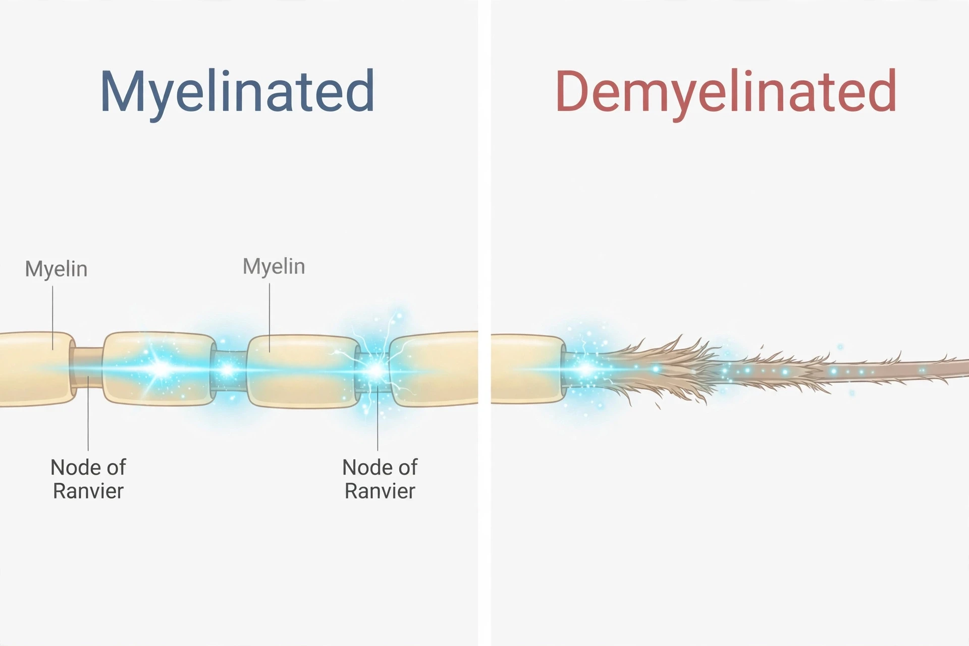
Strip the myelin away, and the jumping stops. The signal must now travel continuously along the exposed axon, leaking current as it goes, arriving late or not arriving at all. That is the physical basis of almost every symptom of multiple sclerosis. A visual signal that arrives late is a blurred image. A motor signal that arrives late is a clumsy step. A signal that never arrives is a numb patch of skin or a muscle that does not answer. The disease is, at its core, a failure of timing.
2.3 Myelin does more than insulate
Insulation is myelin's best-known job, but it is not its only one. Oligodendrocytes also provide metabolic support to the axons they wrap, supplying them with fuel — for example lactate, delivered through a transporter called monocarboxylate transporter 1 — that the axon needs to stay alive and keep working. This supporting role is easy to overlook, and it changes how we should think about the damage. Losing myelin does not only slow the signal. It also starves the axon of support, and that loss of support contributes to the axon degenerating even when conduction still looks intact on the surface. The evidence for this metabolic role is still building, but the idea it carries is important: myelin is not just a passive wrapper. It is part of a living partnership between the oligodendrocyte and the axon, and when the partnership breaks, the axon itself is at risk.
That risk is the bridge between the early, relapsing phase of the disease and the later, progressive phase. Early on, the main problem is demyelination, and some of it can be repaired. Later, the problem becomes the axons themselves, and axons, once cut, do not grow back in the central nervous system. This is why the same disease can look so different at different stages, and why so much of the research now focuses on protecting axons rather than only calming inflammation.
2.4 When myelin is lost, signals slow, scramble, or stop
Myelin is essential to maintain conduction velocity, and its loss can lead to significantly slower conduction or conduction block. That is the hinge of the whole disease. Slower conduction means messages arrive late. Scrambled conduction means they arrive distorted. Conduction block means they do not arrive at all. And because the pattern of demyelination differs from person to person, and from month to month within one person, the symptoms shift and change in ways that can be baffling without this picture in mind.
There is one more piece of the picture, and it is the most serious. While demyelination is the classic hallmark of multiple sclerosis, axonal and neuronal injury are important aspects of the disease too, and they are gaining more recognition. Permanent neurological disability is caused by axonal transection — by nerve fibres being cut. A demyelinated fibre can, in principle, be remyelinated, and the signal can recover. A transected fibre cannot. That is why a relapse can end with a full recovery, and why it can also end with a deficit that never quite lifts. The myelin can be rebuilt. The axon, once severed, cannot.
To understand how nerves normally carry their signals, and what saltatory conduction looks like in a healthy fibre, How the Nervous System Works covers neurons, action potentials, and the wiring of the brain and spinal cord in detail. What follows here is what happens when the insulation on that wiring is attacked.
3. The Immune Attack: How Multiple Sclerosis Damages Myelin and Nerves
3.1 The blood–brain barrier — the gate that fails
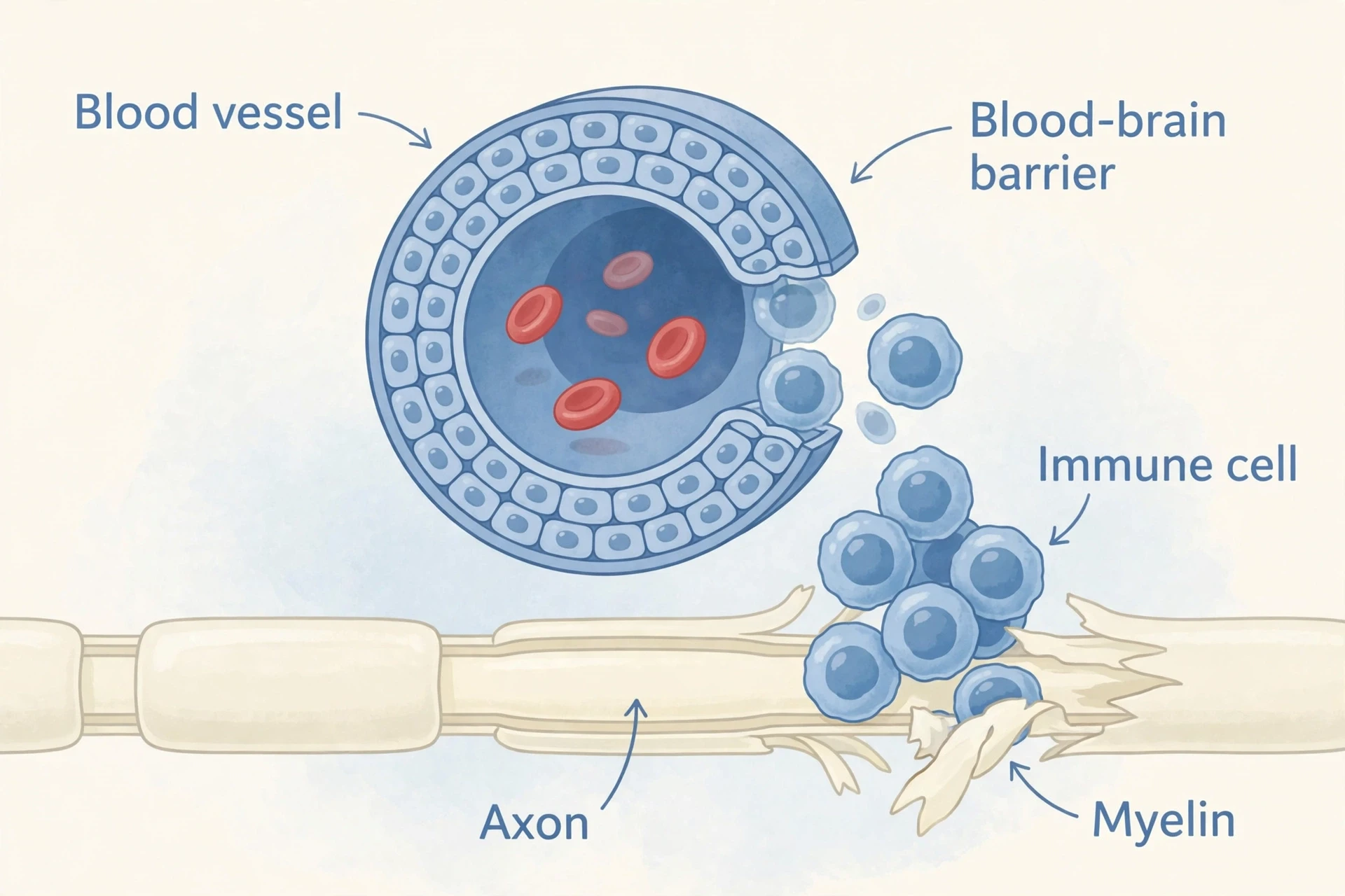
The brain and spinal cord are not open to the bloodstream. They are guarded by the blood–brain barrier, a selective gate formed by the tightly joined cells lining the blood vessels of the central nervous system. Most immune cells cannot cross it. That is by design: the brain is a delicate organ, and an immune response inside it can do as much harm as the threat it is fighting. The barrier lets nutrients and a few selected molecules through, and keeps the rest out.
In multiple sclerosis, the barrier is compromised. Activated T cells, B cells, and monocytes cross into the perivascular space — the narrow gap around blood vessels inside the brain — and gather there in clusters called perivascular cuffs. From that foothold, they move into the surrounding white matter and begin the attack on myelin.
The crossing itself is a precise molecular manoeuvre. Activated T cells use surface molecules called LFA-1 and VLA-4 to grip matching molecules, ICAM-1 and VCAM-1, on the endothelial cells that line the brain's blood vessels. It is a handshake between the immune cell and the vessel wall, and once the grip is made, the cell squeezes through. That handshake is not a detail for specialists only: it is the target of one of the most effective classes of treatment, which works by blocking exactly that grip. Understanding the crossing is understanding where the disease can be interrupted.
For the wider story of how immune cells move, recognise targets, and mount a response, How the Human Immune System Works gives the background. What follows is what that system does when it turns against myelin.
3.2 The cells doing the damage
The attack is not the work of one cell type. T cells, B cells, macrophages, and microglia are all implicated in starting and sustaining the inflammation and demyelination of multiple sclerosis, and each plays a different part.
The acute MS plaque — the fresh lesion — is typified by robust inflammatory infiltration plus demyelination. Around the blood vessels, the perivascular cuffs contain lymphocytes, predominantly T cells, along with monocytes and macrophages. Among the macrophages are lipid-loaded "foamy" macrophages, swollen with the fatty debris of the myelin they have eaten. These cells do not merely stand by while myelin falls apart. They actively strip it from axons, peeling the insulation away and carrying it off in pieces.
CD8-positive T lymphocytes, the immune system's killers, can damage axons directly. They do it in several ways: by releasing cytotoxic granules that punch holes in their targets, by triggering Fas-mediated apoptosis — a programmed self-destruct signal — in the cells they touch, by releasing cytokines such as TNF-alpha that poison the neighbourhood, and by directly transecting axons, cutting the fibres themselves. Macrophages and microglia, the brain's resident immune cells, add their own damage by releasing proteases and reactive nitrogen species that injure oligodendrocytes and further disrupt the blood–brain barrier. The barrier breach lets more cells in, and the cycle feeds itself.
It is a coordinated assault, not a single blow. That is one reason the disease is so hard to stop with a single drug, and why treatments aim at different parts of the process: keeping cells out of the brain, depleting the B cells that drive the response, or trapping lymphocytes where they cannot do harm. The biology of the attack maps directly onto the logic of the treatments, and section 9 follows that map class by class.
3.3 The B-cell side: antibodies and antigen presentation
For many years, T cells were treated as the main story of multiple sclerosis, and B cells were cast in a supporting role. That view has changed. B cells contribute in two ways, and both matter.
First, B cells produce oligoclonal IgG antibodies — the antibodies found in the cerebrospinal fluid of most people with multiple sclerosis — that bind to myelin and activate complement, a cascade of immune proteins that punches holes in whatever the antibodies have marked. Second, B cells act as antigen-presenting cells: they pick up fragments of myelin, display them on their surface, and activate T cells against them. In this second role, the B cell is not a foot soldier but a recruiter, widening the attack by bringing more T cells into it.
This is now an established mechanism, supported by more than one line of evidence, and it is the reason that therapies which deplete B cells have become some of the most effective treatments available. It also explains a diagnostic clue: the oligoclonal bands found in the spinal fluid are the fingerprint of those B cells at work inside the central nervous system, producing antibodies that are present in the fluid around the brain and spinal cord but not in the blood. Section 8 returns to those bands, because they are one of the most useful laboratory findings in diagnosis.
3.4 Th1, Th17, and the brakes that fail
Not all T cells push in the same direction. Two subsets drive the inflammation forward. Th1 cells release interferon-gamma, and Th17 cells release interleukin-17, and both propagate the inflammatory response inside the central nervous system. They recruit more immune cells, activate macrophages, and keep the lesion hot.
Other subsets act as brakes. Th2 cells and regulatory T cells suppress inflammation, calming the response and limiting the damage. In a healthy immune system, the accelerators and the brakes stay in balance: a response flares when it is needed and settles when the job is done. In multiple sclerosis, that balance is lost. The accelerators dominate, the brakes fail to hold, and the inflammation continues long after it should have stopped.
This imbalance is not a metaphor. It is a measurable shift in the kinds of T cells found in the blood and spinal fluid of people with the disease. It is also one reason treatments that nudge the immune response — from a Th1 pattern toward a Th2 pattern, for example — can help. The immune system is not simply "overactive" in multiple sclerosis. It is misdirected, and the direction matters as much as the strength.
3.5 From acute plaque to scar: gliosis and axonal loss
A lesion changes over time. The acute plaque is a place of active inflammation: immune cells pouring in, myelin being stripped, the blood–brain barrier leaking. If the inflammation settles, the lesion may partly heal. But a chronic plaque looks different. Its borders and centre are filled with reactive astrocytes — star-shaped support cells of the brain that have swollen and multiplied in response to the injury. This is astrogliosis, or glial scarring: the brain's attempt to wall off the damage and stabilise the tissue.
The glial scar is a double-edged structure. On one side, it limits the spread of the lesion, containing the damage so that it does not keep growing. On the other, it blocks tissue regeneration, mainly through the reactive astrocytes and microglia themselves and through the inhibitory molecules they lay down in the extracellular matrix around them. The scar is a wall that keeps the fire from spreading, and also a wall that keeps the repair crews out. That tension — protection against repair — runs through the whole later course of the disease, and it is one of the reasons chronic lesions are so much harder to heal than fresh ones.
Meanwhile, the axons inside the lesion are being lost. As noted in section 2, a cut axon is gone for good, while stripped myelin can be rebuilt. In the early years of the disease, most of the injury is demyelination, and recovery is common. Over time, the balance shifts toward axonal loss, and recovery becomes partial. This is the biological reason behind the slow accumulation of disability that defines the progressive forms of the disease, and it is why protecting axons has become as important a goal as calming inflammation.
3.6 Remyelination — and why it often fails
The brain does try to repair itself. Remyelination is the process by which oligodendrocyte precursor cells — OPCs, the stem-like cells that can become new oligodendrocytes — migrate to a demyelinated area, multiply, and mature into new myelin-making cells, wrapping fresh sheaths around the bare axons. It is a real and measurable process, and it is one of the most hopeful facts in the biology of multiple sclerosis.
It is also limited. After inflammation subsides, approximately twenty percent of plaques remyelinate to form what pathologists call a "shadow plaque" — a patch of thinner, paler myelin, visible under the microscope as a ghost of the original sheath. Remyelination is more common in early multiple sclerosis and less likely in old, chronic lesions. The new myelin is thinner than the original, and the conduction it restores is slower than before, but it is better than no myelin at all. A shadow plaque is a partial victory.
Why does remyelination fail in the rest? The answer is not what most people would guess. Failure in chronic multiple sclerosis does not depend mainly on a lack of precursor cells. Premyelinating oligodendrocytes are present in chronic lesions, but they fail to remyelinate the dystrophic, swollen axons inside them. The axons themselves are too damaged to accept new myelin, and the reactive astrocytosis and glial scar may physically block the precursor cells from reaching them. The repair crew is on site, but the road is blocked and the wiring too damaged to reconnect.
That finding reframes the goal of future treatment. Replacing lost oligodendrocytes is not enough. The axons must be protected early, the scar must be made permissive to repair, and the precursor cells must be helped to finish the job. Until that happens, remyelination remains what it is today: a real but partial repair, most likely early in the disease, and least likely exactly where it is needed most.
"While ongoing debate lingers over the autoimmune nature of MS, it is well established that the immune system directly participates in the destruction of myelin and nervous cells." — Wu and Alvarez, PMC3080042
Multiple sclerosis sits in the wider family of conditions in which the immune system attacks the body's own tissues. Autoimmune Diseases Explained sets out how that failure of self-tolerance works across diseases, and why the same immune mechanisms can produce such different illnesses depending on the tissue they target.
4. Lesions on MRI: What the Scan Actually Shows
4.1 Bright spots: T2/FLAIR and gadolinium
On magnetic resonance imaging, multiple sclerosis is characterised by focal lesions with a typical shape and distribution in the brain, the spinal cord, and the optic nerve. The most familiar image is the T2-weighted or FLAIR scan, on which lesions appear as bright white spots against the darker grey of healthy brain tissue. FLAIR — fluid-attenuated inversion recovery — suppresses the signal from cerebrospinal fluid, which makes lesions sitting beside the fluid-filled ventricles much easier to see. These sequences detect both active and inactive lesions, so a single scan can show the history of the disease written across the brain: fresh plaques and old scars side by side.
A second kind of scan adds the dimension of time. On gadolinium-enhanced T1 sequences, active lesions "light up" — they enhance, glowing bright — because the gadolinium contrast leaks through a disrupted blood–brain barrier into the inflamed tissue. Enhancement means active inflammation, happening now. It typically lasts a few weeks, and then fades as the barrier heals and the inflammation settles. A scan that shows both enhancing and non-enhancing lesions at the same time is therefore showing two moments in the disease at once: one lesion that is active today, and another that flared months or years ago. That contrast is one of the pillars of diagnosis, and section 8 explains how doctors use it.
4.2 Dawson's fingers and the four characteristic locations
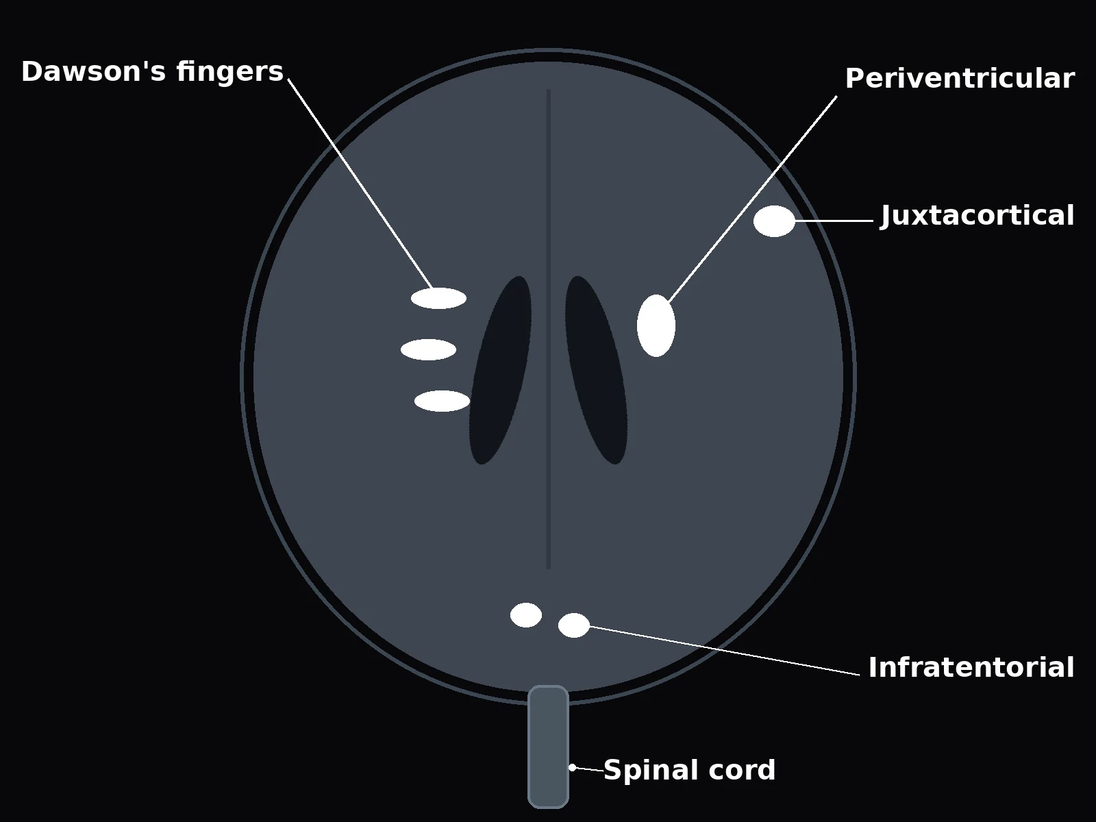
The classic brain finding in multiple sclerosis has a name that sounds like a person, because it is one. Dawson's fingers are ovoid periventricular lesions — patches of demyelination sitting beside the lateral ventricles, the fluid-filled cavities at the centre of the brain — oriented perpendicular to the ventricles, like fingers pointing outward. They represent demyelination along the perivenular white-matter tracts, the small veins that drain the tissue around the ventricles. When a radiologist sees Dawson's fingers, the pattern is hard to mistake for anything else, because few other diseases lay their lesions out in that shape, in that place.
Typical lesions in multiple sclerosis are round or ovoid, relatively well defined, and generally at least three millimetres on their longest axis. Size alone proves nothing, but the combination of shape, sharpness, and location builds the picture. The four characteristic locations used in the 2017 McDonald criteria are the periventricular region beside the ventricles, the cortical and juxtacortical region at the surface of the brain, the infratentorial region — the brainstem and cerebellum, below the main mass of the brain — and the spinal cord. A lesion in two or more of those four places, in the right clinical setting, counts as dissemination in space: evidence that the disease has struck in more than one part of the central nervous system.
4.3 Spinal cord lesions — short, peripheral, usually cervical
Lesions in the spinal cord have their own signature. In multiple sclerosis, they are typically short-segment — spanning less than two vertebral segments — peripheral, sitting in the dorsal or lateral columns at the edge of the cord, and most often cervical, in the neck. That pattern is diagnostically useful because it differs from the main mimic. In neuromyelitis optica, a related but distinct disease, cord lesions typically extend across three or more segments and sit centrally in the cord. The contrast is a clue, not a verdict: a short peripheral lesion points toward multiple sclerosis, and a long central one points away from it. But no single feature decides a diagnosis on its own, and the distinction is one reason spinal cord imaging is part of the work-up.
The location of a cord lesion also predicts its symptoms, and this is where the scan connects to the person's experience. A lesion in the cervical cord can disturb sensation and movement in the arms and legs and interfere with bladder control. A lesion higher up, in the brainstem, can disturb balance and coordination. The map of the lesion is, in a real sense, the map of the symptoms, and section 5 follows that map in detail.
4.4 What MRI cannot prove
For all its power, MRI does not diagnose multiple sclerosis on its own. MRI findings are not pathognomonic — they must be interpreted in the appropriate clinical setting, and only after a better explanation has been excluded. White spots on a brain scan have many causes. Small-vessel disease, migraine, and normal ageing can all produce them. A scan that looks like multiple sclerosis in a person with no compatible symptoms is a puzzle, not a diagnosis. And a scan that looks normal does not entirely rule the disease out, though it makes it much less likely: abnormal MRI scans are found in about ninety percent of people with a definite diagnosis of multiple sclerosis.
I remember the first time I saw an MRI lesion from multiple sclerosis, not as a patient but as a reader staring at a published scan. A bright white oval sat beside a ventricle, neat and almost tidy, and my first thought was how small it looked for the trouble it could cause. That is the thing to hold on to. A lesion is a patch of missing insulation a few millimetres across. It proves that demyelination happened there. It does not prove, by itself, that the person has multiple sclerosis, or how disabled they will become, or what will happen next. It is one piece of evidence, and it only becomes a diagnosis when it is combined with the person's history, their examination, and the exclusion of everything else that could explain it.
MRI is the most sensitive paraclinical tool available for diagnosis, with a sensitivity of about ninety percent or more. Brain MRI should be obtained in all patients in whom multiple sclerosis is being considered, unless MRI is impossible, and spinal cord MRI adds another site where dissemination can be shown, sometimes allowing a diagnosis with fewer brain lesions. But sensitivity is not certainty. The scan shows where myelin has been lost. The diagnosis is a judgement about why, and that judgement belongs to the whole clinical picture.
For the anatomy behind these locations — where the ventricles sit, what the brainstem and cerebellum do, and how the spinal cord is organised — Human Brain Anatomy and Functions gives the map that makes the scan readable.
5. Symptoms of Multiple Sclerosis — and the First Symptoms to Know
5.1 The first symptoms — what usually appears first
The first symptoms of multiple sclerosis are rarely dramatic. In clinical series, the most common initial symptoms are reported roughly as follows: changes in sensation in the arms, legs, or face in about thirty-three percent; complete or partial vision loss from optic neuritis in about twenty percent; weakness in about thirteen percent; double vision in about seven percent; unsteadiness when walking in about five percent; and balance problems in about three percent. About fifteen percent of people have more than one symptom when they first seek medical attention. These figures come from clinical series rather than modern population epidemiology, so they should be read as "roughly" — a guide to what is common, not a precise forecast for any individual.
What those figures hide is how easy the first attack is to miss. Initial attacks are often transient, mild, or even without symptoms, and they are self-limited: they come, they last a few weeks, and they fade. They often do not prompt a visit to a doctor at all, and they are sometimes only identified in retrospect, once the diagnosis is made after further attacks and the person looks back and realises that the numb hand two years ago, or the week of blurred vision, was the beginning. That retrospective recognition is one of the most common stories in multiple sclerosis, and it explains why the time from first symptom to diagnosis can stretch to months or years.
The most common early symptoms overall, as summarised by the MSD Manual, are tingling, numbness, pain, burning, and itching in the arms, legs, trunk, or face; dim or blurred vision; and weakness and clumsiness. None of these is specific to multiple sclerosis. Tingling can come from a trapped nerve. Blurred vision can come from a dozen causes. What marks them out, in hindsight, is the pattern: symptoms that appear over hours or days, last weeks, and then improve — and then return, in a different place, months later.
I know that pattern from the small side of it. A few years ago I noticed my foot catching on the edge of a step, once, then again a week later, and I put it down to tiredness and a pair of worn shoes. It stopped, and I forgot about it. That is not a diagnosis, and I am not suggesting it was one. But it taught me how easily an early symptom hides in ordinary life. A foot catches. Vision dims for a week. A hand tingles and then does not. Each event on its own looks like nothing. It is only the repetition, in different places, that turns a collection of small oddities into a pattern worth investigating. For people who go on to be diagnosed with multiple sclerosis, that is often how the story begins: not with a crisis, but with a series of small things that did not quite add up.
5.2 Optic neuritis — the presenting sign in about one in five
Optic neuritis is inflammation of the optic nerve, the cable that carries visual signals from the eye to the brain, and it is the presenting sign of multiple sclerosis in about twenty percent of cases. Up to half of people with the disease will develop an episode at some point. It typically begins with pain in one eye, often worse when the eye moves, followed by blurry or dimmed vision that develops over hours to days. The vision is at its worst within a week or two, and then it gradually improves over weeks. Colours may look washed out. The centre of vision may go grey or dark. It is frightening, and it is also, in most cases, recoverable: the inflammation settles, the myelin partly repairs, and the vision comes back, though sometimes not quite to where it was.
Optic neuritis matters beyond its own symptoms because it is so visible — to the person, and to the doctor. An eye that has gone dim in a young adult, with pain on movement and a swollen or pale optic disc, is a clear signal that something has attacked the optic nerve. And because the optic nerve is part of the central nervous system, wrapped in myelin made by oligodendrocytes, it is a natural first target for the disease. The 2024 revision of the McDonald criteria recognised this by adding the optic nerve as a fifth site where lesions can count toward diagnosis, a change I cover in section 8.
5.3 Symptoms by lesion location
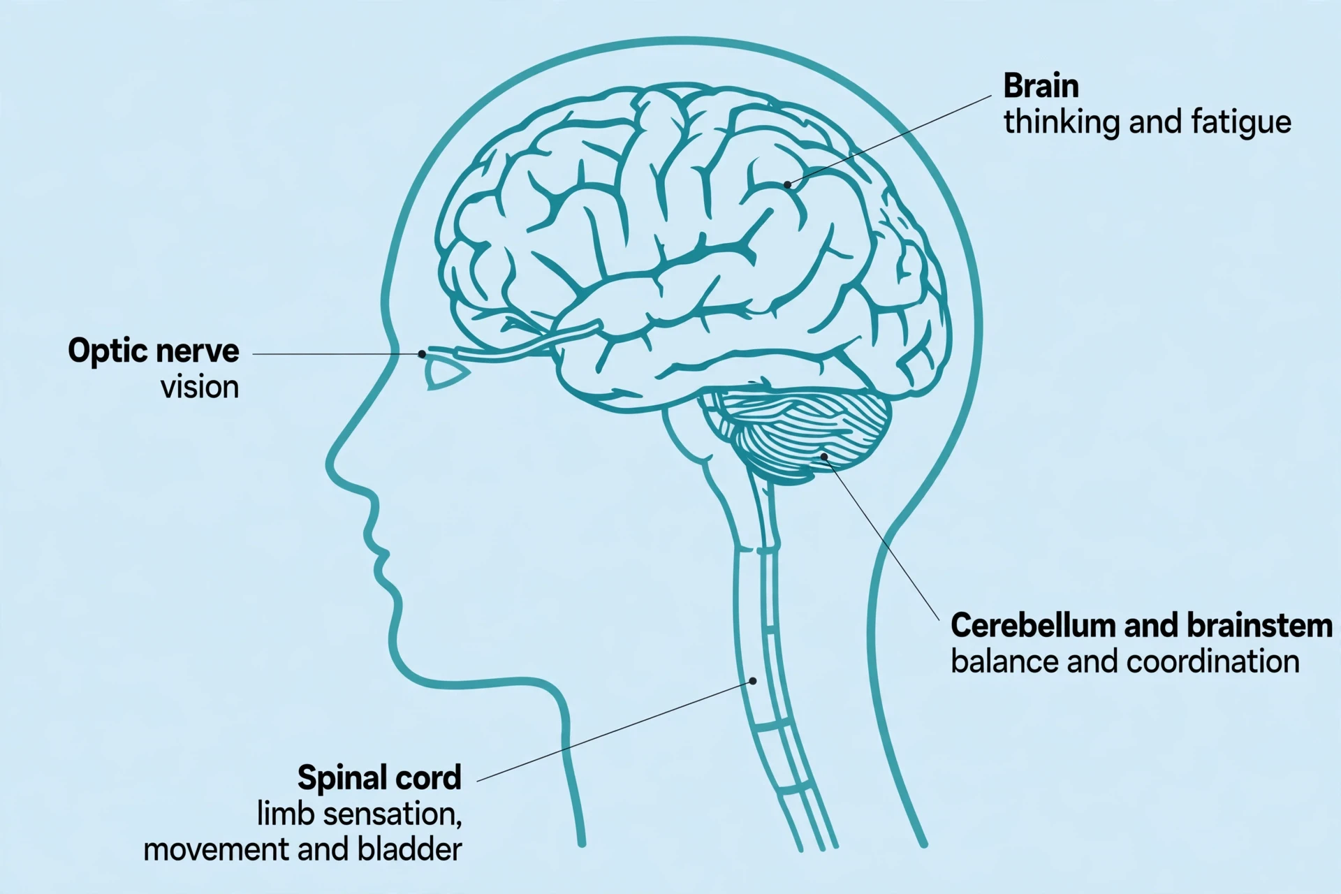
Symptoms depend on lesion location, because each lesion interrupts a specific pathway. An optic-nerve lesion affects vision. A spinal-cord lesion affects limb sensation and movement, and bladder control. A cerebellar or brainstem lesion affects balance and coordination. This is the organising principle of the whole symptom list: one process, many expressions, depending on where the damage lands.
Cerebellar and brainstem involvement produces ataxia — a loss of coordination — along with tremor, vertigo and dizziness, slurred or scanning speech, and double vision from internuclear ophthalmoplegia, a failure of the eyes to move together because the fibres connecting their control centres have been demyelinated. Vertigo is the presenting symptom in about five percent of people, and dizziness affects about half at some stage. These are the symptoms that make walking feel uncertain and the world feel as if it is tilting, and they are among the most disabling, because balance is something we only notice once it is gone.
Lesion location | Typical symptoms | Why that location causes them |
|---|---|---|
Optic nerve | Blurred or dimmed vision in one eye, loss of colour vision, pain on eye movement (optic neuritis) | The optic nerve carries all visual signals from one eye to the brain; demyelination slows or blocks that signal |
Spinal cord (cervical) | Numbness, tingling or weakness in arms and legs; Lhermitte's sign; bladder and bowel problems; the "MS hug" | The cord carries sensory and motor fibres for the whole body below the lesion, plus the pathways controlling bladder and bowel |
Cerebellum and brainstem | Ataxia and poor coordination, tremor, vertigo and dizziness, slurred or scanning speech, double vision | The cerebellum coordinates movement and balance; the brainstem houses the centres for eye movement, speech and balance |
Cerebrum (brain) | Cognitive slowing, memory and attention problems, fatigue, mood changes | Cortical and deep white-matter lesions disrupt the networks that carry out thinking, processing speed and mood regulation (fatigue is a general symptom of the disease, not tied to one location) |
5.4 The invisible symptoms: fatigue, cognition, mood, bladder and bowel
Some of the most disabling symptoms of multiple sclerosis are the ones no one can see. Fatigue is one of the most common and disabling of all, reported by almost eighty percent of people with the disease at some time. It is not ordinary tiredness. It is an overwhelming exhaustion, disproportionate to the effort that produced it, and not relieved by ordinary rest — often called lassitude. A person can wake after a full night's sleep and still feel as if they have run a long distance. That kind of fatigue is hard to explain to others, and hard to plan around, because it does not follow the rules of exertion and recovery that healthy people know. Sleep itself can be part of the problem, and Science of Sleep and Dreams explains how sleep, fatigue, and the brain's recovery processes interact.
Cognitive impairment affects about forty to sixty-five percent of people with multiple sclerosis, with the range depending on how it is classified and tested. The domains most affected are information processing speed, memory, executive function, and attention. In plain terms: thinking gets slower, holding a thought in mind gets harder, and multitasking becomes exhausting. This is the symptom often described as brain fog, and Brain Fog covers the science of that experience and what helps focus. For the underlying machinery of memory itself — how memories are encoded, consolidated, and retrieved — see How the Human Brain Forms Memories.
Bladder problems appear in seventy to eighty percent of people with multiple sclerosis, with some estimates putting lower urinary tract symptoms at eighty to ninety percent. A 2024 Dutch survey of 1,082 people found bladder problems in ninety-one percent and bowel problems in seventy-three percent. Bowel problems overall affect about thirty-nine to seventy-three percent, and nearly half of people with the disease say bowel problems interrupt their daily life. These are among the most common symptoms of the disease and among the least talked about, which is precisely why they deserve a plain statement here: they are a direct consequence of spinal cord lesions interrupting the pathways that control the bladder and bowel, and they are a normal part of the disease, not a personal failing.
Mood is part of the picture too. Depression is the main emotional symptom of multiple sclerosis, and it is more common than in the general population. It is not only a reaction to living with a chronic disease, though that alone would be reason enough. The lesions themselves, particularly in the circuits that regulate mood, contribute directly. Anxiety and Depression Explained covers the science of mood, brain chemistry, and what helps. The point to carry away from this section is simple: multiple sclerosis is a whole-person, brain-and-body disease. It is not only a disease of walking, and judging its severity by how someone walks misses most of it.
5.5 Lhermitte's sign, the "MS hug," and heat (Uhthoff's phenomenon)
Three named phenomena round out the symptom picture, and each is a small lesson in the physics of demyelinated nerves.
Lhermitte's sign is a brief electric-shock-like sensation that shoots down the neck and spine, sometimes into the arms and legs, when the neck is bent forward. It reflects demyelination in the cervical spinal cord, and it is a common finding in early multiple sclerosis. Neck flexion stretches the cord slightly, and the demyelinated fibres of the dorsal columns, already prone to misfiring, discharge together — though the exact mechanism is not fully established. It lasts seconds, and it is startling rather than dangerous, but it is a useful clue: few other conditions produce it.
The "MS hug" is a patient term for a tight, squeezing or aching sensation around the ribs and torso, caused by spasms of the small muscles between the ribs after spinal-cord demyelination. It is a patient term, not a formal medical sign. It is real to the people who feel it, and it has a real mechanism, but it is not a diagnostic criterion.
Uhthoff's phenomenon is the most instructive of the three. It is the temporary worsening of neurological symptoms when the body overheats — in hot weather, during exercise, with a fever, or in a hot bath or shower or sauna. It occurs in sixty to eighty percent of people with multiple sclerosis, and it can be triggered by a core-temperature rise as small as about half a degree Celsius. The mechanism is the physics of section 2: a demyelinated fibre that is already conducting slowly fails completely when it warms up, because the sodium channels that regenerate the signal close faster at higher temperatures. The effect typically reverses completely once body temperature returns to normal. It is not a true relapse. It is a temporary failure of already-damaged wiring, and it passes. Uhthoff's phenomenon works on a different timescale: it flares in minutes and fades just as fast.
6. Disease Courses: CIS, Relapsing-Remitting, and Progressive Multiple Sclerosis
6.1 Clinically isolated syndrome (CIS) — the first episode
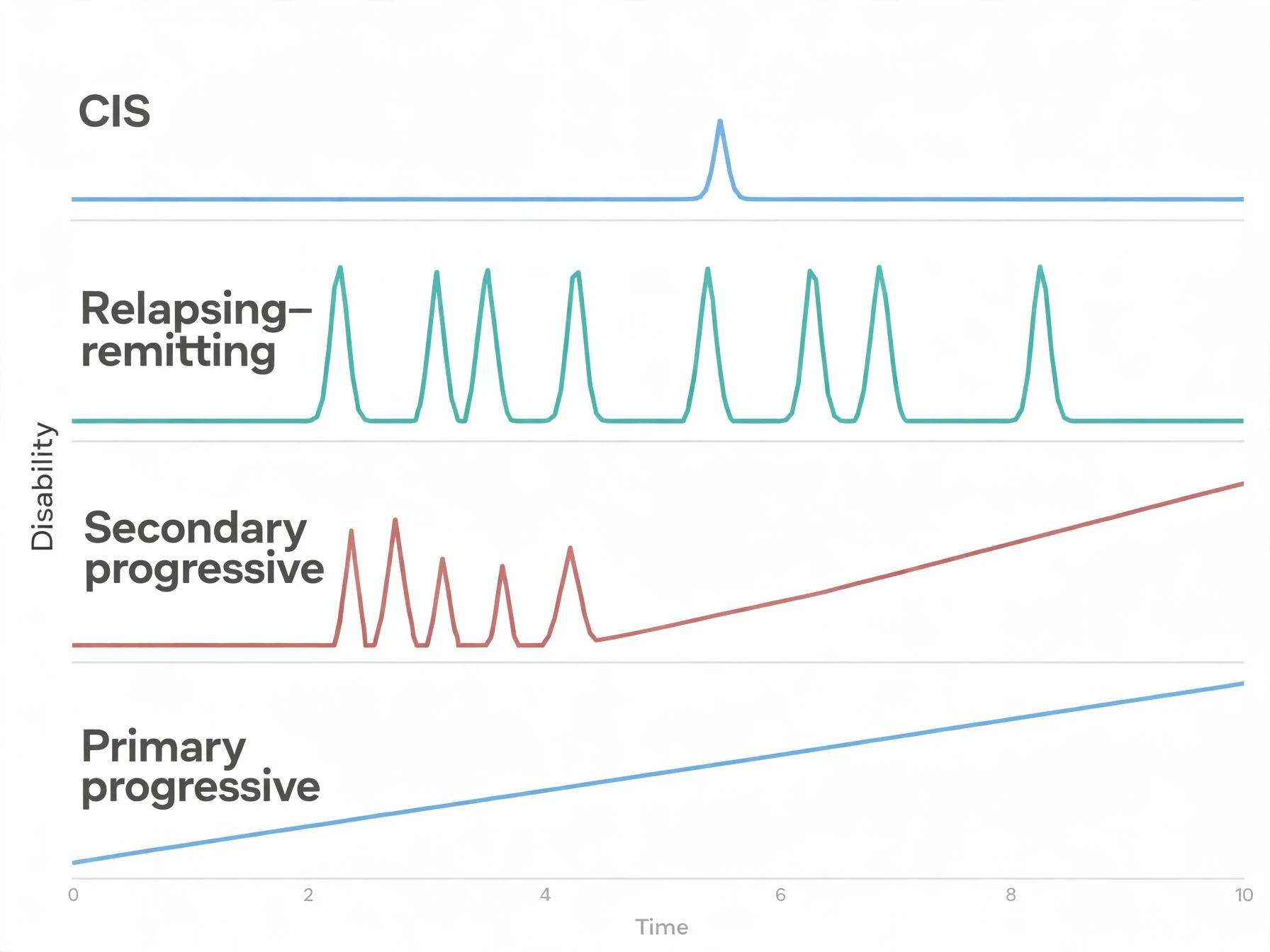
Clinically isolated syndrome, or CIS, is the first episode of neurological symptoms caused by inflammation and demyelination — a single attack, in one place, lasting at least a day. A person with CIS has had one event, and it looks like multiple sclerosis, but the criteria for the disease have not yet been met, because the disease requires evidence of damage in more than one place and at more than one time. CIS is now considered part of the relapsing-remitting phenotype, provided the dissemination-in-time criteria are fulfilled — that is, provided there is evidence that the disease process has been active at more than one point. In practice, many people with CIS and a typical MRI go on to develop multiple sclerosis, and the boundary between the two labels is a matter of how much evidence has accumulated, not a difference in the underlying biology.
6.2 Relapsing-remitting MS (RRMS) — about 85% at diagnosis
About eighty-five percent of people are initially diagnosed with relapsing-remitting multiple sclerosis. The pattern is its name: discrete attacks, called relapses, that develop over days to weeks, followed by complete or partial recovery — remissions — over months or years. A relapse is a new patch of inflammation and demyelination. A remission is the inflammation settling and the myelin partly repairing. Between relapses, the person may feel entirely well, and the disease may seem to have gone away. It has not. The lesions remain on the scan, and the next relapse may strike somewhere else entirely.
The Atlas of MS reporting for 2025 puts the split at about eighty-five percent relapsing-remitting, about twelve percent primary progressive, and about three percent unknown type. Those figures describe the picture at diagnosis. Over time, the picture changes, because a substantial share of people with relapsing-remitting disease gradually move into the next category.
6.3 Secondary progressive MS (SPMS) — when relapses give way to steady decline
Secondary progressive multiple sclerosis is the gradual accumulation of disability following an initially relapsing course. The relapses become less frequent, or stop, and in their place comes a slow, steady worsening: walking gets harder, fatigue deepens, thinking slows, and the decline does not reverse between episodes. It is the phase in which axonal loss, rather than fresh inflammation, becomes the main driver of disability.
In older natural-history data, from before modern disease-modifying therapies, about half of people with relapsing-remitting multiple sclerosis developed secondary progressive disease within about ten to twenty years of diagnosis, and about three-quarters eventually did. Those figures should be read as history: modern rates are lower and uncertain. Earlier diagnosis and earlier treatment have changed the course of the disease for many people, and the old natural-history numbers no longer describe what a newly diagnosed person can expect. But they do describe the direction of travel if the disease is left unchecked, and that is why the secondary progressive phase matters even to someone who has just been diagnosed.
6.4 Primary progressive MS (PPMS) — steady from the start
Primary progressive multiple sclerosis affects about ten to fifteen percent of people with the disease. In this form, disability increases steadily from onset, without distinct relapses. There is no cycle of attack and recovery. There is a slow, continuous accumulation of disability, often affecting walking and balance first, because the spinal cord is frequently involved early.
The people affected differ from those with relapsing-onset disease. The median age of onset is about forty years, against about thirty for relapsing-onset multiple sclerosis. And the female-to-male ratio is near one to one, unlike relapsing-remitting disease, where women outnumber men by about three to one. That difference is a clue to the biology: primary progressive disease seems to involve less of the episodic inflammation that drives relapses, and more of the slow neurodegeneration that drives progression. It is the hardest form to treat, because the treatments that calm relapses have less to work on when there are no relapses to calm — though one drug, ocrelizumab, was the first shown to slow progression in primary progressive disease, as section 9 explains.
6.5 Relapse vs progression — and PIRA
The distinction between a relapse and progression is the most important concept in this section, because it shapes both prognosis and treatment. A relapse is a new or worsening neurological symptom lasting at least twenty-four hours, separated from a previous attack by at least thirty days, and occurring in the absence of fever or infection. The fever rule matters: an infection or a hot day can temporarily worsen symptoms, as Uhthoff's phenomenon shows, and that is not a relapse. A true relapse is new inflammation, and it lasts weeks.
Progression is different. It is a steady, relapse-independent accumulation of disability, measured over months and years rather than days and weeks. And in recent years, researchers have recognised a specific form of it: progression independent of relapse activity, known as PIRA. PIRA is now recognised as a major driver of disability even in early relapsing-remitting multiple sclerosis. In the Swiss Multiple Sclerosis Cohort, fifteen percent of people with CIS or relapsing-remitting disease experienced PIRA within six years, and PIRA accounted for sixty-two percent of confirmed disability progression events in that group. The message is that disability in multiple sclerosis is not only the sum of relapses. Much of it accumulates silently, between attacks, without any dramatic event to mark it. That is why modern treatment aims not only to prevent relapses but to slow the quiet, steady progression underneath them.
The older "progressive-relapsing" category, which once described people with progressive disease punctuated by relapses, was removed in the current classification. The classification from Lublin and colleagues, published in Neurology in 2014 (volume 83, pages 278 to 286), defines the core phenotypes as relapsing-remitting disease and progressive disease — primary and secondary progressive — with activity and progression as modifiers. A person can have relapsing disease that is active, or progressive disease that is not, and the modifiers allow the description to follow the person rather than forcing the person into a box.
Course | Onset pattern | Relapses | Progression | Share | Typical age at onset |
|---|---|---|---|---|---|
Clinically isolated syndrome (CIS) | Single first episode | One episode so far | Not yet established | First presentation | 20 to 40 years |
Relapsing-remitting MS (RRMS) | Discrete attacks | Yes — over days to weeks, then recovery over months or years | Between relapses, generally stable early on | About 85% at diagnosis | About 30 years (median) |
Secondary progressive MS (SPMS) | Follows an initially relapsing course | Become less frequent or stop | Yes — gradual, steady accumulation of disability | About half of RRMS within a decade or two before modern DMTs | Develops years after RRMS onset |
Primary progressive MS (PPMS) | Steady from onset | No distinct relapses | Yes — steady from the start | 10 to 15% | About 40 years (median) |
7. What Causes Multiple Sclerosis? Genes, EBV, and the Environment
7.1 No single cause
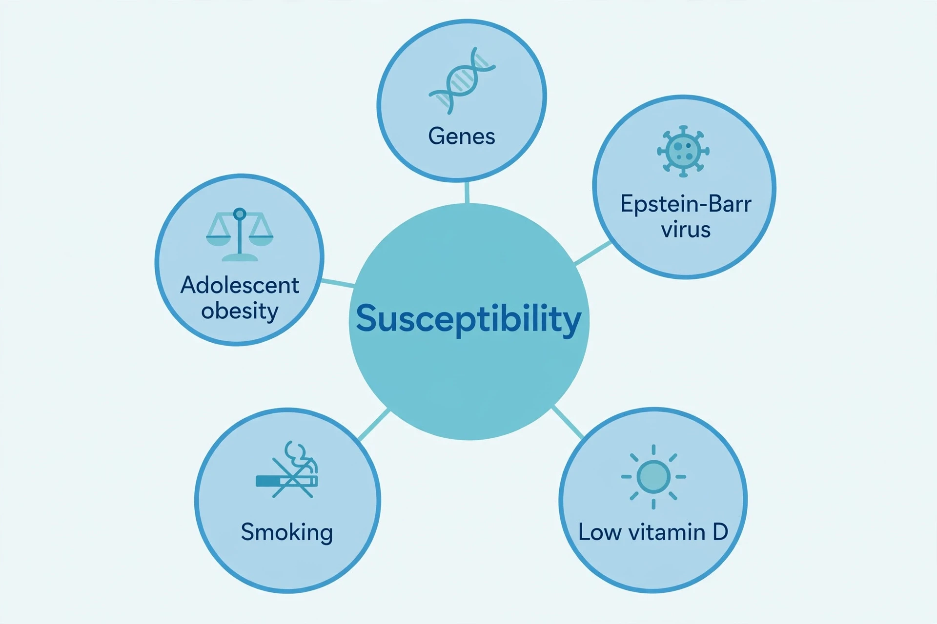
The cause of multiple sclerosis is unknown. That is the position of the MSD Manual and the National MS Society, and it is the right place to start, because everything that follows can otherwise be misread. No single cause has been found. What has been found is a combination: genetic susceptibility and environmental triggers, interacting in ways that are still being worked out. Each risk factor in this section raises the odds. None of them, on its own, explains the disease. A reader who keeps that sentence in mind will not be misled by any single finding, however striking it sounds.
7.2 Genetics — HLA-DRB115:01 and 233 variants
Genes matter, but they are not destiny. The strongest single genetic risk factor for multiple sclerosis is a variant of a gene called HLA-DRB115:01, part of the HLA system that teaches the immune system to distinguish self from not-self. Carrying at least one copy of this variant confers an approximately threefold increase in risk — an odds ratio of about 2.9 to 3.1, and about 3.5 in one large merged dataset — and in people who carry two copies the risk is higher still — an odds ratio of about 11.5, against about 3.0 for a single copy. That is a large effect for a single gene variant, and it points directly at the immune system: the gene that most raises the risk of multiple sclerosis is a gene that controls how the immune system sees the body's own tissues.
Other variants pull in the opposite direction. HLA-A02:01 is protective against multiple sclerosis, with an odds ratio of about 0.72 to 0.73 — carrying it lowers the risk by roughly a quarter. And the picture is far wider than these two genes. Genome-wide association studies have identified 233 common genetic variants associated with multiple sclerosis, thirty-two of them in the HLA region and two hundred and one outside it. The variants outside the HLA region point mostly at immune function, particularly the cell-mediated immune mechanisms that T cells use.
But the arithmetic is humbling. Collectively, genetic factors explain only part of overall disease risk: estimates of heritability run from roughly a third to roughly three-quarters, and the genome-wide variants found so far explain only about a fifth of that heritability. Environmental and lifestyle factors are the major contributors. Having a first-degree relative with multiple sclerosis raises the risk — the absolute risk for a child of a person with the disease is about one to five percent — and identical-twin concordance is about twenty-five to thirty-one percent — an approximate figure from older studies, not modern population data. The twin figure is the most telling of all: identical twins share all their genes, yet if one twin develops multiple sclerosis, the other usually does not. Genes load the gun. Something else pulls the trigger.
For the background on how genes, inheritance, and variation work, The Human Genome Explained and Genetics Explained cover the ground, from the three billion letters of DNA to the way individual variants shift risk.
7.3 Epstein-Barr virus — the 32-fold finding, with the necessary-but-not-sufficient nuance
The most striking single finding in multiple sclerosis research in recent years concerns a virus almost everyone carries. Epstein-Barr virus, or EBV, is the virus that causes glandular fever, and it infects about ninety to ninety-five percent of all adults, usually in childhood or adolescence, after which it stays in the body for life, dormant in B cells.
In 2022, Bjornevik and colleagues published a study in Science that changed the conversation. They followed more than ten million United States military personnel over twenty years, from 1993 to 2013, and identified 955 who developed multiple sclerosis. The risk of multiple sclerosis increased thirty-two-fold after EBV seroconversion — after the blood first showed evidence of EBV infection — and it did not increase after infection with other viruses, including cytomegalovirus, a similar herpesvirus used as a control. All but one of the 801 cases with adequate serum samples had been infected with EBV before the onset of multiple sclerosis, and seroconversion preceded onset by a median of seven and a half years. The virus came first. The disease followed, years later.
That is a remarkable result, and it demands a careful reading. A 2026 review in The Lancet Neurology, by Lünemann and Münz, concludes that the evidence now strongly supports EBV infection as, in most cases, necessary for multiple sclerosis to develop. Necessary, but not sufficient. Almost everyone carries EBV, and only a tiny fraction develop multiple sclerosis. If EBV alone caused the disease, the disease would be as common as the virus. It is not. The study was also observational: it cannot prove causation the way a randomised trial could, and the cohort was young and mostly male military personnel, which is not a cross-section of the general population.
So what might the mechanism be? The leading hypothesis is molecular mimicry. Antibodies against the EBV protein EBNA1 may cross-react with GlialCAM, a protein in oligodendrocytes and astrocytes that supports myelin. The immune system, trained to attack the virus, mistakes a piece of the brain's own support machinery for the enemy. That is a hypothesis, not an established fact, and I present it as one. It is plausible, it fits the data, and it is being tested. It is not yet proven.
The practical consequence is a new direction for prevention research. If EBV is necessary for most multiple sclerosis, then a vaccine against EBV — or a treatment that controls the virus — might one day prevent the disease in people at risk. That work is in its early stages, and no such vaccine exists for routine use. For how vaccines train the immune system in general, see How Vaccines Work. For the biology of viruses and how they evolve, see Virus Evolution Explained and Microbiology Explained.
7.4 Vitamin D and latitude — association, not proof
Multiple sclerosis is more common farther from the equator — in Canada, the northern United States, and northern Europe — than it is in the tropics. That latitude gradient is one of the oldest observations in the epidemiology of the disease, and it has held up: the gradient in the United States persists in the Wallin 2019 data. Low vitamin D, and low sun exposure, is one of the most consistent environmental predictors of the disease, alongside EBV, smoking, and adolescent obesity.
The link is plausible. Vitamin D is made in the skin when sunlight falls on it, and it plays a role in regulating the immune system. People who live far from the equator get less sun, make less vitamin D, and — the hypothesis runs — have an immune system that is more prone to attacking myelin. But the link is an association, and association is not proof. Whether vitamin D supplements prevent multiple sclerosis, or slow it once it has begun, is not established. The plain version is this: low vitamin D is linked to higher risk, and that is all the evidence currently supports. No supplement claim follows from it, and no reader should take one away. For the symptoms and treatment of low vitamin D itself, see Vitamin D Deficiency.
7.5 Smoking and adolescent obesity — modifiable risk factors
Two risk factors stand out because they are, in principle, within a person's control — though "modifiable" should never be read as "blameworthy," and the disease is not anyone's fault.
Smoking significantly increases the risk of developing multiple sclerosis, particularly primary progressive disease, according to the National MS Society, and it is associated with faster disability progression and more MRI brain lesions. That is a qualitative statement: the direction is clear, and no reliable percentage is available to put on the size of the effect.
Adolescent obesity carries a clearer number. Being obese at age eighteen more than doubled the later risk of multiple sclerosis in women, in the Harvard Nurses' Health Studies — 238,000 women followed for forty years, published in Neurology in November 2009. The mechanism is not fully settled, but obesity in adolescence is a state of chronic low-grade inflammation, and it interacts with the immune system during the years when it is still being calibrated. For the science of weight gain, appetite regulation, and why diets fail, see Obesity Explained.
7.6 How risk factors interact — and who gets MS
Risk factors do not act alone. Smoking, EBV infection, and adolescent obesity interact with HLA risk genes, with substantially increased risk in people who carry predisposing HLA variants. The picture is not a list of separate risks added together. It is a web, in which a genetic variant that changes how the immune system sees myelin, a virus that trains the immune system against a lookalike protein, a vitamin D level shaped by latitude, and a history of smoking or adolescent obesity all push in the same direction. Epigenetics Explained covers how the environment changes the way genes are expressed without changing the genes themselves — the mechanism by which a life lived in a particular place, with particular exposures, leaves its mark on the immune system.
That web also helps explain who gets multiple sclerosis. The female predominance — about three to one in round terms — is not explained by any single factor. It is the net result of genes, hormones, and environment interacting differently in women and men. And the age pattern — onset usually between twenty and forty, mean age at diagnosis thirty-two — reflects a disease that needs time to accumulate its triggers, but strikes while the immune system is still at its most active.
8. How Multiple Sclerosis Is Diagnosed: MRI and the McDonald Criteria
8.1 No single test
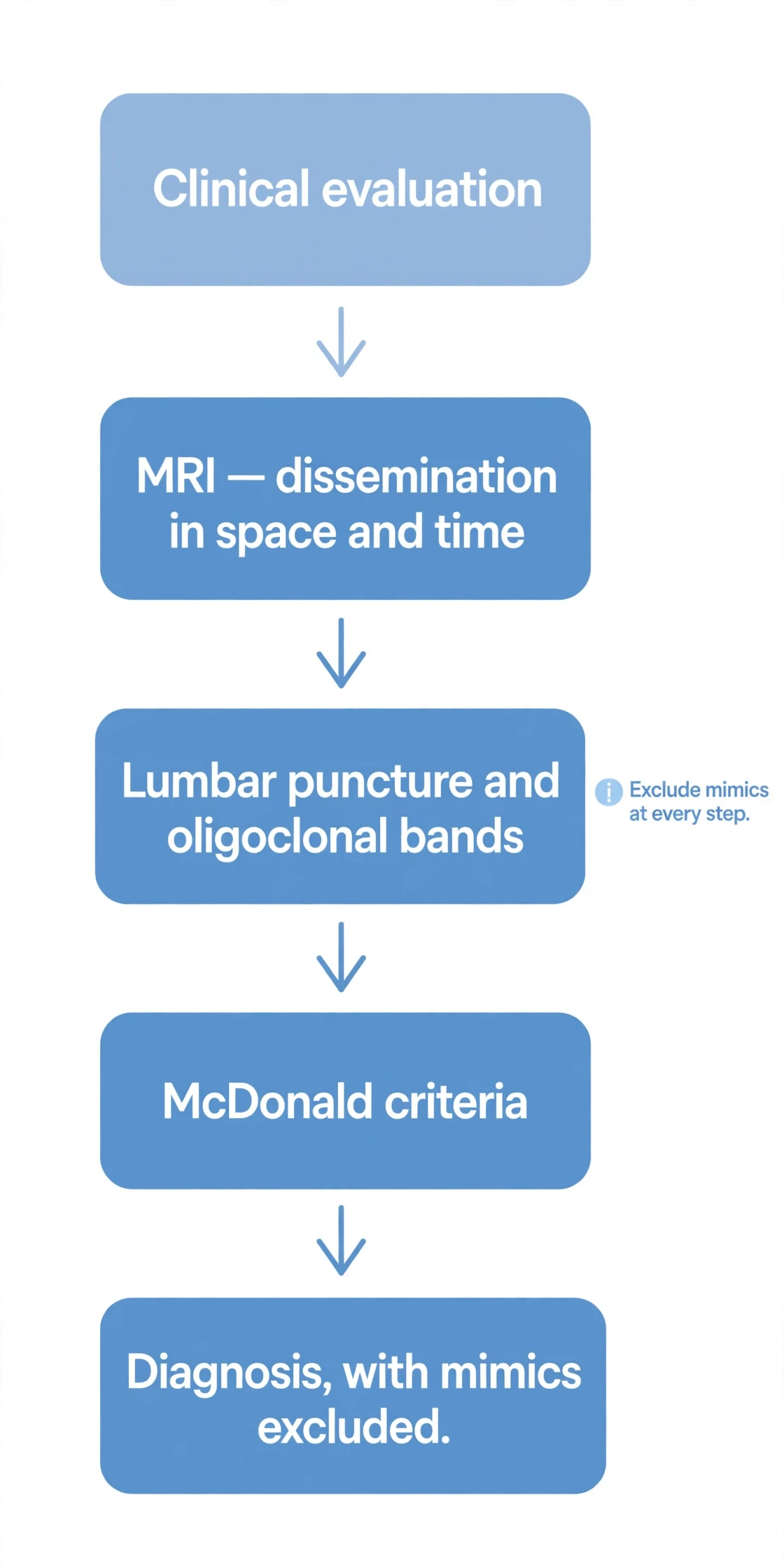
There is no single definitive test for multiple sclerosis. No blood test proves it. No scan proves it on its own. Diagnosis combines clinical evaluation, a neurological examination, MRI, and — where needed — analysis of the cerebrospinal fluid, under a set of rules called the McDonald criteria, while excluding alternative diagnoses. That last phrase, "while excluding alternative diagnoses," is part of the criteria, not an afterthought. A diagnosis of multiple sclerosis is a judgement that the evidence fits the disease and fits nothing else better.
This can be frustrating for a person waiting for an answer. A test that says yes or no would be simpler. But the biology does not allow one. Multiple sclerosis is defined by a pattern — damage in more than one place, at more than one time — and a pattern can only be shown by assembling several pieces of evidence and seeing whether they fit together. The rest of this section explains how those pieces are assembled.
8.2 The McDonald criteria 2017 — the widely used standard
The McDonald criteria, in their 2017 revision by Thompson and colleagues, published in The Lancet Neurology in 2018 (volume 17, pages 162 to 173), are the widely used standard for diagnosis. The core idea is simple. Multiple sclerosis is diagnosed when dissemination in space and dissemination in time are demonstrated, clinically or by MRI, with no better explanation. Dissemination in space means lesions in more than one part of the central nervous system. Dissemination in time means lesions that appeared at different times. A disease that strikes in many places, at many times, is a disease of the whole central nervous system, not a single event.
The 2017 revision changed three things from the 2010 version. First, in a patient with a typical clinically isolated syndrome and dissemination in space already demonstrated, cerebrospinal-fluid-specific oligoclonal bands can substitute for dissemination in time — allowing the diagnosis to be made at the first visit, without waiting for a second attack. Second, symptomatic lesions can count toward dissemination in space and time, where previously only silent lesions counted. Third, cortical lesions — lesions in the grey matter at the surface of the brain — can demonstrate dissemination in space. Each change had the same effect: an earlier, more certain diagnosis, without lowering the standard of proof.
Element | What it requires | How it is shown |
|---|---|---|
Dissemination in space (DIS) | At least one T2-hyperintense lesion in at least 2 of 4 CNS areas | Periventricular, cortical or juxtacortical, infratentorial (brainstem and cerebellum), spinal cord |
Dissemination in time (DIT) — route 1 | Simultaneous gadolinium-enhancing and non-enhancing lesions | A single MRI scan showing one active lesion and one older lesion at the same time |
Dissemination in time (DIT) — route 2 | A new T2 or gadolinium-enhancing lesion on follow-up MRI | A repeat scan showing a lesion that was not there before |
Dissemination in time (DIT) — route 3 | Cerebrospinal-fluid-specific oligoclonal bands | Lumbar puncture showing bands in the spinal fluid but not in paired blood serum |
Primary progressive pathway | At least 1 year of disability progression independent of relapses, plus at least 2 of 3 supporting findings | Dissemination in space by MRI; dissemination in time by MRI or positive spinal-fluid oligoclonal bands; typical spinal-cord or brain lesions |
2024 changes (footnote) | Optic nerve added as a fifth DIS site; DIT can be bypassed if typical lesions are present in at least 4 of the 5 sites; central vein sign and paramagnetic rim lesions formally integrated | Montalban et al., Lancet Neurology 2025; see section 8.3 |
8.3 The 2024 revision — the newest development, not yet universal practice
A newer revision exists. The 2024 revision of the McDonald criteria, by Montalban and colleagues, published in The Lancet Neurology in 2025 (volume 24, pages 850 to 865), adds the optic nerve as a fifth dissemination-in-space site, alongside the four from 2017. It also allows dissemination in time to be bypassed entirely if typical lesions are present in at least four of the five sites. It formally integrates two newer MRI markers — the central vein sign and paramagnetic rim lesions — to improve specificity, helping doctors distinguish multiple sclerosis lesions from the white spots caused by other conditions.
The 2024 criteria are the newest publication, so 2017 should not be presented as 'the latest' without noting them. But it is equally important not to overstate their status. The 2017 criteria remain the widely used standard. How widely clinics have adopted the 2024 revision in routine practice is not yet clear.
8.4 MRI, lumbar puncture, and evoked potentials
MRI is the most sensitive paraclinical tool for diagnosing multiple sclerosis. Brain MRI should be obtained in everyone in whom the disease is being considered, unless MRI is impossible. Spinal cord MRI adds a dissemination-in-space site, and it can enable a diagnosis with fewer brain lesions — an important point for people whose brain scan is nearly, but not quite, diagnostic on its own.
Lumbar puncture — a spinal tap — examines the cerebrospinal fluid that bathes the brain and spinal cord. In multiple sclerosis, the concentration of IgG, the main class of antibody, is raised in more than ninety percent of patients. More specifically, oligoclonal bands — distinct bands of IgG that appear in the spinal fluid but not in a paired sample of blood serum — indicate that antibodies are being made inside the central nervous system itself. They are present in the large majority, roughly eighty-five to ninety-five percent, of people with established multiple sclerosis, though the figures in the literature range widely: sixty to seventy percent in clinically isolated syndrome, up to ninety percent in established disease, and about ninety-five percent in some teaching sources. Two cautions matter. Oligoclonal bands neither prove multiple sclerosis on their own, nor rule it out when they are absent. They are one piece of evidence, and they are most useful when combined with the MRI and the clinical picture — which is exactly how the 2017 criteria use them, as a substitute for dissemination in time.
Evoked potentials measure slowed conduction directly. Electrodes record the brain's response to a stimulus — a flashing pattern for the visual pathway, a click for the auditory pathway, a small electrical pulse for the somatosensory pathway — and the time the signal takes to arrive reveals whether the fibres carrying it are demyelinated. About seventy-five percent of people with multiple sclerosis have abnormal visual evoked potentials, whether or not they have ever had optic neuritis, and more than eighty percent have abnormal somatosensory evoked potentials. Evoked potentials are used less often than they once were, because MRI has largely taken over their role, but they remain a useful way of showing that a pathway is damaged even when the person has no symptoms from it — silent evidence of dissemination in space.
8.5 Ruling out the mimics
"No better explanation" is part of the McDonald criteria, and it is where diagnosis becomes hardest. Several conditions can mimic multiple sclerosis, and they must be ruled out before the label is applied. They include neuromyelitis optica spectrum disorder, a related disease that attacks the optic nerves and spinal cord; MOG antibody disease, another antibody-driven demyelinating condition; vasculitis, inflammation of the blood vessels; lupus, a systemic autoimmune disease; vitamin B12 deficiency, which can damage the spinal cord in a pattern that resembles multiple sclerosis; Lyme disease; sarcoidosis; and tumours. The point for the reader is simpler: a diagnosis of multiple sclerosis is not only a matter of finding lesions. It is a matter of finding lesions, showing the pattern, and making sure nothing else explains them.
9. Treatment for Multiple Sclerosis: What Disease-Modifying Therapies Do — and Do Not Do
9.1 There is no cure — and three aims of treatment
There is no cure for multiple sclerosis. That sentence belongs at the start of any discussion of treatment, because everything else is measured against it. What treatment can do falls into three broad aims. First, disease-modifying therapies, or DMTs, reduce relapses and slow progression. Second, treatment of acute relapses — usually corticosteroids — shortens the attack and speeds recovery, though it does not change the long-term course. Third, symptom management and rehabilitation help with the day-to-day consequences: fatigue, spasticity, bladder problems, pain, and mobility, through medication, physiotherapy, and other support.
Nothing in this section is a recommendation for any individual. Treatments differ in their benefits, their risks, and their suitability for different people and different forms of the disease, and the choice among them is a decision for a person and their own neurologist, made with full knowledge of their own history. What follows is an educational map of the classes and their mechanisms — how each one works, in general terms, and what it cannot do.
9.2 Before 1993 there were no DMTs — the first drug
The DMT era began with the approval by the United States Food and Drug Administration of interferon beta-1b in 1993 — the first drug shown to modify the course of multiple sclerosis. Before 1993, there were no disease-modifying therapies at all. Doctors could treat relapses and manage symptoms, but they had nothing that changed the course of the disease itself.
In the published trial, interferon beta-1b cut the annual relapse rate by thirty-four percent at two years, in 372 people with relapsing-remitting multiple sclerosis. Glatiramer acetate followed, approved in late 1996 and reaching patients in 1997. Those two drugs, modest by today's standards, were a turning point: for the first time, multiple sclerosis was a disease with a treatment that altered its course, and the three decades since have been a story of more drugs, acting in more ways, with greater effect. There are now more than twenty FDA-approved DMTs. Counts differ — some list twenty-three approved medicines, others over twenty-five including newer agents — so 'more than twenty' is the safe figure.
9.3 DMT classes by mechanism
Each class of DMT works at a different point in the immune attack described in section 3, and the mechanism is the best way to understand them.
Interferons, beta-1a and beta-1b, have broad immunomodulatory effects. They inhibit the production of pro-inflammatory T-cell cytokines, calming the immune response across the board. Their efficacy is moderate by modern standards, but they were the first, and they established the principle that the course of the disease could be changed.
Glatiramer acetate is a mixture of four amino acids that mimics myelin proteins. It shifts the immune response from a pro-inflammatory Th1 pattern toward an anti-inflammatory Th2 pattern, and it promotes regulatory T cells — the brakes described in section 3. Its exact molecular mechanism is unknown, which is worth stating plainly: a drug can be shown to work without every step of its action being understood.
Fumarates, including dimethyl fumarate and related drugs, activate the Nrf2 antioxidant-response pathway, with anti-inflammatory and immunomodulatory effects. They reduce immune-cell activation and lower lymphocyte counts, damping the response from the inside.
S1P receptor modulators — fingolimod, siponimod, ozanimod, and ponesimod — trap lymphocytes in the lymph nodes by blocking their egress. Lymphocytes that cannot leave the lymph nodes cannot reach the blood, and cannot cross into the central nervous system. The drug does not kill the cells. It keeps them where they cannot do harm.
Natalizumab is a monoclonal antibody against alpha-4 integrin. It binds VLA-4, the molecule T cells use to grip the blood-vessel wall, and blocks their migration across the blood–brain barrier into the central nervous system. Approved in 2004, it is a higher-efficacy therapy, and its mechanism is a direct answer to the crossing described in section 3.1: if the handshake between the T cell and the vessel wall is blocked, the cell cannot get in.
Anti-CD20 monoclonal antibodies — ocrelizumab, ofatumumab, and ublituximab — deplete CD20-positive B cells. They answer the B-cell story of section 3.3: remove the cells that make the antibodies and recruit the T cells, and the attack weakens. Ocrelizumab was the first DMT also shown to slow progression in primary progressive multiple sclerosis, which made it a landmark for the form of the disease that had previously had no effective treatment.
Cladribine is a purine analogue that inhibits DNA synthesis in proliferating lymphocytes, depleting them by apoptosis — programmed cell death — and it is given as short pulsed courses, after which the immune system rebuilds itself. Teriflunomide inhibits dihydroorotate dehydrogenase, an enzyme needed for pyrimidine synthesis, and so reduces lymphocyte proliferation. Alemtuzumab, an antibody against CD52, depletes both T and B cells, and is also a pulsed immune-reconstitution therapy: a short course, then the immune system rebuilds, ideally in a less self-reactive form.
DMT class | Mechanism | What it does | What it does not do |
|---|---|---|---|
Interferons (beta-1a / beta-1b) | Broad immunomodulation; inhibit pro-inflammatory T-cell cytokines | Moderately reduces relapses | Does not cure MS; does not repair lost myelin |
Glatiramer acetate | Amino-acid mixture mimicking myelin proteins; Th1 to Th2 shift; promotes regulatory T cells | Reduces relapses; exact molecular mechanism unknown | Does not cure MS; does not restore lost function |
Fumarates | Activate the Nrf2 antioxidant-response pathway | Reduce immune-cell activation and lymphocyte counts | Does not cure MS; does not repair lost myelin |
S1P receptor modulators | Trap lymphocytes in lymph nodes by blocking egress | Reduce lymphocytes in blood and therefore in the CNS | Does not cure MS; effect lasts only while the drug is taken |
Natalizumab (anti-alpha-4 integrin) | Binds VLA-4; blocks leukocyte migration across the blood–brain barrier | Higher efficacy in reducing relapses and new lesions | Does not cure MS; does not repair lost myelin |
Anti-CD20 monoclonal antibodies | Deplete CD20-positive B cells | Reduce relapses and new lesions; ocrelizumab also slows progression in primary progressive MS | Does not cure MS; does not restore lost function |
Cladribine | Purine analogue; inhibits DNA synthesis in proliferating lymphocytes | Short pulsed courses with durable immune reconstitution | Does not cure MS; does not repair lost myelin |
Teriflunomide | Inhibits dihydroorotate dehydrogenase (DHODH) | Reduces lymphocyte proliferation | Does not cure MS; does not restore lost function |
Alemtuzumab (anti-CD52) | Depletes T and B cells | Short pulsed courses with immune reconstitution | Does not cure MS; does not repair lost myelin |
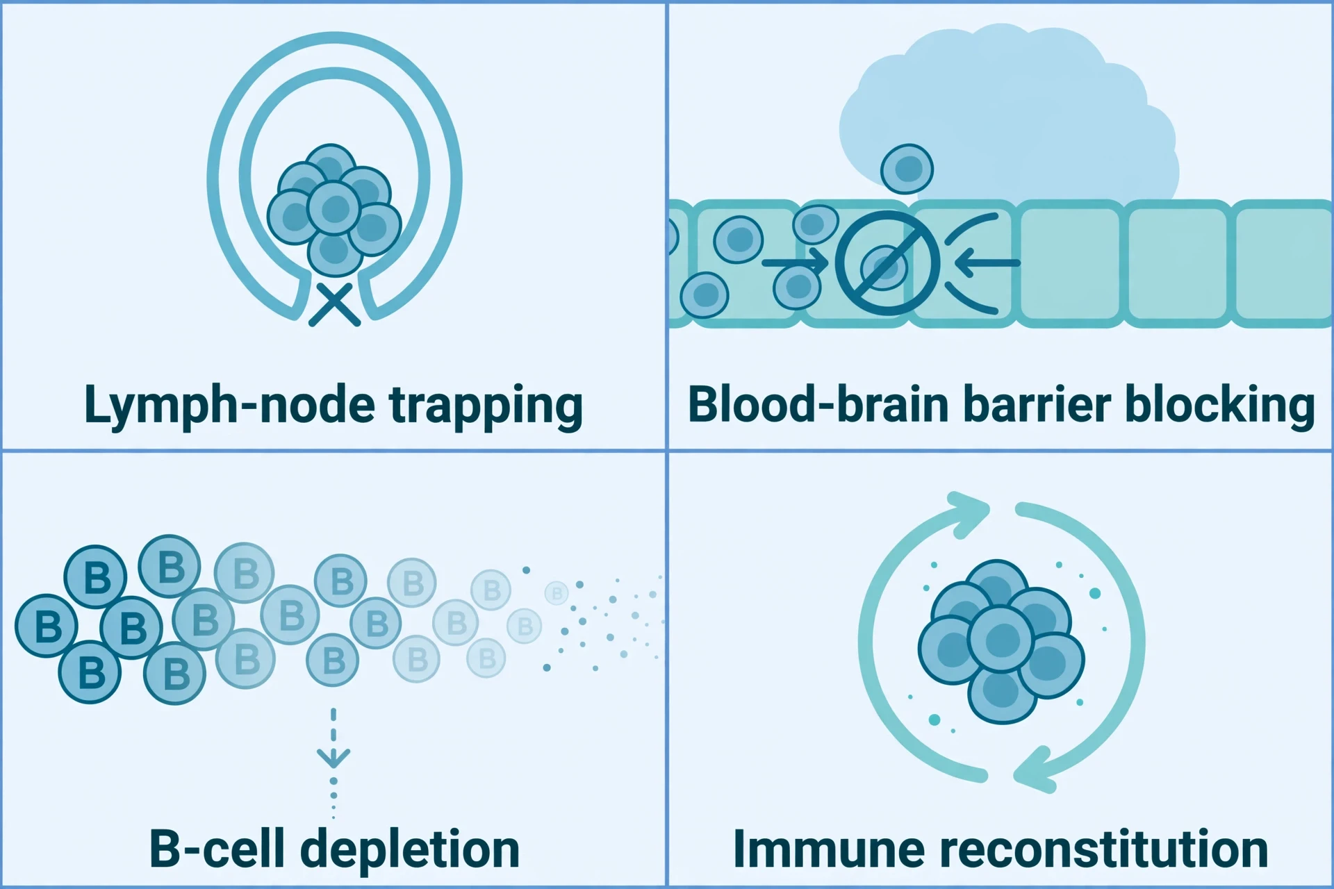
9.4 What DMTs do not do
The limits are as important as the mechanisms. Disease-modifying therapies do not cure multiple sclerosis. They do not repair myelin already lost, and they do not restore function already lost. What they do is reduce the frequency of relapses, reduce new MRI lesions, and — for some of them — slow the accumulation of disability. That is a great deal, and it is not everything. A person on an effective DMT can still have relapses, and can still accumulate disability over time, more slowly than they would have without treatment.
There is a second distinction that matters. Continuous therapies work only while they are being taken: stop the drug, and its effect stops with it. Pulsed therapies — cladribine and alemtuzumab — are given as short courses, after which the immune system rebuilds itself, and the effect can persist long after the last dose. Both approaches have their place, and the choice between them depends on the person, the form of the disease, and the balance of benefit and risk. That balance is a clinical decision, and this article does not make it.
9.5 Why early treatment matters
Neurologists stress starting a DMT early, and staying on one that controls both clinical and MRI disease, to avoid relapses and progression. The reasoning follows from everything in sections 2 and 3. Every relapse is a patch of demyelination, and every patch carries a risk of axonal loss that cannot be undone. The earlier the inflammation is controlled, the less damage accumulates, and the better the long-term outlook. That is a general educational statement about the direction of the evidence, not advice to any individual about when to start or which treatment to choose. Those decisions belong to a person and their neurologist, and they depend on far more than this article can cover.
10. Prognosis and Living with Multiple Sclerosis
10.1 Life expectancy — near-normal, with a ~7-year gap on average
Most people with multiple sclerosis have a near-normal life expectancy. The National MS Society figure quoted across sources puts the average gap at about seven years shorter than the general population, with published estimates ranging from about two to about fourteen years. The gap is steadily shrinking, as diagnosis comes earlier and disease-modifying therapies improve.
Multiple sclerosis itself is rarely fatal. Most people with the disease ultimately die of the same common conditions as the general population: heart disease, stroke, and cancer. What shortens lifespan is complications — infections, cardiovascular disease, and the consequences of advanced disability — rather than the neurological disease itself. Secondary health conditions such as heart disease, diabetes, and high blood pressure often affect lifespan in multiple sclerosis more than the neurological disease does, which is why general health care matters as much as multiple-sclerosis-specific care. In short, the disease affects quality of life more than longevity, and with appropriate treatment, life expectancy essentially mirrors that of people without the disease.
These are population figures. They describe averages across large groups, and they say nothing about any individual person. No one can be told, from a population average, how long they will live or how disabled they will become. The figures are useful for understanding the disease in general, and useless for predicting a single life. Both of those things are true at once.
10.2 Mobility and disability — the numbers with their timeframes
Mobility is the question most people ask first, and it deserves a straight answer with its timeframes stated. Around two-thirds of people with multiple sclerosis are able to walk without a wheelchair twenty years after diagnosis. Three out of four can get around without a wheelchair, according to the National MS Society. Around forty-five percent of people with the disease are not severely affected by it.
Each of those figures measures something different: walking without a wheelchair at twenty years, getting around without a wheelchair in general, and being "not severely affected." They are compatible, and they are not interchangeable, and no single lifetime wheelchair percentage captures them — a single number would imply a certainty the evidence does not support. What the figures together say is this: many people with multiple sclerosis never need a wheelchair, many use a cane or another aid at some point, and a minority need a wheelchair. The disease is not a sentence to a wheelchair, and it is not a guarantee of walking unaided either. It is a risk, spread unevenly, and shaped by the form of the disease, the timing of treatment, and factors no one fully understands.
10.3 RRMS vs PPMS outlook
Relapsing-remitting multiple sclerosis generally has a better long-term outlook than primary progressive multiple sclerosis. That follows from the biology: a disease driven by episodic inflammation can be treated with drugs that calm inflammation, while a disease driven by slow, steady neurodegeneration has less for those drugs to work on. Early diagnosis and immediate treatment are consistently associated with better long-term outcomes, as a general statement about the evidence. The association is real, and it is one of the strongest arguments for taking the disease seriously from the start. It is also, again, a statement about groups, not about any individual.
10.4 Exercise, rehabilitation, and heat
Living with multiple sclerosis is not only a matter of drugs. Regular exercise, physiotherapy, and rehabilitation are standard parts of living with the disease, improving strength, balance, fatigue, and mood. That may sound surprising for a condition in which movement can be difficult, but the evidence supports it: the body that is used stays stronger, the balance that is practised stays steadier, and the fatigue that is met with graded activity is more manageable than the fatigue that is met with rest alone. How Exercise Changes the Body covers the science of how exercise reshapes muscle, heart, brain, and mood — the same systems that multiple sclerosis strains.
Heat is the practical problem. Uhthoff's phenomenon, described in section 5, means that exercise in a hot room, or on a hot day, can temporarily worsen symptoms. Heat sensitivity during exercise is managed with cooling strategies, such as pre-cooling before activity, exercising in a cool environment, and using cooling vests or cold drinks. The point is not to avoid exercise. It is to manage the temperature, so that the exercise can happen.
I learned that lesson on an ordinary summer afternoon, not in a clinic. I had been out in the heat for an hour, and when I came inside and stood under a hot shower, my legs felt heavier than they had all day, and my thinking went woolly at the edges. It passed within half an hour of cooling down, and nothing was damaged. That is only an analogy, not the phenomenon itself — I do not have multiple sclerosis — but it gives a sense of what a flare feels like: alarming, and then gone, leaving nothing behind but the memory of it. For a person with multiple sclerosis, the same thing happens with demyelinated fibres that are already conducting slowly: a small rise in temperature pushes them over the edge, and cooling brings them back. It is not a relapse. It is the physics of damaged wiring, and once you understand it, it is manageable — a reason to carry water and plan for shade, not a reason to stay inside.

Living with the disease also means living with its less visible parts — the fatigue that rest does not fix, the cognitive slowing, the bladder problems, and the low mood that the lesions themselves can cause. Those are not separate from the disease. They are the disease, and they deserve the same attention as walking. The practical message of this section is simpler. Multiple sclerosis is a long-term condition, and a long-term condition is managed day by day: with treatment, with exercise, with cooling strategies, with attention to general health, and with an understanding of what is happening inside the nervous system that makes the bad days explicable and the good days worth using well.
11. A Short History of Multiple Sclerosis
11.1 Before Charcot — lesions seen, disease unnamed
Before multiple sclerosis had a name, it had a picture. In the early nineteenth century, the British pathologist Robert Carswell and the French pathologist Jean Cruveilhier described and illustrated the lesions of the disease in the brain and spinal cord, but neither identified it as a separate disease. Cruveilhier, in 1841, was the first to correlate the scars in the brain with the symptoms the person had shown in life — a connection that now seems obvious, and was then a new way of thinking about disease.
The earliest case often cited is older still. Lidwina of Schiedam, a Dutch girl who fell while skating in 1396 and developed progressive disability, is often described as the earliest identifiable case of multiple sclerosis. That is retrospective speculation, not a diagnosis. No one in the fourteenth century could have known what caused her illness, and the story should be read as a curiosity, not as evidence. The first descriptions in Britain came from William Moxon in 1873, and in the United States from Edward Seguin in 1878. By then, the disease was beginning to be recognised as something distinct, and it needed a person to say so clearly.
11.2 Charcot, 1868 — sclérose en plaques
That person was Jean-Martin Charcot, who first recognised multiple sclerosis as a distinct disease in 1868, in lectures at the Salpêtrière hospital in Paris. He named it sclérose en plaques — sclerosis in plaques — and he arrived at the diagnosis by correlating clinical symptoms with the plaques of demyelination found after death. That method, the anatomo-clinical method, was his great contribution: observe the patient in life, examine the brain after death, and join the two together.
Charcot's triad — the three signs he used to recognise the disease — were nystagmus, an involuntary flickering of the eyes; intention tremor, a shaking that appears when reaching for something; and scanning, or telegraphic, speech, in which words come out in separated bursts. That triad was the standard of diagnosis for almost a century. It is a reminder of how far the field has come: a disease once diagnosed by three signs at the bedside is now diagnosed by MRI scans, spinal fluid analysis, and criteria that count lesions in five regions of the nervous system.
Charcot's name is attached to more than multiple sclerosis. He also described Parkinson's disease, and the tremor that bears his triad's second element has its counterpart in that disease. Parkinson's Disease Explained covers the dopamine, tremor, and movement of that condition, and the contrast is instructive: two diseases, both described by the same man, one driven by an immune attack on myelin and the other by the loss of dopamine-producing cells.
11.3 Criteria, MRI, and the first treatment
The history after Charcot is a history of making the diagnosis more precise. The first diagnostic criteria came from Schumacher in 1965, then Poser in 1983, then McDonald in 2001, revised in 2005, 2010, 2017, and 2024. Each revision brought the diagnosis earlier and made it more certain, and the 2017 and 2024 versions are the ones in use and under discussion today.
Magnetic resonance imaging revolutionised diagnosis. For the first time, doctors could see the lesions in a living person, and they could see that multiple sclerosis is constantly active even when symptoms abate — that the disease does not switch off between relapses, but continues to lay down new lesions in silence. In 1981, Young and colleagues published the first MRI scans of the brain in a person with multiple sclerosis (Lancet 1981;2:1063–1066). The effect was transformative: MRI turned a disease diagnosed by clinical signs into a disease that could be seen.
The other turning point was treatment. The National MS Society in the United States was founded in 1946 by Sylvia Lawry, and the patient organisations that grew from that era kept research alive through decades when there was no treatment to offer. Then, in 1993, interferon beta-1b became the first disease-modifying therapy, cutting relapses by about a third in its trial (see section 9). That single result, modest as it looks today, changed the disease from an untreatable condition into a treatable one, and the thirty years since have been a story of more treatments, acting in more ways, with greater effect.
12. Common Mistakes and Misconceptions About Multiple Sclerosis
I disagree with the habit of treating multiple sclerosis as a single story — the story of a wheelchair, or a death sentence, or a disease that only affects the body. The disease is none of those things, and the misconceptions below persist because they are simple, and the truth is not. Each one is worth correcting plainly, because a wrong idea about multiple sclerosis does real harm: it frightens people unnecessarily, it stops them seeking help, and it makes the people living with the disease feel misunderstood. Here are the eight corrections the evidence supports.
12.1 "MS is contagious" — FALSE
Multiple sclerosis is not infectious and cannot be caught from another person. The National MS Society states it plainly: "MS is not contagious; no one in your family will catch it from you." The Epstein-Barr virus is a risk factor for the disease, and EBV itself is a common virus that spreads between people. That does not make multiple sclerosis contagious. A risk factor is not a cause, and a virus that almost everyone carries cannot be the reason a disease that few people develop is passed from person to person. No one needs to keep their distance from a person with multiple sclerosis, and no family member is at risk of catching it from them.
12.2 "MS means a wheelchair for everyone" — FALSE
Three out of four people with multiple sclerosis can get around without a wheelchair, the National MS Society reports, and about two-thirds can still walk without one twenty years after diagnosis. Many people with the disease never need a wheelchair at all. A wheelchair is a tool that some people use, at some stage, for some journeys — and the figures, with their timeframes stated, show that it is not the fate of everyone, or even of most people. The image of the wheelchair as the inevitable end of the disease is wrong, and it is cruel, because it tells a newly diagnosed person that their future is already decided. It is not.
12.3 "MS is only a physical disease" — FALSE
Multiple sclerosis commonly affects cognition, in forty to sixty-five percent of people, and mood, with depression as the main emotional symptom, as well as fatigue and bladder and bowel function. It is a whole-person, brain-and-body disease. A person can walk well and still be exhausted, or still struggle to hold a thought, or still feel low for reasons that have nothing to do with their attitude and everything to do with lesions in the circuits that regulate mood and thinking. Judging the severity of multiple sclerosis by how someone walks is like judging a book by its cover: it misses the chapters that matter most to the person living with it.
12.4 "Diet cures MS" — FALSE
No specific diet has been found to treat multiple sclerosis or slow its progression. The National MS Society advises a general healthy diet — low in fat, high in fibre — and urges people to ignore "miracle diet" claims. That advice is not a dismissal of food. A healthy diet matters for general health, and general health matters for lifespan in multiple sclerosis, as section 10 explains. But a diet is not a treatment for demyelination, and anyone selling one as a cure is selling something the evidence does not support. The plainer version is less exciting and more useful: eat well, because it helps your health, not because it will cure your disease.
12.5 "MS is fatal" — FALSE as a general statement
Multiple sclerosis is not considered a fatal disease. Most deaths associated with it occur from complications in advanced progressive stages, or from conditions unrelated to the disease, and life expectancy is near-normal, on average about seven years shorter than the general population. That is the general statement, and it is true. The exception belongs alongside the general picture: rare, rapidly progressive cases can be fatal. A disease that is rarely fatal is not a disease that is never fatal, and pretending otherwise would be as wrong as the myth itself. The truth sits in the middle: a serious, lifelong condition that most people live with for a near-normal lifespan.
12.6 "MS is the same as muscular dystrophy" — FALSE
Multiple sclerosis is a disease of demyelination in the central nervous system. Muscular dystrophy is a group of genetic diseases that waste muscle. Different tissue, different cause. The confusion is understandable — both can affect movement, and both are long-term conditions — but the biology could hardly be more different. One is an immune attack on the insulation of the brain and spinal cord. The other is a failure of the muscle fibres themselves. Knowing which is which matters, because the treatments, the prognosis, and the research are entirely different.
12.7 "Modern treatments only mask symptoms" — FALSE
Disease-modifying therapies slow the disease and increase the time between attacks. They modify the course of multiple sclerosis, not just its symptoms. That is the point of the entire DMT era, from interferon beta-1b in 1993 to the present day: the drugs reduce relapses, reduce new MRI lesions, and slow the accumulation of disability. Symptom management is a separate aim of treatment, and it matters, but it is not what DMTs do. To call them symptom-maskers is to misunderstand what thirty years of treatment have achieved.
12.8 "People with MS shouldn't have children / will pass it on" — FALSE
Multiple sclerosis does not affect fertility, and the risk to a child is small — about one to five percent. That figure is worth repeating, because it is the number that replaces a fear with a fact. A child of a person with multiple sclerosis has a slightly higher risk than a child of someone without the disease, and that risk is still small in absolute terms. The decision to have children is a personal one, and it involves many considerations, but the idea that multiple sclerosis makes it impossible, or irresponsible, is not supported by the evidence.
Conclusion — What to Remember About Multiple Sclerosis
I began with a hand that tingled and then came back to life, and with the question of what happens when the signal does not come back. The answer, after twelve sections, is a story in three threads. Myelin is the insulation that lets nerves work at speed, and when it is lost, signals slow, scramble, or stop. The immune attack is the process that strips it away: cells crossing a failing barrier, gathering around vessels, and peeling myelin from axons, with B cells and T cells, macrophages and microglia, each playing a part. And the nerves are the map on which the damage is written: a lesion on the optic nerve dims vision, a lesion in the spinal cord weakens a leg, a lesion in the brainstem steals balance, and the same disease looks different in every person because the map is different in every person.
Three facts are worth carrying away. First, there is no single cause. Genes load the risk, Epstein-Barr virus appears necessary in most cases without being sufficient, and vitamin D, smoking, and adolescent obesity each push the odds in the same direction — but no one of them explains the disease, and the cause, in the full sense, remains unknown. Second, diagnosis is criteria-based, not a single test. MRI, spinal fluid analysis, and the McDonald criteria assemble a pattern — lesions scattered across the central nervous system, appearing at different times, with no better explanation — and that pattern is the diagnosis. Third, treatments modify the course of the disease but do not cure it. They reduce relapses, reduce new lesions, and slow disability, and they cannot repair myelin already lost or restore function already gone.
The note to end on is that the outlook has improved, steadily, for a generation. Earlier diagnosis, the 2017 criteria and their 2024 revision, and more than twenty disease-modifying therapies have changed what it means to hear the words "multiple sclerosis." The disease is still serious, still lifelong, and still uncertain in its course for any individual. But it is no longer the untreatable condition it was before 1993, and the gap in life expectancy — about seven years on average, and shrinking — is a measure of how far the field has come. Understanding the myelin, the immune attack, and the nerves does not change the diagnosis. It changes what a person can do with it: ask better questions, recognise what is happening and why, and meet the disease with knowledge instead of fear.
Frequently Asked Questions
What is multiple sclerosis?
It is a long-term disease of the central nervous system — the brain, the spinal cord, and the optic nerves — in which the immune system attacks myelin, the insulation around nerve fibres, disrupting the signals they carry. The damage produces a broad spectrum of neurological symptoms, depending on where the lesions sit, from blurred vision and numbness to weakness, fatigue, and problems with balance, memory, and bladder control. It is a major cause of disability in young adults, and it affects about 3.1 million people worldwide.
What causes multiple sclerosis?
Scientists have found no single cause. It is understood as a combination of genetic susceptibility and environmental triggers. The strongest genetic risk factor is the HLA-DRB115:01 variant, which roughly triples the risk, and genome-wide studies have found 233 variants associated with the disease — though genes explain only part of overall risk. Environmental factors include Epstein-Barr virus infection, which increased the risk thirty-two-fold in a large cohort study, low vitamin D and latitude, smoking, and adolescent obesity. These factors interact with each other and with genes, and none of them alone causes the disease.
What is myelin?
Myelin is a lipid-rich insulating sheath that wraps around axons, the fibres that carry electrical signals away from a nerve cell. In the central nervous system it is made by cells called oligodendrocytes, and in the peripheral nervous system by Schwann cells. Myelin is interrupted at intervals by gaps called nodes of Ranvier, and the signal jumps from node to node in a process called saltatory conduction, travelling at tens of metres per second rather than the roughly one metre per second of an unmyelinated fibre. Myelin also provides metabolic support to the axon, so losing it can contribute to the axon degenerating even when conduction still looks intact.
What are the first symptoms of MS?
The first symptoms are usually changes in sensation, optic neuritis, or weakness. In clinical series, the pattern looks like this: sensory changes in the arms, legs, or face in about a third of people; optic neuritis, with pain in one eye and blurred or dimmed vision, in about a fifth; weakness in about thirteen percent; double vision in about seven percent; and unsteadiness or balance problems in a smaller share. About fifteen percent have more than one symptom at first presentation. Early attacks are often transient, mild, and self-limited, and they are sometimes only recognised in retrospect, once the diagnosis is made after further attacks.
Is multiple sclerosis curable?
No, there is no cure for multiple sclerosis. Disease-modifying therapies reduce the frequency of relapses, reduce new MRI lesions, and slow the accumulation of disability, but they do not repair myelin already lost and do not restore function already lost. Treatment has three aims: disease-modifying therapies to change the course of the disease, treatment of acute relapses, usually with corticosteroids, and symptom management and rehabilitation. The first disease-modifying therapy was approved in 1993, and there are now more than twenty of them, acting by different mechanisms on the immune system.
Is multiple sclerosis fatal?
It is rarely fatal in itself. Life expectancy is near-normal for most people with the disease — on average about seven years less, though published estimates run from about two years to about fourteen. Most people with multiple sclerosis ultimately die of the same common conditions as everyone else — heart disease, stroke, and cancer — and it is complications, rather than the neurological disease itself, that shorten lifespan. Rare, rapidly progressive cases can be fatal, and that exception belongs alongside the general picture.
Is multiple sclerosis contagious?
No, multiple sclerosis is not contagious. You cannot catch it from someone who has it, and it cannot be passed to family members. Epstein-Barr virus, a common virus that infects about ninety to ninety-five percent of adults, is a risk factor for the disease, but that does not make multiple sclerosis itself contagious. EBV is a risk factor, not a cause: a virus that almost everyone carries cannot explain a disease that only a small fraction of people develop.
How is multiple sclerosis diagnosed?
There is no single test for multiple sclerosis. Diagnosis brings together clinical evaluation, a neurological examination, MRI, and — where needed — spinal-fluid analysis, under the McDonald criteria, while excluding alternative diagnoses. The 2017 criteria, the widely used standard, require dissemination in space and dissemination in time: lesions in more than one part of the nervous system, appearing at different times, with nothing else explaining them. A 2024 revision adds the optic nerve as a fifth site and allows dissemination in time to be bypassed in some cases, and it is the newest revision rather than yet universal practice. MRI is the most sensitive tool, and oligoclonal bands in the spinal fluid can substitute for dissemination in time.
Sources
King R et al. Global prevalence of multiple sclerosis surpasses 3 million: 2024 update of the MSIF Atlas of MS. Multiple Sclerosis Journal 2026;32(13):1365–1374. https://doi.org/10.1177/13524585261479954
MS International Federation. Over 3 million people now live with MS worldwide. 11 September 2026. https://www.msif.org/news/2026/09/11/over-3-million-with-ms/
Walton C et al. Rising prevalence of multiple sclerosis worldwide: Insights from the Atlas of MS, third edition. Multiple Sclerosis Journal 2020;26:1816–1821. https://doi.org/10.1177/1352458520970841
Wallin MT et al. The prevalence of MS in the United States: A population-based estimate using health claims data. Neurology 2019;92:e1029–e1040. https://doi.org/10.1212/WNL.0000000000007035
National MS Society. Study: Nearly 1 million people live with MS in the United States. https://www.nationalmssociety.org/news-and-magazine/momentum-magazine/research-and-science/study-nearly-1-million-live-with-ms-in-us
Bjornevik K et al. Longitudinal analysis reveals high prevalence of Epstein-Barr virus associated with multiple sclerosis. Science 2022;375:296–301. https://doi.org/10.1126/science.abj8222
Lünemann JD, Münz C. Epstein–Barr virus and multiple sclerosis: from associations to mechanisms to potential therapies. Lancet Neurol 2026;25:755–763. https://doi.org/10.1016/S1474-4422(26)00177-8
Thompson AJ et al. Diagnosis of multiple sclerosis: 2017 revisions of the McDonald criteria. The Lancet Neurology 2018;17:162–173. https://doi.org/10.1016/S1474-4422(17)30470-2
Montalban X et al. Diagnosis of multiple sclerosis: 2024 revisions of the McDonald criteria. The Lancet Neurology 2025;24:850–865. https://doi.org/10.1016/S1474-4422(25)00270-4
GPnotebook. McDonald criteria for diagnosis of multiple sclerosis — summary of changes between 2017 and 2024 guidance. https://gpnotebook.com/en-AU/pages/neurology/mcdonald-criteria-for-diagnosis-of-multiple-sclerosis-ms-summary-of-changes-between-2017-and-2024-guidance
McDonald WI et al. Recommended diagnostic criteria for multiple sclerosis: Guidelines from the International Panel on the diagnosis of multiple sclerosis. Annals of Neurology 2001;50:121–127. https://doi.org/10.1002/ana.1032
Lublin FD et al. Defining the clinical course of multiple sclerosis: The 2013 revisions. Neurology 2014;83:278–286. https://doi.org/10.1212/WNL.0000000000000560
Sawcer S et al. (International Multiple Sclerosis Genetics Consortium and Wellcome Trust Case Control Consortium 2). Genetic risk and a primary role for cell-mediated immune mechanisms in multiple sclerosis. Nature 2011;476:214–219. https://doi.org/10.1038/nature10251
Barrie W et al. Elevated genetic risk for multiple sclerosis emerged in steppe pastoralist populations. Nature 2024;625:321. https://doi.org/10.1038/s41586-023-06618-z
Christogianni A et al. Temperature sensitivity in multiple sclerosis: An overview of its impact on sensory and cognitive symptoms. Temperature 2018. https://doi.org/10.1080/23328940.2018.1475831
Dutch bladder and bowel survey in multiple sclerosis. PLoS One 2024;19:e0310691. https://doi.org/10.1371/journal.pone.0310691
Lipska J. Analysis of potentially modifiable risk factors of multiple sclerosis. Aktualności Neurologiczne 2024;24:59–63. https://doi.org/10.15557/AN.2024.0009
MSD Manual (Consumer Version). Multiple Sclerosis (MS). https://www.msdmanuals.com/home/brain-spinal-cord-and-nerve-disorders/multiple-sclerosis-ms-and-related-disorders/multiple-sclerosis-ms
Pirko I and Noseworthy JH. Demyelinating disorders of the central nervous system. Textbook of Clinical Neurology 2007. https://pmc.ncbi.nlm.nih.gov/articles/PMC7158368/
Wu GF and Alvarez E. The immuno-pathophysiology of multiple sclerosis. Neurologic Clinics 2011. https://pmc.ncbi.nlm.nih.gov/articles/PMC3080042/
Atlas of MS reporting: relapsing-remitting 85%, primary progressive 12%, unknown 3%. https://www.atlasofms.org/
Primary progressive multiple sclerosis: clinical features and epidemiology. PMC4329850. https://pmc.ncbi.nlm.nih.gov/articles/PMC4329850/
HLA-DRB115:01 and genetic risk in multiple sclerosis. PMC10739394. https://pmc.ncbi.nlm.nih.gov/articles/PMC10739394/
HLA-DRB115:01 odds ratio table. PMC2892470. https://pmc.ncbi.nlm.nih.gov/articles/PMC2892470/table/pone-0011296-t002/
Wikipedia. Multiple sclerosis. https://en.wikipedia.org/wiki/Multiple_sclerosis
Wikipedia. Signs and symptoms of multiple sclerosis. https://en.wikipedia.org/wiki/Signs_and_symptoms_of_multiple_sclerosis
Wikipedia. Uhthoff's phenomenon. https://en.wikipedia.org/wiki/Uhthoff's_phenomenon
Wikipedia. Myelin sheaths. https://en.wikipedia.org/wiki/Myelin_sheaths
Healthline. Multiple sclerosis prognosis and life expectancy. https://www.healthline.com/health/multiple-sclerosis/prognosis-and-life-expectancy
National MS Society. Do lifestyle choices affect MS? https://www.nationalmssociety.org/news-and-magazine/momentum-magazine/research-and-science/do-lifestyle-choices-affect-ms
National MS Society. MS facts, myths, and misconceptions. https://www.Nationalmssociety.org/news-and-magazine/momentum-magazine/the-ms-experience/ms-facts-vs-myths
Women's Health Associates. Multiple sclerosis in women: statistics. https://www.womenshealthassoc.com/insights/multiple-sclerosis-women-statistics
University of Utah, Spencer S. Eccles Health Sciences Library. Multiple sclerosis: laboratory findings. https://library.med.utah.edu/kw/ms/lab.html
WikiDoc. Multiple sclerosis MRI. https://www.wikidoc.org/index.php/Multiple_sclerosis_MRI
Doctors Clinic Dubai. MRI for multiple sclerosis. https://doctorsclinicdubai.ae/blog/mri-for-multiple-sclerosis
European Society of Radiology (ECR 2018 poster). Spinal cord lesions in multiple sclerosis. https://epos.myesr.org/poster/esr/ecr2018/C-1845/findings%20and%20procedure%20details
MS Trust. Types of MS. https://mstrust.org.uk/sites/default/files/files/ms-info-health-professionals-section1-types-ms.pdf
MS Trust. History of MS. https://mstrust.org.uk/a-z/history-ms
Encyclopedia.pub. Fatigue and cognitive impairment in multiple sclerosis. https://encyclopedia.pub/entry/44133
BlackDoctor. True or false: myths about multiple sclerosis. https://blackdoctor.com/quiz-true-or-false-myths-about-multiple-sclerosis/
The Free Library. The ten biggest myths about MS. https://www.thefreelibrary.com/The+ten+biggest+myths+about+MS.-a019802752
MS Society UK. Six myths about MS. https://www.mssociety.org.uk/support-and-community/community-blog/6-myths-about-ms-south-asian-community
Healio. Multiple sclerosis treatment options: assessment and treatment. https://www.healio.com/clinical-guidance/multiple-sclerosis/treatment-options-assessment-and-treatment
Healio. History of MS includes dramatic advances in care but unmet needs remain. https://www.healio.com/news/neurology/20201229/qa-history-of-ms-includes-dramatic-advances-in-care-but-unmet-needs-remain
Nature. The history of disease-modifying therapy in multiple sclerosis. https://www.nature.com/articles/d42859-018-00023-7
MS and Neuro. Multiple sclerosis: treatment aims. https://www.msandneuro.com/centers/multiple-sclerosis
MedCompanions. Multiple sclerosis disease-modifying therapy. https://www.medcompanions.org/multiple-sclerosis/dmt/
NurseRegistry. Is multiple sclerosis fatal? https://www.nurseregistry.com/blog/is-multiple-sclerosis-fatal/
OrganizerTone. Navigating multiple sclerosis: life expectancy and influencing factors. https://organizertone.com/en/navigating-multiple-sclerosis-understanding-life-expectancy-and-influencing-factors-3090997/
OrganizerTone. Multiple sclerosis: what neurologists say about life expectancy and prognosis. https://organizertone.com/en/navigating-multiple-sclerosis-what-neurologists-say-about-life-expectancy-and-prognosis-3496914/
News-Medical. Multiple sclerosis history. https://www.news-medical.net/health/Multiple-Sclerosis-History.aspx
PMC. Lidwina of Schiedam and the history of multiple sclerosis. https://www.ncbi.nlm.nih.gov/pmc/articles/PMC1142241
American Academy of Neurology. Charcot's triad and the history of multiple sclerosis (Reyes, medical student essay). https://aan.com/siteassets/home-page/education-and-research/research/award-winners/scientific-award-winners/2023-winners/reyes_zabrina_medical_student_essay.pdf
MS Trust. MS hug. https://mstrust.org.uk/a-z/ms-hug
Achilles Neurology. Early signs of multiple sclerosis. https://achillesneurology.com/articles/early-signs-of-multiple-sclerosis
ShareCare. MS fatigue and sleep apnea. https://askmd.sharecare.com/learn/sleep/sleep-apnea/ms-fatigue-could-be-sleep-apnea/
Australian Balance and Dizziness Clinics. Multiple sclerosis clinician cheat sheet. https://australianbalanceanddizzinessclinics.com.au/assets/pdfs/Multiple_Sclerosis_Clinician_Cheat_Sheet_VP.pdf
Endeavor Health. Multiple sclerosis. https://www.endeavorhealth.org/articles/multiple-sclerosis
2020 Detroit. Uhthoff's phenomenon. https://www.2020detroit.com/uhthoffs-phenomenon/
TrialFriend. Multiple sclerosis trials. https://www.trialfriend.com/trials/multiple-sclerosis
Spokane Journal. Analyzing causes of MS: adolescent obesity and the Nurses' Health Studies. https://www.spokanejournal.com/articles/10229-analyzing-causes-of-ms
Health Dispatch. MS and Epstein-Barr virus. https://www.health-dispatch.com/articles/ms-epstein-barr/
The Infected Neuron. Eradicate multiple sclerosis with an EBV vaccine (molecular mimicry hypothesis). https://theinfectedneuron.substack.com/p/eradicate-multiple-sclerosis-with-ebv-vaccine
Springer Medizin. A role for the Epstein-Barr virus in multiple sclerosis aetiology. https://www.springermedizin.de/a-role-for-the-epstein-barr-virus-in-multiple-sclerosis-aetiolog/23111964
Springer Medizin. Treatment of patients with multiple sclerosis transitioning between disease courses. https://www.springermedizin.de/treatment-of-patients-with-multiple-sclerosis-transitioning-betw/24116654
ScienceDirect. Remyelinization. https://www.sciencedirect.com/topics/immunology-and-microbiology/remyelinization
ScienceDirect. Relapsing-remitting multiple sclerosis. https://www.sciencedirect.com/topics/neuroscience/relapsing-remitting-multiple-sclerosis
Schreiner TG, Romanescu C, Popescu BO. The Blood–Brain Barrier—A Key Player in Multiple Sclerosis Disease Mechanisms. Biomolecules 2022;12:538. https://doi.org/10.3390/biom12040538
MDPI. The glial scar in central nervous system injury. https://www.mdpi.com/2073-4409/13/12/1024
Japanese Society of Clinical Neurology. T-cell migration across the blood–brain barrier. https://www.jstage.jst.go.jp/article/clinicalneurol/53/11/53_898/_article/-char/en
PubMed. T cells, B cells, macrophages and microglia in multiple sclerosis. https://pubmed.ncbi.nlm.nih.gov/7749893/
MedicalCriteria. 2017 McDonald criteria. https://medicalcriteria.com/web/2017-mcdonald/
A novel disease specific scale to characterize the symptoms and impacts of fatigue in US adults with relapsing multiple sclerosis: A real-world study. Multiple Sclerosis and Related Disorders 2022. https://www.msard-journal.com/article/S2211-0348(22)00152-3/fulltext
ECG Waves. McDonald criteria 2017: primary progressive pathway. https://ecgwaves.com/tools/mcdonald-criteria-2017
GearUpToFit. Multiple sclerosis diagnosis. https://gearuptofit.com/health/multiple-sclerosis-diagnosis/
Greer JM. Autoimmune T-cell reactivity to myelin proteolipids and glycolipids in multiple sclerosis. Multiple Sclerosis International 2013. https://doi.org/10.1155/2013/151427
De Pittà M. Myelin and saltatory conduction. arXiv:1708.00534 (2017). https://arxiv.org/abs/1708.00534
Science Forums. Oligodendrocyte metabolic support of axons. https://www.science-forums.com/can-oligodendrocytes-regenerate-myelin-in-the-brain-mechanisms-timelines-and-age-3f38b75751d15bcaee7a
RemNote. Multiple sclerosis pathophysiology: immunology and histopathology study deck. https://www.remnote.com/learn/science/neuroscience/multiple-sclerosis-pathophysiology-immunology-histopathology-study-deck
Isobe N et al. Genetic risk variants in African Americans with multiple sclerosis. Neurology 2013;81:219–227. https://pubmed.ncbi.nlm.nih.gov/23771490/
Olsson T, Barcellos LF, Alfredsson L. Interactions between genetic, lifestyle and environmental risk factors for multiple sclerosis. Nature Reviews Neurology 2017;13(1):25–36. https://doi.org/10.1038/nrneurol.2016.187
Genetics Education Canada. Multiple sclerosis. https://www.geneticseducation.ca/index.php?option=com_content&view=article&id=436
Analyses of cerebrospinal fluid in the diagnosis and monitoring of multiple sclerosis. Journal of Neuroimmunology 2009. https://www.sciencedirect.com/science/article/abs/pii/S0165572809003440
MS Society UK. Life expectancy and MS. https://www.mssociety.org.uk/about-ms/what-ms/life-expectancy-and-ms
Young IR, Hall AS, Pallis CA, Legg NJ, Bydder GM, Steiner RE. Nuclear magnetic resonance imaging of the brain in multiple sclerosis. Lancet 1981;2(8255):1063–1066. https://doi.org/10.1016/S0140-6736(81)91273-3