
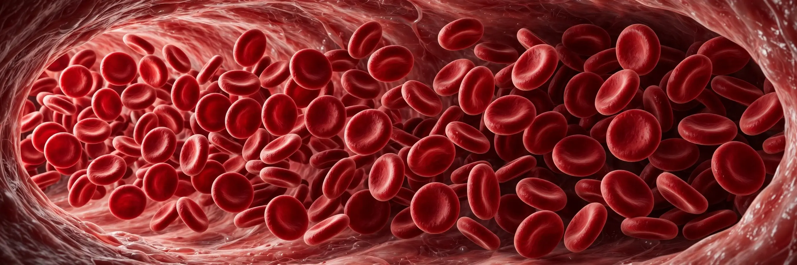
The first time I gave blood, I watched the bag fill and felt a small, irrational worry: was I losing something I could not get back? The nurse smiled and told me my body would replace the fluid within a day and the red cells within weeks — a small, first lesson in how blood works. She was right, of course. I walked out feeling exactly the same as I had walked in, lighter by about half a litre and richer by one new piece of knowledge — that blood is not a fixed supply. It is a living, self-renewing tissue, rebuilt constantly, cell by cell, litre by litre, for as long as we live.
That is the question this article answers: how blood works. Not just what blood is made of, but how each part does its job, how a cut stops bleeding, why blood is red, why your blood type matters, and what happens when the system breaks down. Blood carries oxygen from your lungs to your toes, heat from your muscles to your skin, hormones from one organ to another, and waste back to the kidneys and liver for disposal. It seals its own leaks, fights its own infections, and keeps its own chemistry balanced within a narrow band. All of this happens inside about five litres of fluid that circulates through your body roughly once a minute, every minute, for your entire life.
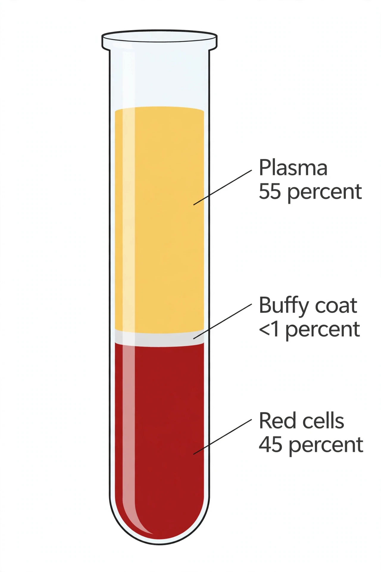
It is worth saying at the start what blood is not. It is not a simple red liquid. It is a tissue — a connective tissue, in the same family as bone and cartilage — made of living cells suspended in a fluid matrix. Cut yourself, and you see only the red part. Spin a tube of blood in a centrifuge, and the truth separates into layers: a straw-coloured fluid on top, a thin pale band in the middle, and a deep red mass at the bottom. Each layer has its own cells, its own proteins, and its own job. Understanding how blood works means understanding all three layers, and how they cooperate.
This article follows the blood itself. It starts with the recipe — what blood is made of and in what proportions. Then it takes each ingredient in turn: plasma, the liquid river; red blood cells, the oxygen fleet; white blood cells, the patrol; and platelets, the repair crew. Then it follows the two great performances blood is famous for: clotting, the cascade that turns liquid into a solid seal, and circulation, the endless journey around the body. After that come blood groups and transfusion, the diseases that strike blood itself, and finally blood donation — the supply that cannot be stockpiled, because every bag of donated blood has an expiry date. Nothing here is medical advice for any individual. It is an explainer, built on published research and the guidance of health authorities, so that the next time you see your own blood — in a test tube, on a scraped knee, or in a donation bag — you will know exactly what you are looking at.
1. What Blood Is — A Liquid Tissue That Never Stops Moving
If you prick your finger, a bead of blood wells up, bright red and startlingly quick. It looks simple. It is anything but. That single drop contains roughly five million red blood cells, several thousand white blood cells, and hundreds of thousands of platelets, all floating in a fluid packed with proteins, salts, sugars, hormones, and waste products. Multiply that drop by about five litres, and you have the full picture: a moving organ, distributed through the whole body, doing a dozen jobs at once.
Blood is a connective tissue. That surprises people, because we think of tissues as solid — muscle, skin, bone. But anatomists classify blood as connective tissue because it fits the definition: cells suspended in an extracellular matrix. The matrix here is not solid collagen but liquid plasma, and the cells are not fixed in place but carried along in the flow. Blood is the only tissue that moves through the body as part of its normal function, and it is the only one that visits every other tissue. Your brain, your liver, your skin, your bones — all of them depend on blood arriving on schedule, and all of them send their waste back out through it.
The numbers give a sense of scale. An average adult carries about five litres of blood, roughly seven to eight percent of body weight. Men typically carry five to six litres, women four to five, because blood volume scales with body size. The heart pumps about five litres every minute at rest, so the entire blood volume completes a circuit of the body in roughly a minute. Over a day, that adds up to about 7,200 litres pumped — more than fourteen hundred times the volume of blood you actually own. The blood in your body right now will pass through your heart more than a thousand times today, and it will do the same tomorrow, and the day after, for as long as you live.
Blood does five things at once, and it is worth naming them before going deeper. First, transport: it carries oxygen from the lungs to the tissues, carbon dioxide back to the lungs, nutrients from the gut to the cells, and waste from the cells to the kidneys and liver. Second, regulation: it keeps body temperature even by carrying heat from active muscles to the skin, and it keeps the body's chemistry balanced, holding blood pH in the narrow range of 7.35 to 7.45. Third, protection: white blood cells hunt infections, platelets and clotting factors seal leaks, and antibodies patrol for invaders. Fourth, communication: hormones travel in blood from the gland that makes them to the organ that must respond. Fifth, clotting itself: blood can change state, from liquid to gel, at exactly the place and moment a vessel is damaged, and then dissolve the clot again once the repair is done.
That last ability — changing from liquid to solid and back — is the most remarkable thing blood does, and it is the one most people never think about. Your blood must stay liquid inside healthy vessels, flowing freely for decades without clotting. Yet the moment a vessel wall breaks, it must clot within minutes. It must clot fast enough to save you from bleeding, but not so eagerly that it clots inside an intact vessel and blocks the flow. Getting that balance right, every day, is one of the body's hardest jobs. When the balance fails in one direction, people bleed. When it fails in the other, they develop clots where no clot belongs. Much of this article is about how the body walks that line.
Blood also keeps the body's chemistry steady in ways that are easy to miss. Its pH — the measure of how acidic or alkaline it is — is held between 7.35 and 7.45, a narrow band, by a set of buffers in the plasma and inside the red cells. The bicarbonate buffer is the most important: carbon dioxide from the tissues dissolves in the blood, becomes bicarbonate, and is carried to the lungs, where it turns back into carbon dioxide and is breathed out. Breathing faster blows off more carbon dioxide and raises the pH; breathing slower does the opposite. The kidneys adjust the balance over hours and days, holding on to bicarbonate or excreting it as needed. It is a system with two controls — the lungs for speed, the kidneys for endurance — and it is why a blood gas test, which measures pH, carbon dioxide, and oxygen together, can say so much about a person's lungs, kidneys, and metabolism in a single reading. The same blood that carries oxygen also carries the body's acid-base balance, and the same organs that filter the blood also tune its chemistry.
A small warning before the details begin. Blood is one of the most measured things in medicine — a blood test can report dozens of values — and it is easy to treat those numbers as a verdict on your health. They are not, on their own. A single low or high value means little without context, and this article will not tell you what your own results mean. What it will do is explain the machinery behind the numbers, so that when a doctor talks about haemoglobin, haematocrit, or platelets, you know what those words refer to and why they matter.
2. The Recipe: What Blood Is Made Of
If you draw blood into a tube and spin it in a centrifuge, the contents separate by density into three layers, and the recipe becomes visible. I remember the first time I saw this in a teaching laboratory: a tube of blood that had looked uniformly red in the hand came out of the centrifuge striped like a tiny flag. The top layer, a clear straw-yellow fluid, filled more than half the tube. Below it sat a thin, pale grey-white band, barely a millimetre thick. And at the bottom, packed tight, a deep red mass. Those three layers are the whole story of blood, in miniature.
The top layer is plasma, about 55 percent of the blood's volume. The bottom layer is red blood cells, about 44 to 45 percent. The thin band in the middle is the buffy coat — white blood cells and platelets together, less than one percent of the volume. Those proportions are so stable that doctors use them as a measurement: the fraction of blood volume taken up by red cells is called the haematocrit, and it normally sits between 41 and 50 percent in men and 36 and 44 percent in women. A haematocrit well above or below that range tells a doctor that something in the recipe has changed — too many red cells, too few, or too much or too little plasma.
The numbers in a single drop are hard to picture, so it helps to count them. One microlitre of blood — a cube one millimetre on each side, far smaller than a drop — contains about five million red blood cells, about seven thousand white blood cells, and about 250,000 platelets. Scale that up to the five litres in an adult, and the totals become almost absurd: roughly 25 trillion red blood cells, tens of billions of white blood cells, and well over a trillion platelets, all moving through the same vessels. Red cells outnumber white cells by about a thousand to one, which is why blood looks red and not white, and why a single white cell is hard to find in a drop under a microscope.
Component | Share of blood volume | Count per microlitre | Main job |
|---|---|---|---|
Plasma | About 55% | — (fluid) | Carries cells, proteins, nutrients, hormones and waste |
Red blood cells | About 44–45% | 4.2–6.1 million | Carry oxygen to tissues and carbon dioxide back |
White blood cells | Under 1% (with platelets) | 4,000–11,000 | Defend against infection and disease |
Platelets | Under 1% (with white cells) | 150,000–400,000 | Plug leaks and start clotting |
The recipe is not fixed for life. It shifts with age, altitude, illness, and even hydration. Lose water through sweating or diarrhoea and plasma volume falls, so the cells become a larger share of a smaller total — the blood thickens, and the haematocrit rises. Spend weeks at high altitude and the kidneys sense the thinner air and release a hormone that tells the bone marrow to make more red cells, raising the oxygen-carrying capacity. Become pregnant and plasma volume expands faster than red cell production, so the blood dilutes and the haematocrit falls — a normal change that can look like anaemia on a test if the doctor does not know the context. Blood is a recipe that the body adjusts constantly, and the adjustments themselves are part of how it works.
Blood volume itself is regulated, not left to chance. When volume falls — after bleeding, sweating, or vomiting — the kidneys release renin, setting off a chain that produces aldosterone, a hormone that tells the kidneys to hold on to salt and water. The pituitary releases antidiuretic hormone, which tells the kidneys to conserve water directly. Thirst rises, and drinking restores the volume. When volume is too high, the heart releases a hormone called atrial natriuretic peptide, which tells the kidneys to excrete more salt and water. The system is a loop of sensors and hormones, and it is why blood volume stays close to five litres in a healthy adult even as water intake and loss vary from day to day.
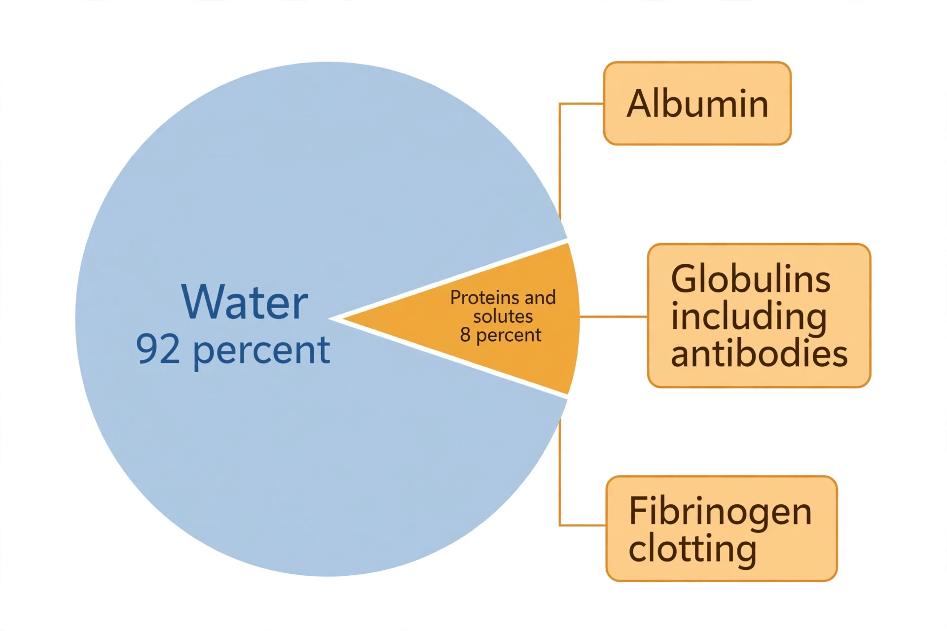
There is one more ingredient that does not appear in the centrifuge layers, because it is dissolved everywhere: the proteins, salts, and small molecules carried in plasma. Plasma is about 92 percent water, and the remaining 8 percent is a crowded mixture — albumin, antibodies, clotting factors, hormones, glucose, salts, and waste products like urea. That 8 percent is where much of blood's chemistry happens, and it is the subject of the next section.
Blood is a fluid tissue — a connective tissue whose intercellular substance is liquid, with its cells suspended and carried about in the circulation. That is the standard anatomy-textbook picture, from Gray's Anatomy to modern physiology.
Understanding how blood works starts with this picture: a liquid tissue, about 55 percent plasma and 45 percent cells, constantly adjusted, constantly moving, and constantly rebuilt. The next four sections take the ingredients one at a time, beginning with the river itself.
3. Plasma: The Straw-Coloured River
Plasma is the part of blood most people never see, because it is hidden behind the red. Yet it is the largest part of blood by volume, and without it the cells would have nowhere to go. To understand how blood works at the chemical level, start here: plasma is about 92 percent water, and the rest is a dissolved load of proteins, salts, sugars, fats, hormones, and waste. It is straw-yellow in colour — you can see it in a tube of blood left to settle, or in the bag of plasma a donor gives — and it is the river in which everything else travels.
The most abundant protein in plasma is albumin, made by the liver, and it does two jobs that are easy to undervalue. First, albumin acts as a carrier, binding to hormones, fatty acids, drugs, and other molecules that do not dissolve well in water, and ferrying them through the bloodstream. Second, and more importantly, albumin holds water inside the blood vessels. Proteins are too large to leak easily through the vessel walls, so they create an osmotic pull — sometimes called oncotic pressure — that draws water back into the vessels and keeps it there. When albumin levels fall, as they can in liver disease or malnutrition, fluid leaks out of the vessels into the tissues, and the ankles and abdomen swell. The swelling is not a problem of too much water in the body. It is a problem of water in the wrong place, because the protein that should have held it in the vessels is missing. The liver makes most of the plasma proteins, including albumin and fibrinogen, which is why liver disease shows up so quickly in blood tests — for the full story of the organ that does the making, see How the Liver Works.
The second group of plasma proteins is the globulins. Some globulins carry things — one carries iron through the blood, another carries thyroid hormones, another carries copper. But the most famous globulins are the antibodies, also called immunoglobulins, made by white blood cells. Antibodies are the guided missiles of the immune system: each one is shaped to recognise one specific invader, and once it binds, it marks the invader for destruction. When you recover from an infection, the antibodies against that germ stay in your plasma for months or years, ready to respond faster if the germ returns. When you receive a vaccine, the antibodies it trains your body to make end up here, in the plasma, waiting. The immune system is often described as cells, but much of its memory lives in this fluid.
The third group is the clotting factors, and the most important of them is fibrinogen. Fibrinogen is a large, soluble protein that floats in plasma doing nothing at all — until it is needed. When a vessel is damaged, an enzyme called thrombin cuts fibrinogen into fibrin, a sticky, insoluble protein that forms long threads. Those threads weave into a mesh that traps blood cells and seals the wound. Fibrinogen is the raw material of every clot, and section 8 will follow its transformation in detail. For now, the point is that plasma carries the clotting system in dissolved, inactive form, ready to switch on in seconds.
Plasma protein | Share of plasma protein | Made in | Main job |
|---|---|---|---|
Albumin | About 55–60% | Liver | Holds water in vessels (osmotic pressure); carries hormones and fatty acids |
Globulins | About 35–40% | Liver and white blood cells | Carry iron, hormones and fats; antibodies fight infection |
Fibrinogen | About 4–7% | Liver | Raw material of clots — converted to fibrin when bleeding starts |
Plasma also carries the body's small cargo: glucose for energy, amino acids for building proteins, fatty acids for fuel, salts — sodium, potassium, calcium, chloride, bicarbonate — that keep the chemistry of every cell working, and waste products such as urea and creatinine on their way to the kidneys. It carries hormones from the glands that make them to the tissues that must respond, and it carries heat from working muscles to the skin, where it can escape. The water in plasma is not just a solvent. It is the medium of exchange for the whole body, and the reason blood can do so many jobs at once is that plasma can carry so many different things at the same time.
There is a useful distinction that confuses people the first time they meet it: plasma and serum are not the same thing. Plasma is the fluid you get when you prevent blood from clotting — it still contains fibrinogen and the other clotting factors. Serum is the fluid left behind after blood has been allowed to clot — the fibrinogen has been used up making the clot, so serum has no clotting factors. Laboratories choose one or the other depending on the test. When a doctor orders a blood test, the tube they use decides whether the lab works with plasma or serum, and the difference matters for which measurements are reliable.
Plasma can also be donated on its own. In a plasma donation, a machine draws blood, spins out the plasma, and returns the red cells to the donor, so the donor can give more often than with whole blood. Donated plasma is used to make medicines for people whose blood cannot clot properly, for burn victims, and for people with immune deficiencies — the antibodies and clotting factors in one person's plasma become treatment for another. It is a reminder that plasma is not just packaging for the cells. It is a medicine in its own right.
4. Red Blood Cells: The Oxygen Fleet
If plasma is the river, red blood cells are the fleet that sails on it. They are the most numerous cells in the body — about 25 trillion of them, roughly 84 percent of all the cells you own — and they have exactly one job: to carry oxygen from the lungs to the tissues, and to help carry carbon dioxide back. They do that job so well that the rest of the body takes it for granted, until the fleet thins out and the whole system feels it.
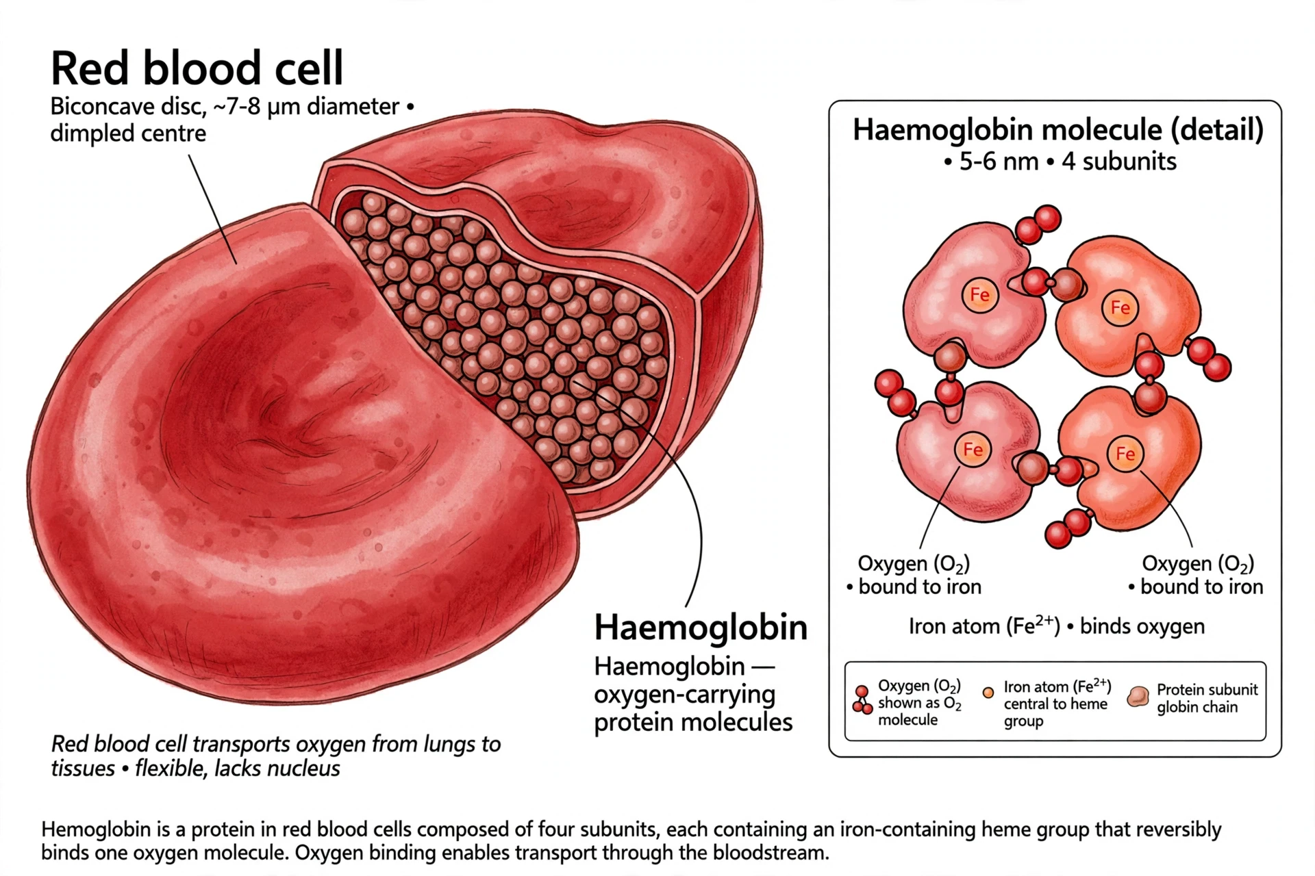
A red blood cell is a strange object by the standards of a cell. It has no nucleus, no mitochondria, and almost no internal machinery at all. During its development in the bone marrow, it throws away its nucleus and its organelles, leaving a bag of haemoglobin wrapped in a flexible membrane. The reason is space: without a nucleus, the cell can pack in more haemoglobin — about 250 million molecules per cell — and without mitochondria, it does not burn the oxygen it is supposed to deliver. The trade-off is that a red blood cell cannot repair itself, cannot divide, and cannot make new proteins. It is born with everything it will ever have, and it wears out in about 120 days.
The shape matters as much as the contents. A red blood cell is a biconcave disc — a disc pinched in on both sides, like a doughnut with the hole filled in. That shape gives it a large surface area for its volume, so oxygen can move in and out quickly, and it lets the cell fold and squeeze through capillaries that are narrower than the cell itself. Capillaries can be as narrow as five micrometres across, while a red cell is about seven to eight micrometres wide. The cell deforms, slides through single file, and springs back to shape on the other side. A rigid cell could not do this, and the flexibility is part of what makes the system work — and part of what fails in diseases where red cells stiffen.
The haemoglobin inside is the molecule that does the actual carrying. Each haemoglobin molecule is made of four subunits, and each subunit holds a haem group — a ring-shaped structure with a single iron atom at its centre. Oxygen binds to that iron atom, one molecule of oxygen per haem group, four per haemoglobin. In the lungs, where oxygen is plentiful, haemoglobin loads up. In the tissues, where oxygen is scarce, it lets go. The binding is reversible and sensitive to conditions: in active muscle, where carbon dioxide is high and the blood is slightly more acidic, haemoglobin releases oxygen more readily — exactly where it is needed most. About 97 percent of the oxygen in blood travels bound to haemoglobin; only about 3 percent dissolves directly in plasma.
Red cells are made in the bone marrow, in a process called erythropoiesis, and the scale of production is hard to grasp. The marrow makes about two million new red cells every second — roughly 200 billion a day — to replace the ones that wear out. That is the body's largest single manufacturing operation, bigger than the production of any other cell type. It takes about seven days for a new red cell to mature, and the process is controlled by a hormone called erythropoietin, or EPO, made mainly by the kidneys. When oxygen levels fall — at high altitude, in lung disease, or after blood loss — the kidneys release more EPO, and the marrow speeds up. This is why the kidneys matter to blood: they are the sensor that tells the marrow how many red cells to make. For a fuller picture of how the kidneys sense and regulate the body's fluids, see How the Kidneys Work.
Haemoglobin carries carbon dioxide as well as oxygen, though by a different route. Only a small share of carbon dioxide travels bound to haemoglobin itself. Most of it — about 70 percent — travels as bicarbonate, dissolved in the plasma: carbon dioxide from the tissues enters the red cell, an enzyme called carbonic anhydrase converts it to bicarbonate, and the bicarbonate moves out into the plasma for the journey to the lungs, where the reaction reverses and the carbon dioxide is breathed out. The red cell is therefore a chemical workshop as well as a delivery van, converting one gas into a soluble form for transport and back again. Before birth, the system runs on a different haemoglobin altogether: foetal haemoglobin, built with gamma chains instead of beta chains, binds oxygen more tightly than adult haemoglobin, which lets the foetus pull oxygen across the placenta from the mother's blood. After birth, foetal haemoglobin is gradually replaced by the adult form — adult haemoglobin becomes the predominant type at about six months of age, and reaches adult levels by about a year — one more quiet handover in a system that is always being rebuilt.
Old red cells are removed by macrophages, mostly in the spleen and liver. The spleen is the quality-control station: red cells must squeeze through its narrow passages, and old, stiff cells get caught and dismantled. The iron from their haemoglobin is recycled — the body is careful with iron, because it is hard to absorb from food and easy to lose — and the rest of the haem molecule is broken down into bilirubin, the pigment that colours bile and gives a fading bruise its yellow-green tint. About one percent of red cells are replaced every day, so the entire fleet turns over roughly three times a year.
When the fleet fails, the whole body notices. Anaemia — too few red cells, or too little haemoglobin inside them — is one of the most common conditions in the world, affecting about 1.92 billion people, and it is covered in section 12. The signs are those of a supply line falling short: tiredness, breathlessness, a pale face, a heart working harder to push thinner blood faster. The blood is still red, and it still flows, but it carries less oxygen per litre, and every tissue feels the difference.
5. Why Blood Is Red — and Why Veins Only Look Blue
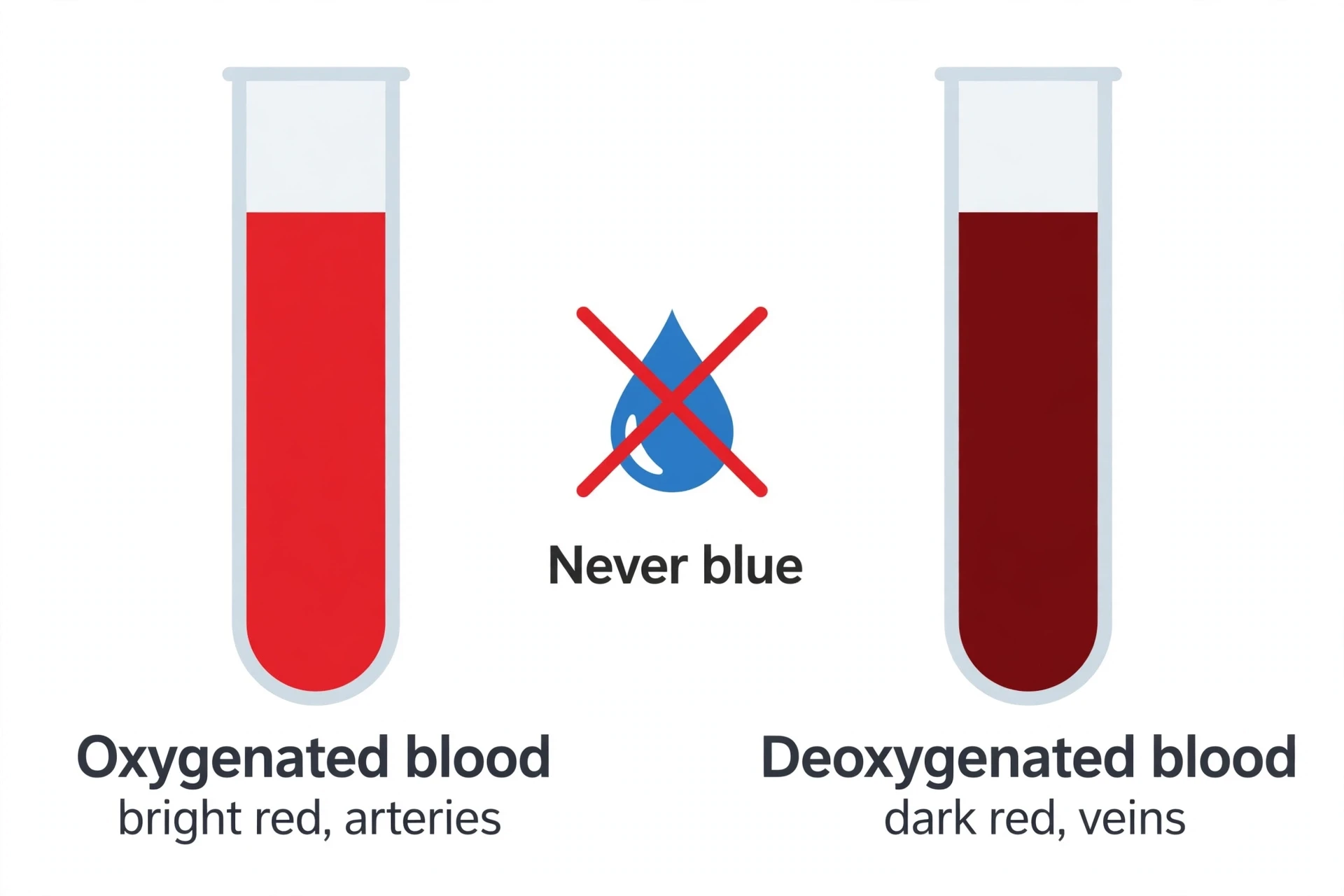
Blood is red because of haemoglobin, and haemoglobin is red because of the way it handles light. The haem group at the heart of each haemoglobin subunit absorbs blue and green light strongly and reflects red and orange light back to the eye. That is the whole story in one sentence, and it is worth pausing on, because it corrects one of the most persistent myths in biology: that blood is blue inside your veins and only turns red when it meets air. It is not. Human blood is always red. The shade changes, but the colour does not.
Oxygenated blood — blood that has just left the lungs and is travelling through the arteries — is a vivid, bright cherry red. The haemoglobin is loaded with oxygen, and the molecule's shape in that state absorbs green light in two clean bands, leaving a strong red reflection. Deoxygenated blood — blood that has delivered its oxygen and is returning through the veins — is a darker, deeper red, sometimes described as maroon or burgundy. Without oxygen, haemoglobin changes shape, and it absorbs more light across the spectrum, including more red light, so the colour darkens. Darker, but still unmistakably red. If you have ever had blood drawn from a vein, you have seen it: the blood in the tube is dark red, not blue, and it was dark red inside your vein a moment before.
So why do veins look blue through the skin? The answer is in the skin, not in the blood. Light has to travel through skin and fat to reach a vein, and then back out to your eye. Blue light does not penetrate as far into tissue as red light; it scatters and reflects back sooner. Red light penetrates deeper, reaches the vein, and is absorbed by the dark, oxygen-poor blood inside. What comes back to your eye is shifted towards the blue end of the spectrum, because more of the red has been absorbed on the way. It is an optical effect — the same kind of effect that makes a deep swimming pool look blue from above. The blood inside the vein is dark red. The blue is something your eye constructs from the light that survives the journey.
Most people assume the blue-vein idea came from textbooks, and in a sense it did: anatomy diagrams have used red for arteries and blue for veins for generations, purely as a visual shorthand to help students tell the two circuits apart. The colour was a convention, not a description, but generations of students took it literally. The diagram did its job too well.
There are two other colour stories worth knowing. The first is carbon monoxide. Carbon monoxide binds to the iron in haem at about 200 times the strength of oxygen, and once it is bound, oxygen cannot take its place. Because carbon monoxide keeps haemoglobin in its bright red, oxygen-loaded shape, the blood of a person with carbon monoxide poisoning stays cherry red — sometimes making the victim look flushed or rosy-cheeked even as they are starved of oxygen. It is one of the reasons carbon monoxide poisoning is so dangerous: the usual signs of oxygen starvation, including a bluish tinge to the lips, may never appear.
The second story is that blood is not always red in nature. Horseshoe crabs, squid, and octopuses carry oxygen with a different molecule, haemocyanin, which uses copper instead of iron at its centre. When haemocyanin binds oxygen, it turns blue — a vivid, unmistakable blue, visible in the blood itself, not just in the skin above it. Their blood really is blue. Ours is not, and the difference comes down to one atom: iron in our haemoglobin, copper in their haemocyanin.
6. White Blood Cells: The Patrol
If red cells are the fleet, white blood cells are the patrol — fewer in number, slower to arrive, and far more varied in what they do. There are only about 4,000 to 11,000 white blood cells in each microlitre of blood, roughly one white cell for every thousand red cells, and they make up less than one percent of blood volume. But they are the cells that decide whether a cut becomes an infection, whether a cold stays a cold, and whether a cancer cell is caught early or missed. The immune system lives in the blood, and white blood cells are its moving parts.
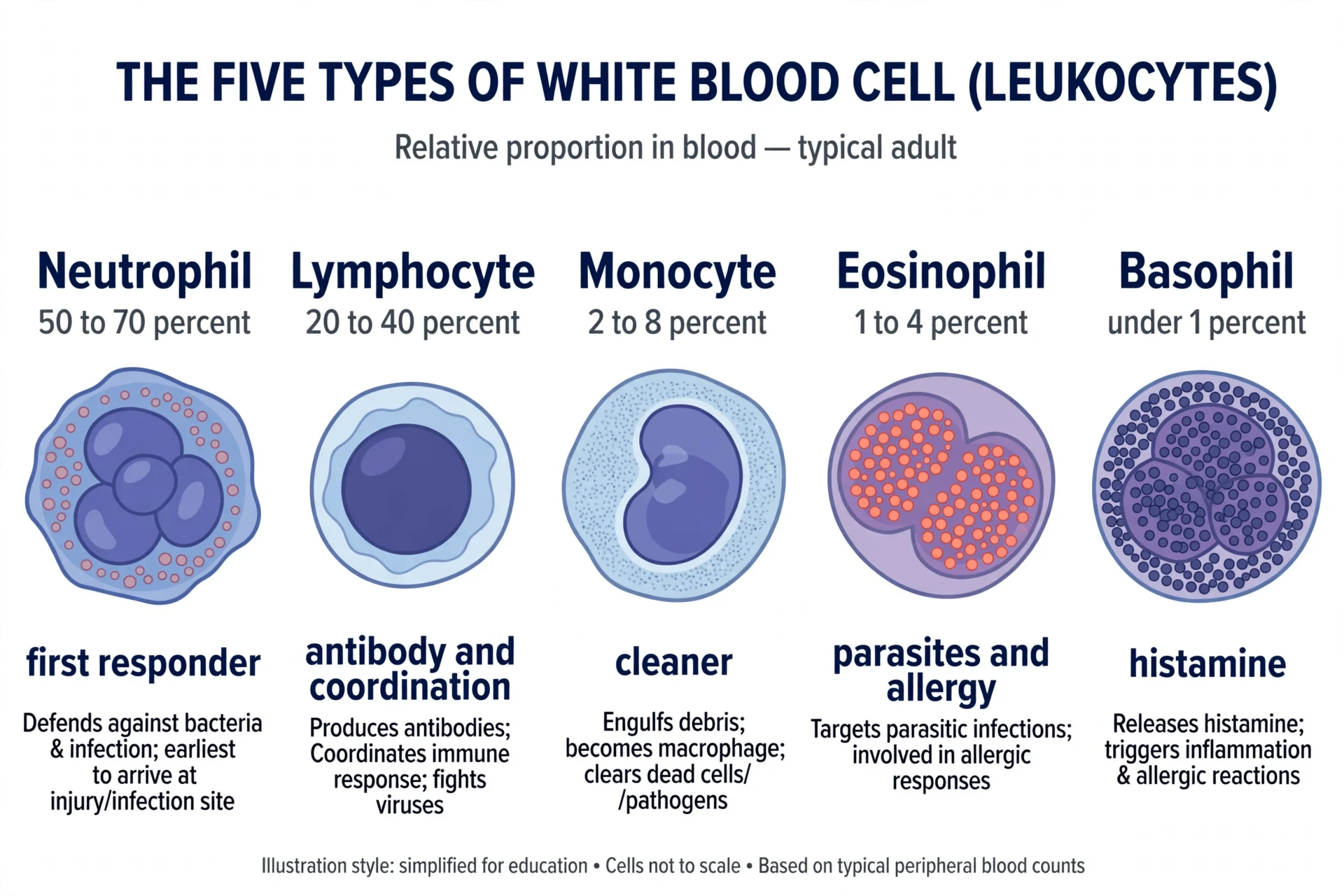
There are five types of white blood cell, and they divide into two families. The first family is the rapid responders, the cells that arrive first and act without needing to recognise a specific germ. The second family is the specialists, the cells that learn to recognise one particular invader and remember it for years. Both families travel in the blood, and both can leave it — squeezing between the cells of a vessel wall in a process called diapedesis — to reach tissues where they are needed.
The most numerous white cells are the neutrophils, making up 50 to 70 percent of all white blood cells. They are the first responders. When tissue is damaged or infected, chemical signals call neutrophils to the site, and they arrive within minutes to hours, engulfing bacteria and debris. A neutrophil lives only hours to a few days, and it dies doing its job — the pus in an infected wound is mostly dead neutrophils, along with the bacteria and debris they consumed. Their numbers rise sharply during a bacterial infection, which is why a white blood cell count is one of the first things a doctor checks when infection is suspected.
The second most numerous are the lymphocytes, 20 to 40 percent of white cells. These are the specialists, and they come in two main kinds. B cells make antibodies — the guided missiles described in section 3 — and after an infection, some B cells become memory cells that remember the invader for years. T cells do the close work: some kill infected cells directly, others coordinate the whole response. Lymphocytes are the cells that make vaccination work, because a vaccine trains them to recognise a germ without the danger of the real infection. For the full story of how these cells learn, remember, and coordinate, see How the Human Immune System Works and How Vaccines Work. When the immune system turns on the body's own tissues instead — attacking myelin in the nervous system, for example — the result is a disease like Multiple Sclerosis Explained, where the same cells that defend the body become the ones doing the damage.
The third type is the monocyte, 2 to 8 percent of white cells. Monocytes are the largest white blood cells, and they are the long-lived cleaners. When they leave the blood and enter the tissues, they grow into macrophages — large cells that engulf dead cells, debris, and invaders, and that present fragments of what they have eaten to the lymphocytes, teaching the specialists what to attack. The macrophages of the spleen that dismantle old red blood cells are monocytes that settled there. So are the cells that clear away the debris after an injury.
The last two types are the smallest groups, and both deal with the body's overreactions. Eosinophils, one to four percent, specialise in fighting parasites — worms too large for a neutrophil to swallow — and they are also involved in allergic reactions, where they gather in tissues and release chemicals that cause swelling and itching. Basophils, under one percent, are the rarest white cells; they carry granules of histamine, the chemical that drives the redness, swelling, and itching of an allergic response. In an allergy, the problem is not the germ. It is the patrol overreacting to something harmless, and basophils and eosinophils are the cells doing the overreacting.
White blood cell | Share of white cells | Lifespan | Main job |
|---|---|---|---|
Neutrophil | 50–70% | Hours to a few days | First responder — engulfs bacteria and debris |
Lymphocyte | 20–40% | Weeks to years | B cells make antibodies; T cells kill infected cells and coordinate |
Monocyte | 2–8% | Days in blood, months as macrophages | Becomes a macrophage — cleans up dead cells and debris |
Eosinophil | 1–4% | Days to weeks | Fights parasites; involved in allergic reactions |
Basophil | Under 1% | Days | Releases histamine; drives allergic inflammation |
White blood cells are made in the bone marrow, like red cells, but their production is tuned by different signals, and some of them finish their education elsewhere. Lymphocytes are born in the marrow, but T cells mature in the thymus — a small organ behind the breastbone that is large in childhood and shrinks with age — while B cells mature in the marrow itself. Both then settle in the lymph nodes, the spleen, and the tissues, waiting for the invader they were built to recognise. The blood is their highway between these stations, and a lymphocyte may circulate for years, passing through node after node, before it ever meets its match. Infection triggers the release of growth factors that push the marrow to make more neutrophils, which is why the white cell count rises in infection. Leukaemia is what happens when that production goes wrong: a white blood cell turns cancerous and multiplies without control, flooding the blood with useless copies and crowding out the red cells and platelets. It is a disease of the patrol itself, and it is covered in section 13. Understanding how blood works means understanding that the cells in it are not all made in the same place, and not all of them stay in the blood — the blood is the road, not the home.
7. Platelets: The Repair Crew
Platelets are the smallest of the formed elements, and the strangest. They are not cells at all, but fragments of cells — pieces broken off from giant bone marrow cells called megakaryocytes. A single megakaryocyte can shed one to three thousand platelets, each one a disc-shaped fragment about two to three micrometres across, with no nucleus and a short life of seven to ten days. There are 150,000 to 400,000 of them in every microlitre of blood, and they spend their short lives waiting for a wall to break.
A healthy blood vessel keeps its platelets quiet. The inner lining of the vessel, the endothelium, is smooth and slippery, and it releases chemicals that stop platelets from sticking. But the moment the lining is damaged, the situation changes completely. The break exposes collagen — the tough protein that forms the vessel wall's structure — and a large protein called von Willebrand factor, which acts like a grappling hook. Platelets in the flowing blood snag on the von Willebrand factor through a receptor called glycoprotein Ib, and the moment they stick, they change.
An activated platelet is a different creature from a resting one. It changes shape, sprouting spiky projections. It empties its granules, releasing adenosine diphosphate (ADP), serotonin, and thromboxane A2 — chemical signals that call more platelets to the scene and make them sticky. And it switches on a new receptor on its surface, glycoprotein IIb/IIIa, which grabs fibrinogen and uses it as a bridge to link one platelet to the next. Within a minute or two, a growing pile of platelets has formed a soft plug over the break. This is primary haemostasis: the first, fast seal.
The plug alone is not enough. It is soft, it can be washed away by the flow, and it does not last. It needs to be reinforced, and that is the job of the clotting cascade — the subject of the next section. But the platelet plug is what buys the time, and it is what you see when a small cut stops bleeding quickly: the platelets did their work before the cascade had finished.
Platelets also do quiet maintenance that nobody notices. The vessel lining is constantly suffering tiny injuries — small gaps open between its cells as part of normal wear — and platelets patch them without any bleeding ever becoming visible. People with very low platelet counts bruise easily and bleed from the gums and nose, not because their vessels are weaker, but because the daily patching has stopped. The repair crew works around the clock, and its absence is felt before its presence is ever noticed.
Platelet numbers can go wrong in both directions. Too few platelets — thrombocytopenia — can be caused by the marrow making too few, by the immune system destroying them (immune thrombocytopenia), or by the spleen trapping too many. The result is bruising, nosebleeds, bleeding gums, and, in severe cases, bleeding inside the body. Too many platelets — thrombocytosis — can be a reaction to inflammation, iron deficiency, or surgery, or it can be a bone marrow disorder in which the marrow makes too many, raising the risk of clots. Both directions are diagnosed with a platelet count and a look at the rest of the blood, and both are reminders that the repair crew, like everything else in blood, is kept within a range — and that the range itself is part of how the system stays safe.
8. How Blood Clots: The Cascade, Step by Step
A clot is a controlled accident. Blood must turn from liquid to solid at exactly the right place, at exactly the right time, and then stop — no further, no longer. The system that does this is called haemostasis, and it works in three overlapping stages: the vessel narrows, the platelets plug the gap, and the clotting cascade builds a fibrin mesh to lock the plug in place. Then, once the vessel has healed, a second system dissolves the clot and restores the flow. Each stage has its own machinery, and each can fail in its own way.
The first stage begins within seconds of injury. The smooth muscle in the damaged vessel wall contracts, narrowing the vessel and slowing the flow of blood through the break. This is the vascular spasm, and it is a reflex: the vessel clamps down on itself. It is not enough on its own, and it does not last long, but it reduces the bleeding while the platelets arrive. If you have ever pressed on a cut and watched the bleeding slow, you have helped this stage along with your own fingers.
The second stage is the platelet plug, described in the previous section: platelets stick to exposed collagen and von Willebrand factor, activate, recruit more platelets, and pile up into a soft plug. For a small break in a small vessel, the plug alone can be enough. For anything larger, it needs reinforcement, and that is where the cascade begins.
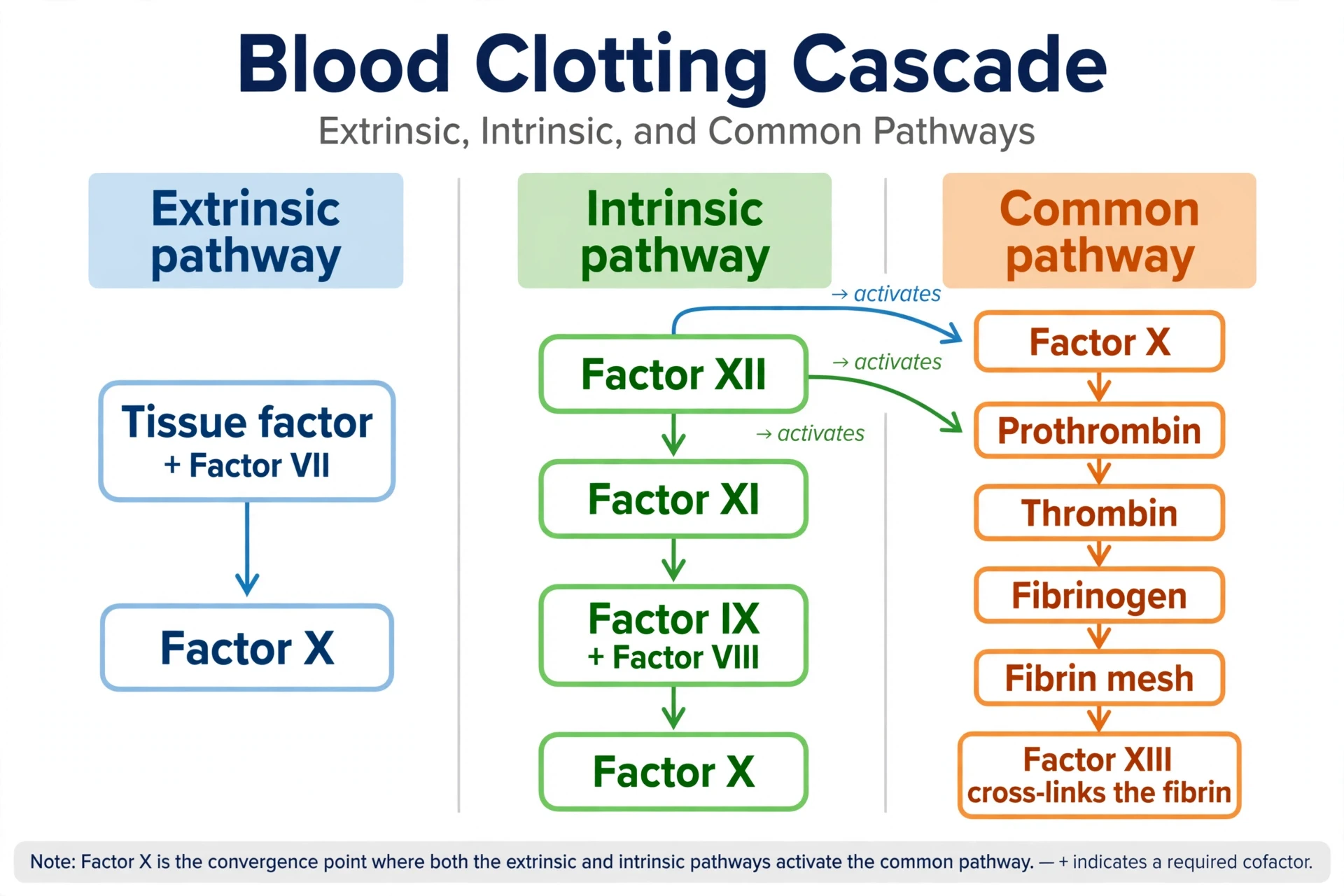
The clotting cascade is a chain of activations. The clotting factors are proteins that float in plasma in an inactive form, each one waiting to be switched on by the one before it. When the first factor activates, it activates the next, which activates the next, and the signal grows at every step — a small trigger producing a large result. The cascade is usually drawn as three pathways that converge, and the drawing is worth understanding, because it explains the blood tests doctors use.
The extrinsic pathway is the fast one, and it is the one that starts clotting in the body. When tissue outside the vessel is damaged, it releases a protein called tissue factor (Factor III). Tissue factor binds to Factor VII, which is already present in the blood in small amounts in its active form, and the pair switches on Factor X. The extrinsic pathway is called extrinsic because tissue factor comes from outside the bloodstream — from the damaged tissue itself.
The intrinsic pathway is the long one, and every component it needs is already present inside the blood — hence intrinsic. It starts when blood comes into contact with a negatively charged surface, such as exposed collagen or a glass test tube. Factor XII activates on contact, then activates Factor XI, which activates Factor IX, which — together with its cofactor, Factor VIII — activates Factor X. The intrinsic pathway is slower than the extrinsic one, but it amplifies the signal, producing far more activated Factor X than the extrinsic route alone.
Both pathways converge on the common pathway. Activated Factor X joins its cofactor, Factor Va, on the surface of an activated platelet, with calcium ions present, to form a complex called prothrombinase. Prothrombinase cuts prothrombin — Factor II, a protein made by the liver — into thrombin, Factor IIa. Thrombin is the central enzyme of the whole system. It does three things at once: it activates more platelets, it switches on the cofactors (Factors V, VIII, and XI) that accelerate its own production, and it converts fibrinogen into fibrin.
Fibrin is the end of the chain. Thrombin cuts two small pieces off each fibrinogen molecule, and the remaining pieces — fibrin monomers — join together spontaneously into long, thin strands. Those strands weave through the platelet plug, trapping red cells and platelets in a mesh. Then thrombin activates Factor XIII, which cross-links the strands, welding the mesh into a firm, stable clot. A clot is, in the end, a net of fibrin with blood cells caught in it — and the dark reddish-brown scab on a scraped knee is that net, dried out, sitting on the skin.
The cascade needs two helpers that are easy to overlook. The first is calcium. Several steps in the cascade require calcium ions, which is why blood drawn into a tube with a calcium-binding chemical will not clot at all — the cascade is starved of the calcium it needs. The second is vitamin K. The liver needs vitamin K to make four of the clotting factors — II, VII, IX, and X — and a person short of vitamin K bleeds more easily. This is why newborn babies, whose guts have not yet built up the bacteria that make vitamin K, are given a vitamin K injection at birth, and why the blood-thinning drug warfarin works by blocking vitamin K. This is how blood works at the level of a single cut: a local trigger, a chain of activations, a mesh of fibrin, and then a careful dismantling once the job is done.
But a cascade that only accelerates would be a disaster. Once a clot has formed, the body must stop it from growing and, later, take it apart. The brakes are proteins that circulate in the blood alongside the clotting factors: antithrombin, which switches off thrombin and Factor Xa, and the protein C and protein S system, which switches off Factors Va and VIIIa. When the vessel has healed, the clot is dissolved by fibrinolysis: an enzyme called plasmin, activated from its precursor plasminogen by tissue plasminogen activator, cuts the fibrin mesh into pieces that the body clears away. The scab falls off because the clot beneath it has been dismantled from the inside, not because it dried out.
Doctors read this system with two blood tests. The prothrombin time (PT), reported as the INR, measures the extrinsic and common pathways: it adds tissue factor to a plasma sample and times how long fibrin takes to form. The activated partial thromboplastin time (aPTT) measures the intrinsic and common pathways: it adds a contact activator and times the same endpoint. A long PT points to a problem in the extrinsic route; a long aPTT points to the intrinsic route; a long result in both points to the common pathway, or to a shortage of fibrinogen itself. The two tests are, in effect, a way of asking the cascade which of its arms is broken.
One last correction is worth making. The three-pathway diagram describes the test tube better than the body. In the body, the current understanding — the cell-based model — is that tissue factor starts clotting, producing a small amount of thrombin, and that thrombin then activates platelets and the cofactors, so that the platelet surface becomes the factory where the thrombin burst happens. The old diagram is still useful, because it matches the tests, but the body is less tidy than the diagram. The cascade is not three separate roads to a clot. It is one process, running on the surface of platelets, with tissue factor as the spark and thrombin as the amplifier.
9. The Journey: How Blood Moves Around the Body
Blood does not move on its own. It is pushed, and the pump is the heart — a fist-sized muscle that beats about 100,000 times a day and never rests. At rest, the heart moves about five litres of blood every minute, which means the whole blood volume passes through the heart in about a minute, and over a day the heart moves about 7,200 litres. During hard exercise, the output can rise to 20 to 25 litres a minute in a healthy adult, and to more than 35 litres a minute in an elite endurance athlete. The blood you own is small. The blood your heart moves is enormous, because the same five litres goes round again and again.
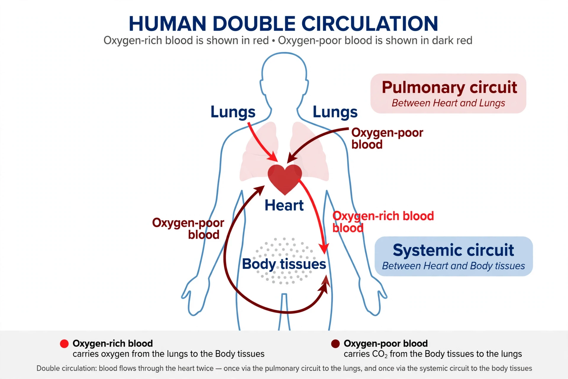
The journey is a double loop. The right side of the heart pumps blood to the lungs, where it drops off carbon dioxide and picks up oxygen. That oxygen-rich blood returns to the left side of the heart, which pumps it out to the whole body — the brain, the gut, the muscles, the skin — and the oxygen-poor blood returns through the veins to the right side again. Two circuits, one pump, running in parallel. The pulmonary circuit is short and low-pressure; the systemic circuit is long and high-pressure, because it must push blood to the furthest corners of the body. That is why the left ventricle, which drives the systemic circuit, has much thicker muscle than the right.
For the full mechanics of the pump itself — the chambers, the valves, the electrical timing — see How the Human Heart Works. What matters here is what the journey does to the blood, and what the blood does on the journey.
The blood leaves the heart through the aorta, the body's largest artery, and branches into smaller and smaller arteries, then arterioles, then capillaries — the smallest vessels, with walls a single cell thick and a diameter of five to ten micrometres, barely wide enough for a red blood cell to pass through single file. It is in the capillaries that the real work happens. Oxygen and nutrients diffuse out of the blood and into the tissues; carbon dioxide and waste diffuse in. The capillary walls are thin precisely so that this exchange can happen, and the blood slows down in the capillaries so that there is time for it. In the larger vessels, blood races. In the capillaries, it crawls — and that crawling is the point.
The total length of the body's vessels is often quoted at about 96,000 kilometres — a figure from a 1922 estimate that assumed a heavily muscled body of about 143 kilograms. Modern estimates put the total much lower, at roughly 9,000 to 19,000 kilometres: still an enormous network, but laid end to end it would reach only about a quarter to a half of the way around the Earth, not twice around it. Most of that length is capillaries, and most of the blood at any moment is not in the arteries at all, but in the veins — the low-pressure return roads, which hold about two-thirds of the blood volume. The veins are the body's blood reservoir, and they can tighten or relax to shift blood where it is needed.
Pressure falls along the journey. In the aorta, blood pressure is at its highest, around 120 millimetres of mercury when the heart contracts. By the time blood reaches the capillaries, the pressure has fallen to a fraction of that, and by the veins it is barely above zero. Getting blood back up from the legs to the heart against gravity, with almost no pressure left, is a problem, and the body solves it with three helpers: one-way valves inside the veins that stop blood flowing backwards, the skeletal muscle pump — the contraction of leg muscles during walking, squeezing the veins and pushing blood upward — and the breathing pump, the change in chest pressure with each breath, which draws blood towards the heart. Stand still for a long time and the muscle pump stops; blood pools in the legs, less returns to the heart, and some people feel faint. Walking is, among other things, a way of helping your blood get home.
Blood pressure is measured in the arm, at the brachial artery, and it is reported as two numbers: the systolic pressure, when the heart contracts, and the diastolic pressure, when the heart relaxes between beats. A reading of 120 over 80 is the classic normal value, though what counts as normal shifts with age and with guidance that changes over time. The two numbers describe the pressure the blood exerts on the artery wall, and they are the most direct measure of how hard the heart is working and how much resistance the vessels are offering. High blood pressure — hypertension — is usually silent, which is why it is measured so often: it damages the arteries, the heart, the kidneys, and the brain over years without any symptom to warn of it. Understanding how blood works means understanding that pressure is not just a number on a cuff. It is the force that drives the whole journey, and when it is too high or too low, the journey itself is at risk.
Blood also carries heat. Working muscles generate heat, and blood absorbs it and carries it to the skin, where it can escape. When you exercise, the vessels in your skin dilate and blood flow to the skin rises, which is why your face flushes. When you are cold, those vessels narrow, keeping warm blood in the body's core. The skin is the body's radiator, and blood is the coolant — a system described in detail in How the Skin Works. Exercise changes the whole system, raising cardiac output and redistributing flow to the muscles; see How Exercise Changes the Body for the full picture.
10. Blood Groups: Why Your Type Matters
For centuries, blood transfusion was a gamble. Doctors knew that giving one person's blood to another could save a life — and that it could also kill, suddenly and mysteriously, with the recipient's blood clumping and breaking down. Nobody knew why it worked sometimes and failed others, until a young Austrian pathologist named Karl Landsteiner started mixing people's blood together in test tubes in 1900.
Landsteiner's experiment was simple. He took blood from himself and five colleagues, separated the serum from the red cells, and mixed each person's serum with each person's red cells, in every combination. In some combinations, nothing happened. In others, the red cells clumped together — agglutination — in visible clusters. Landsteiner's insight was that the clumping was not random: it followed a pattern, and the pattern meant that red cells carry markers on their surface, and serum carries antibodies against the markers a person does not have. In 1901 he published his discovery of three blood groups — A, B, and C, the last later renamed O — and in 1902 his colleagues von Decastello and Sturli found the fourth, AB. For the discovery, Landsteiner received the Nobel Prize in Physiology or Medicine in 1930.
"Thanks to Landsteiner's discovery of the blood groups, blood transfusions have come back into use and have saved a great many lives." — Professor G. Hedrén, presenting the 1930 Nobel Prize in Physiology or Medicine to Karl Landsteiner
The markers are molecules called antigens, and they sit on the surface of every red blood cell. Group A blood has A antigens; group B has B antigens; group AB has both; group O has neither. The antibodies are in the plasma, and they are the mirror image: a person with group A blood has anti-B antibodies, a person with group B has anti-A antibodies, a person with group O has both anti-A and anti-B, and a person with group AB has neither. The rule that makes transfusion work is simple: a person's plasma must never meet red cells carrying an antigen it has antibodies against. If it does, the antibodies bind to the red cells, the cells clump, and the clumped cells break apart — a transfusion reaction that can destroy the kidneys and kill.
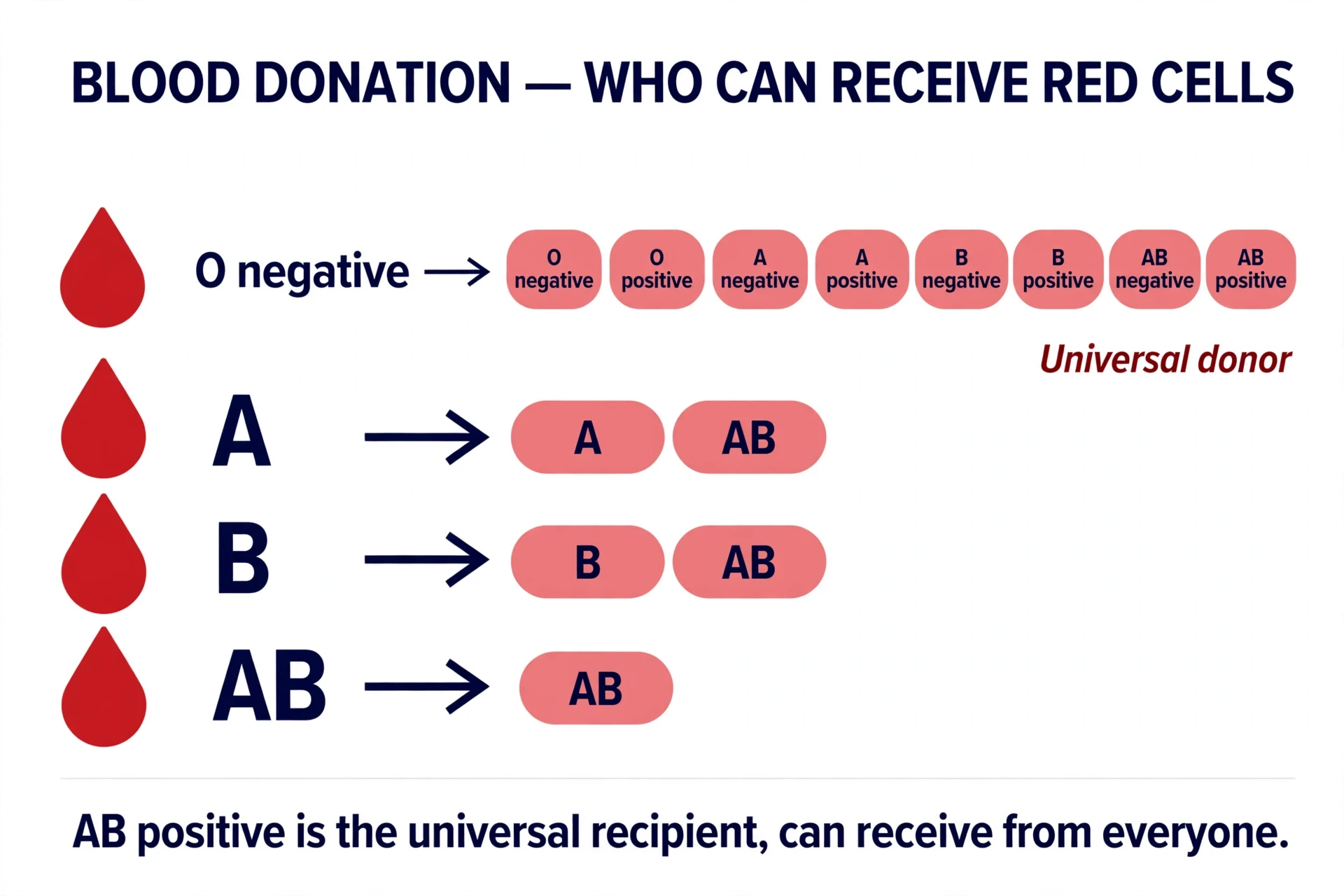
That is why blood types must be matched. A person with group O blood can receive only group O red cells, because their plasma attacks both A and B antigens — but they can donate red cells to anyone, because their red cells carry no antigens to attack, which is why group O negative is called the universal donor. A person with group AB blood can receive red cells from anyone, because their plasma has no anti-A or anti-B antibodies — the universal recipient — but they can donate red cells only to other AB people. In practice, doctors match blood type exactly whenever they can; the universal rules are for emergencies, when there is no time to wait.
There is a second system layered on top of ABO: the Rh factor, named for the rhesus monkey in which it was first studied, discovered by Landsteiner and Wiener in 1940. The Rh factor is another antigen, the D antigen, on the red cell surface. If you have it, you are Rh positive; if not, Rh negative. About 85 percent of white people are Rh positive, and the proportion varies by population. Rh matters most in pregnancy: an Rh-negative mother carrying an Rh-positive baby can develop anti-D antibodies that attack the baby's red cells, in this pregnancy or the next. The problem is prevented by giving the mother anti-D immunoglobulin, which stops her immune system from making the antibodies in the first place — a treatment introduced in 1968 that turned a once-feared complication into a routine injection. For the wider story of pregnancy, see How Pregnancy Works.
Blood group | Antigens on red cells | Antibodies in plasma | Can receive red cells from | Can donate red cells to |
|---|---|---|---|---|
O negative | None | Anti-A, anti-B | O negative | Everyone (universal donor) |
O positive | None | Anti-A, anti-B | O negative, O positive | Rh-positive groups |
A negative | A | Anti-B | A negative, O negative | A and AB groups |
A positive | A | Anti-B | A, O (either Rh) | A and AB (Rh positive) |
B negative | B | Anti-A | B negative, O negative | B and AB groups |
B positive | B | Anti-A | B, O (either Rh) | B and AB (Rh positive) |
AB negative | A and B | None | All Rh-negative groups | AB groups |
AB positive | A and B | None | Everyone (universal recipient) | AB positive only |
ABO and Rh are the two systems that matter most, but they are not the only ones. The International Society of Blood Transfusion recognises 49 blood group systems and more than 360 antigens — Kell, Duffy, Kidd, and others — each capable, in rare cases, of causing a transfusion reaction. Most people will never need to know their Kell status, because ABO and Rh matching covers the common risks, but blood banks test for the rarer antigens when a patient needs repeated transfusions, because each transfusion is a chance for the immune system to learn a new antigen and react to it next time.
The discovery itself is worth a closer look, because it shows how blood works as an identity system — and how a careful experiment can overturn centuries of confusion. Landsteiner was not trying to solve transfusion. He was studying why serum from one person would clump the red cells of another, a curiosity of the laboratory, and he tested blood from only 22 people. From those small numbers, he deduced a system that applied to all of humanity, and he was right. The ABO groups turned out to be inherited in a simple Mendelian pattern, one of the first human traits shown to follow Mendel's laws, and the antigens themselves turned out to be sugar molecules — chains of sugars built onto the red cell surface by enzymes, with the A and B enzymes adding one different sugar each, and the O gene adding none. Blood groups are, at the molecular level, a difference in the sugar coating of your red cells. It is a small chemical difference with large consequences, and it is why a test that takes minutes can decide whether a transfusion saves a life or ends one.
Blood groups are inherited, and they follow simple Mendelian rules: the A and B antigens are dominant, and O is recessive. A child of two group O parents will be group O; a child of an AB parent and an O parent will be A or B. Before DNA testing, blood groups were used in paternity disputes, and they are still used in population genetics to trace migrations — group B is common in Asia, group A in Europe, and group O is nearly universal among Indigenous peoples of the Americas. Your blood group is a small piece of your ancestry, written on the surface of your red cells.
11. Transfusion: Borrowing Somebody Else's Blood
The first successful transfusion of human blood from one person to another was performed in London in 1818 by the obstetrician James Blundell, who gave blood to a woman bleeding after childbirth. She survived. Others did not, and for the next eighty years transfusion remained a desperate measure, used when nothing else was left, because nobody understood why it sometimes killed. Landsteiner's discovery in 1901 changed that: once blood could be typed, it could be matched, and transfusion became a treatment rather than a gamble.
The next step was storage. Blood taken from a donor cannot be used immediately in every case — it must be tested, typed, and kept until a patient needs it. The first hospital blood bank in the United States, a place where blood was collected, stored, and distributed — and the institution that gave the idea its name — was set up in 1937 by Bernard Fantus at Cook County Hospital in Chicago. A blood bank had already been operating in Leningrad since 1932. The idea spread quickly, and during the Second World War, blood banks and large-scale plasma collection made large-scale transfusion possible for the first time. Separating a single donation into red cells for one patient, plasma for another, and platelets for a third — routine component therapy — came later, in the 1960s and 1970s, and that was the beginning of modern transfusion medicine.
Today, a transfusion is almost never whole blood. The donation is spun in a centrifuge and separated into its components, and each patient receives only the part they need. A patient with anaemia gets red cells. A patient with a clotting problem gets plasma or cryoprecipitate, the concentrated clotting factors. A patient with a low platelet count gets platelets. Giving only the needed component is safer, because it exposes the patient to fewer foreign cells and proteins, and it is more efficient, because one donation can help several people.
Before any transfusion, the blood is typed and crossmatched. Typing determines the ABO and Rh group of both donor and recipient. Crossmatching mixes the recipient's plasma with the donor's red cells in the laboratory and checks for clumping — a final test that the two bloods are compatible. The blood is also screened for infections: HIV, hepatitis B and C, syphilis, and others, depending on the country. In high-income countries, screening of donations for the four main infections is routine and close to universal, and the risk of receiving infected blood is very low — but it is never zero, which is why doctors transfuse only when the benefit clearly outweighs the risk.
Transfusion is not free of danger. The most common reactions are mild — a fever or a rash during the transfusion, caused by the recipient's immune system reacting to the donor's white cells. The serious reactions are rare but real: an acute haemolytic reaction, when mismatched blood is given by mistake and the recipient's red cells break down rapidly; transfusion-related acute lung injury, a sudden breathing failure; and circulatory overload, when too much fluid is given too fast to a heart that cannot handle it. These are the reasons transfusion is treated with such care — typed, crossmatched, screened, and monitored — and why the blood bank checks a patient's identity at the bedside, again and again, before the first drop flows. Most transfusion errors are not laboratory errors. They are bedside errors, the wrong bag given to the wrong patient, and the safety systems are built around preventing exactly that.
Blood transfusion is also where the limits of medicine show. There is no artificial blood that replaces all of blood's functions. Researchers have developed haemoglobin-based oxygen carriers — molecules that can carry oxygen without red cells — but none has been approved as a full replacement for donated blood, because blood does more than carry oxygen: it clots, it fights infection, it carries hormones and heat. A bag of red cells is a partial substitute, not a complete one, and for now the only source of human blood is another human.
The search for a substitute has been running for decades, and it is worth understanding why it is so hard. The earliest attempts used simple salt solutions to replace lost volume, which kept blood pressure up but carried no oxygen. Later attempts used purified haemoglobin, free in the plasma — but free haemoglobin is toxic to the kidneys, scavenges nitric oxide from the vessel walls, and causes the vessels to constrict, raising blood pressure. Chemists have tried wrapping haemoglobin in protective coatings, cross-linking it into larger molecules, and growing red cells from stem cells in the laboratory. Lab-grown red cells have been transfused into volunteers in small trials — a genuine milestone — and the first recipients showed no ill effects, though how long the lab-grown cells survive in the body was still being measured at the time of writing. Growing them at the scale of a blood supply, at a cost any health system could afford, remains far away. The difficulty is not a lack of effort. It is that blood is not one product but a system of products, each with its own job, its own lifespan, and its own chemistry, and replacing the system means replacing all of it at once.
12. When Blood Goes Wrong: Anaemia
Anaemia is the most common blood disorder in the world, and one of the most common conditions of any kind. The Global Burden of Disease Study 2021 estimates that about 1.92 billion people — roughly one in four people on Earth — are affected by it. Among children aged six months to five years, about 40 percent are anaemic (2019 data). Among pregnant women, about 37 percent (2019 data; the World Health Organization's 2025 modelled estimate for 2023 is 35.5 percent). Among women of reproductive age, about 30 percent. These are not small numbers, and they are not a problem of one region: anaemia affects rich and poor countries alike, though it is heaviest where diets are poor, malaria is common, and health care is thin.
Anaemia is not a disease in itself. It is a condition — a shortage of red blood cells, or of haemoglobin inside them, so that the blood carries less oxygen than the tissues need. The World Health Organization defines it by haemoglobin levels: below 13 grams per decilitre in men, below 12 in non-pregnant women, and below 11 in pregnant women (10.5 grams per decilitre in the second trimester). The 2024 revision of the WHO guidance updated those cutoffs for the first time in more than fifty years, after a large international study re-examined what counts as normal. The symptoms are the symptoms of a delivery system running short: tiredness that rest does not fix, breathlessness on mild exertion, a pale face and pale inner eyelids, a fast heartbeat, dizziness, and in children, slower growth and learning.
The most common cause, worldwide, is iron deficiency. Iron is the raw material of haem — the part of haemoglobin that holds oxygen — and without enough iron, the marrow cannot make enough haemoglobin. Each red cell ends up smaller and paler than it should be, and the blood carries less oxygen. Iron deficiency has many causes: a diet low in iron, blood loss (heavy menstrual bleeding is a common cause in women, and slow bleeding from the gut is a common cause in older adults), and pregnancy, when the mother's iron must supply both her own blood and the baby's. Iron is also hard for the body to absorb — only a fraction of the iron in food is taken up — and it is lost whenever blood is lost, because the iron is in the red cells.
But iron is only one cause. Anaemia can also come from a shortage of vitamin B12 or folate, the vitamins the marrow needs to make red cells properly. Without them, the marrow produces large, immature red cells that do not work well — a condition called megaloblastic anaemia. It can come from chronic disease: in long-standing inflammation, infection, or kidney failure, the body deliberately holds back iron and slows red cell production, which is one reason people with chronic illness so often feel tired. It can come from the marrow itself failing, after chemotherapy, or in aplastic anaemia, where the marrow stops making blood cells altogether. And it can come from red cells being destroyed faster than they can be replaced — haemolysis — in conditions such as autoimmune haemolytic anaemia, where the immune system attacks its own red cells, or in malaria, where the parasite bursts red cells from the inside.
A second inherited cause deserves its own paragraph, because it affects millions of people and illustrates how blood works at the level of a single gene. Thalassaemia is a group of disorders in which the body makes too little of one of the two globin chains that form haemoglobin — alpha or beta. The haemoglobin that results is unstable, the red cells break down early, and the marrow expands to compensate, sometimes deforming the bones of the face and skull in severe cases. Beta-thalassaemia major, the most severe form, appears in infancy and requires regular blood transfusions for life, along with treatment to remove the iron that accumulates from those transfusions. Like sickle cell disease, thalassaemia is commonest where malaria was once common — the Mediterranean, the Middle East, South and Southeast Asia — and like sickle cell trait, carrying one faulty copy of the alpha-thalassaemia gene appears to protect against severe malaria — though the evidence for beta-thalassaemia is weaker, and a 2026 pooled analysis found no statistically significant protection overall. The geography of both diseases is a map of where malaria used to kill, drawn in the genes of the people who survived it.
Anaemia is diagnosed with a blood test, and treated by treating its cause: iron supplements or diet changes for iron deficiency, B12 or folate for vitamin deficiency, and investigation for the source of any blood loss. What anaemia is not is a condition to diagnose in yourself from tiredness alone. Tiredness has many causes, and a low haemoglobin level has many causes too, and sorting them out is a doctor's job, with blood tests that distinguish one kind of anaemia from another. The point of this section is not to help a reader self-diagnose. It is to explain why a number as ordinary as haemoglobin — a number that appears on almost every blood test — carries so much information about how blood works.
13. When Blood Goes Wrong: Sickle Cell, Haemophilia, and Clots in the Wrong Place
Anaemia is a shortage. The disorders in this section are different: the blood is present in normal amounts, but something in its design has gone wrong. A single changed amino acid makes red cells collapse into sickles. A missing clotting factor makes blood that cannot clot. A clotting system that works too well makes clots where no clot belongs. Each one shows, in its own way, how finely balanced blood has to be.
Sickle cell disease is the most common inherited blood disorder in the world. About 515,000 babies are born with it each year, according to Global Burden of Disease 2021 data published in The Lancet Haematology in 2023 — more than 75 percent of them in sub-Saharan Africa, with the rest spread across South Asia, the Middle East, and, through migration, Europe and the Americas. The cause is a single change in the gene for beta-globin, one of the two protein chains that make up haemoglobin: one amino acid, glutamic acid, is replaced by valine. That one change alters the shape of the haemoglobin molecule, and when the haemoglobin gives up its oxygen, the altered molecules stick together into long, stiff fibres inside the red cell. The cell, normally a flexible disc, is pulled into a crescent or sickle shape.
Sickled cells are rigid, and they do not flow well. They jam in small blood vessels, blocking the flow and starving tissues of oxygen — a sickle cell crisis, which causes severe pain, often in the bones, chest, or abdomen. They also break apart much sooner than normal red cells, lasting days or weeks instead of 120 days, so the person is chronically anaemic. Over time, the repeated blockages damage the spleen, the kidneys, the lungs, and the brain; stroke is a real risk in children with the disease. The condition is inherited: a child must receive the sickle gene from both parents to have the disease, and a child who receives it from only one parent has sickle cell trait — usually without symptoms, but with red cells that can sickle under extreme conditions, such as severe dehydration or very low oxygen.
There is a reason the sickle gene is so common in Africa, and it is one of the clearest examples of evolution shaping blood. Carrying one copy of the sickle gene — sickle cell trait — protects against malaria. The malaria parasite lives inside red cells, and in a person with the trait, infected red cells tend to sickle and are removed by the spleen, and the parasite grows less well inside sickled cells — the exact mechanism is still being worked out. In regions where malaria kills, the protection of one copy outweighs the risk of two, and the gene persists. It is a bargain with a harsh price, and it explains why a disease that shortens life is carried by so many people.
Haemophilia is the opposite problem: blood that cannot clot when it should. Haemophilia A, the most common form, is caused by a shortage of clotting Factor VIII; haemophilia B is caused by a shortage of Factor IX. Both are inherited on the X chromosome, which is why they affect mainly males: a boy needs only one faulty X chromosome to have the disease, while a girl with one faulty X usually has a healthy second copy to compensate. Haemophilia A occurs in about one in 5,000 to 10,000 male births; haemophilia B in about one in 20,000 to 30,000. The World Federation of Hemophilia's Annual Global Survey 2024, published in October 2025, identified about 272,000 people with haemophilia worldwide — about 224,000 with haemophilia A and about 46,000 with haemophilia B — but estimates the true total at about 850,000, of whom about 289,000 have severe disease. Most of the rest are undiagnosed, because the disease is only recognised where testing exists.
Without enough Factor VIII or IX, the intrinsic pathway of the clotting cascade stalls. The platelet plug still forms, but the fibrin mesh that should reinforce it is weak or absent, so bleeding continues — into joints, into muscles, and after injury, for far longer than it should. The classic image of haemophilia is a child with a swollen, painful knee after a minor knock, the joint filling with blood because the clot that should have sealed the small internal bleed never held. Modern treatment replaces the missing factor, given as an injection, and in recent years gene therapy has begun to offer longer-lasting correction for some patients. The disease is no longer the sentence it once was — the royal families of Europe, who carried haemophilia through Queen Victoria's descendants, knew it as a condition with no treatment at all — but it remains a lifelong condition that demands care.
The most common inherited bleeding disorder is neither haemophilia nor sickle cell disease. It is von Willebrand disease, caused by a shortage or defect of von Willebrand factor — the protein that helps platelets grip the vessel wall, described in section 7. About one percent of the population has low von Willebrand factor by laboratory testing, though only about one in 1,000 people has symptoms severe enough to be diagnosed and treated; about one in 10,000 is registered as receiving specialist care for it: easy bruising, nosebleeds, heavy menstrual bleeding, and bleeding after surgery or dental work. It is often missed, because its symptoms are the kind people live with for years before anyone thinks to test for them.
And then there is the opposite failure: blood that clots when it should not. A deep vein thrombosis is a clot that forms in a deep vein, usually in the leg, without any injury to seal. A pulmonary embolism is what happens when part of that clot breaks off, travels to the lungs, and blocks a pulmonary artery. Together they are called venous thromboembolism, and they kill because a clot that belongs in a wound has formed in a vessel instead. The risk factors were described in the nineteenth century by the pathologist Rudolf Virchow, and they still hold: slow blood flow, damage to the vessel wall, and blood that is more likely to clot than usual. Long immobility — a long-haul flight, a hospital stay, a broken leg in a cast — slows the flow. Surgery or injury damages the wall. Cancer, pregnancy, and some medicines make the blood more likely to clot. The treatment is the mirror image of the clotting cascade: drugs that thin the blood, such as heparin and warfarin, which slow the cascade down. For the broader story of how the heart and vessels fail, see Heart Disease Explained.
One more disorder belongs here, because it is a disease of the patrol itself. Leukaemia is a cancer of the white blood cells, in which a single white cell begins to multiply without control, flooding the blood and bone marrow with copies that do not work. The healthy cells are crowded out: red cells fall, causing anaemia; platelets fall, causing bruising and bleeding; and the white cells that remain are too many and too useless to fight infection. Leukaemia is the most common cancer in children, and it is also a disease of adults, with different types at different ages. Acute leukaemia grows fast and needs urgent treatment; chronic leukaemia grows slowly and may be watched for years before treatment begins. Lymphoid leukaemia starts in the lymphocyte line; myeloid leukaemia starts in the line that makes neutrophils, red cells, and platelets. The four combinations — acute lymphoblastic, acute myeloid, chronic lymphocytic, and chronic myeloid — behave differently, are treated differently, and carry different outlooks, but they share one mechanism: the patrol multiplying without orders, and the rest of the blood paying the price. For the wider story of how cancer begins and spreads, see How Cancer Works and Autoimmune Diseases Explained for the immune system's failures in the other direction.
14. Blood Donation: The Supply That Cannot Be Stockpiled
Blood is the one essential medicine that cannot be manufactured, and the one that cannot be stockpiled. That is the simple fact behind the world's blood supply, and it is why blood donation exists. Every bag of blood in a hospital came from a person who sat in a chair and gave it, and every bag has an expiry date. The supply is not a warehouse. It is a river, and it must keep flowing.
The numbers come from the World Health Organization's Global status report on blood safety and availability 2025, based on data from 168 countries for the year 2023 and released in June 2026. About 120.4 million blood donations are collected worldwide each year, and collections rose by nearly 19 percent between 2013 and 2023. Voluntary, unpaid donors now provide more than 85 percent of the total. But the distribution is uneven: high-income countries, home to 15 percent of the world's population, collect 36 percent of all donations. Donation rates vary enormously by income: a median of about 29 donations per thousand people in high-income countries, falling to about 4.5 per thousand in low-income countries — and 55 countries collect fewer than 10 donations per thousand people. In low- and middle-income countries, transfusions are most often given for pregnancy complications and severe childhood anaemia; in high-income countries, they are most often given for cardiovascular and transplant surgery, major trauma, and cancer treatment.
A whole-blood donation is about 470 to 500 millilitres — roughly a pint — and it takes about ten minutes to collect. The fluid comes back within a day and the red cells over the following six to twelve weeks. In the United States, a donor can give whole blood every 56 days, up to six times a year. Platelets can be given every seven days, and plasma every 28 days, because those components are replaced faster. The donation is separated into components, and a single donation can help up to three patients: the red cells to one, the plasma to another, the platelets to a third.
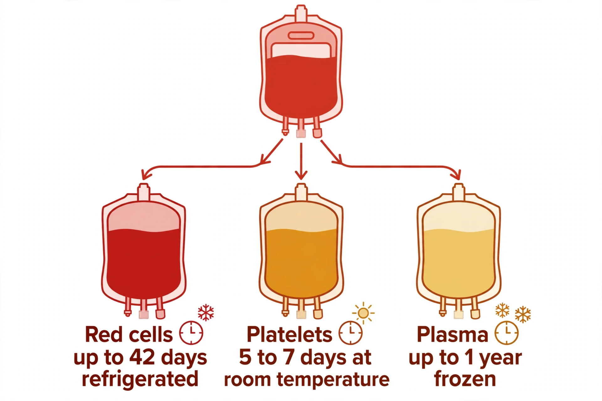
But each component has its own clock, and the clocks are short. Red cells can be refrigerated for up to 42 days. Platelets are normally kept at room temperature, gently shaken, and last only five to seven days. Cold storage was long avoided because cold damages platelets, but the US Food and Drug Administration has licensed cold-stored platelets kept for up to 14 days for patients who are bleeding, and frozen platelets also exist for special uses — so the room-temperature rule is standard practice, not an absolute limit. Plasma can be frozen for up to a year. The consequence is that a hospital cannot build up a large reserve of platelets, no matter how many donors come forward in a good week: the platelets collected on Monday are unusable by the weekend. The entire red-cell inventory of a country turns over roughly nine times a year. Blood is a perishable good, and the only way to keep the supply going is for donors to keep coming back.
Component | How it is stored | Shelf life | Main use |
|---|---|---|---|
Red cells | Refrigerated at 1–6 °C | Up to 42 days | Anaemia, surgery, blood loss |
Platelets | Room temperature, gentle agitation | 5–7 days | Low platelets, cancer treatment, bleeding |
Plasma | Frozen at −18 °C or colder | Up to 1 year | Clotting problems, burns, immune deficiency |
Cryoprecipitate | Frozen | Up to 1 year | Concentrated clotting factors |
The demand never stops. The American Red Cross says someone in the United States needs blood every two seconds — for surgery, for childbirth, for cancer treatment, for trauma, for sickle cell disease and thalassaemia, where patients need regular transfusions for life. About 62 percent of the American population is eligible to donate, and about 3 percent does. In January 2024, the Red Cross declared an emergency blood shortage, with donor numbers at their lowest in twenty years. The gap is not caused by a lack of willing people. It is caused by the arithmetic of a perishable supply and a constant demand: the river must be fed every day, and a single bad week can empty it.
Donated blood is tested before it is used. Every donation is screened for HIV, hepatitis B and C, syphilis, and other infections, and in wealthy countries the screening covers the four main infections in close to all donations. The screening is the reason modern transfusion is as safe as it is, and it is also one of the reasons the supply is fragile in poorer countries, where screening is less complete and the demand is greatest.
Blood cannot be manufactured: the only source of human blood is another human being, and that is why every transfusion begins with a stranger's generosity.
There is a final point about donation that matters more than the numbers. Blood donation is one of the few medical acts in which a healthy person gives something of their own body to a stranger they will never meet, with no benefit to themselves. The system works because enough people do it, regularly, without being paid. The World Health Organization's position is that voluntary, unpaid donation is the safest basis for a blood supply, and the data supports it: countries that rely on paid or family-replacement donors have higher rates of infection in donated blood. The safety of the supply depends, in the end, on the motives of the donor — and on the fact that most donors give because someone, somewhere, will need what they have.
15. Blood Tests: What a Full Blood Count Actually Measures
If you want to see how blood works in practice, look at a full blood count — the most common blood test in medicine, and the one most people will have at some point in their lives. It measures the formed elements directly: how many red cells, how many white cells, how many platelets, how much haemoglobin, and how much of the blood's volume the red cells occupy. It is a snapshot of the fleet, taken in a single tube, and it can reveal anaemia, infection, a bleeding problem, or a bone marrow disorder before any symptom appears.
The first numbers on the report are the counts. The red blood cell count is the number of red cells per microlitre — normally 4.7 to 6.1 million in men and 4.2 to 5.4 million in women. The white blood cell count is normally 4,000 to 11,000 per microlitre, and the platelet count 150,000 to 400,000. A high white cell count usually means infection or inflammation, because the marrow has been told to make more defenders. A low white cell count can mean a viral infection, a marrow problem, or the effect of chemotherapy. A low platelet count means the repair crew is thin, and bruising and bleeding follow. A high platelet count can mean the marrow is overproducing, or that the body is reacting to inflammation or iron deficiency.
The second group of numbers describes the red cells themselves. Haemoglobin is the amount of the oxygen-carrying protein in the blood — normally 14 to 18 grams per decilitre in men and 12 to 16 in women. Haematocrit is the fraction of the blood's volume taken up by red cells — 41 to 50 percent in men, 36 to 44 percent in women. The mean cell volume, or MCV, is the average size of a red cell: small cells point towards iron deficiency, large cells towards B12 or folate deficiency. The mean cell haemoglobin, or MCH, is the average amount of haemoglobin per cell. Together, these numbers let a doctor tell one kind of anaemia from another, which is why a full blood count is the first test ordered when anaemia is suspected.
The third group is the differential — the breakdown of the white blood cells into their five types. A differential reports the percentage and the absolute count of neutrophils, lymphocytes, monocytes, eosinophils, and basophils. A high neutrophil count points towards a bacterial infection. A high lymphocyte count points towards a viral infection. A high eosinophil count points towards a parasite or an allergy. The differential is the test that turns a single white cell count into a story about what kind of immune response is happening, and it is the reason a doctor who sees a high white count always asks which type is high.
Blood tests go beyond the full blood count, of course. A doctor can measure the iron in the blood and the ferritin that stores it, to confirm iron deficiency. They can measure the clotting time — the PT and aPTT described in section 8 — to test the cascade. They can measure blood glucose, cholesterol, kidney function, and liver function, all from the same sample. But the full blood count is the foundation, because it measures the blood itself rather than what the blood carries. It is the test that answers, in numbers, the question this article asks in words: how blood works, right now, in this person, at this moment.
16. Common Mistakes and Misconceptions About Blood
Blood is familiar enough that everyone has an opinion about it, and some of those opinions are wrong in ways that matter. This section collects the most persistent ones.
Blood is blue inside your veins. Covered in section 5, but worth repeating as the first misconception, because it is the most common. Blood is always red. Deoxygenated blood is dark red, not blue. A vein looks blue from the outside because light behaves differently as it passes through skin and the tissue beneath — the red wavelengths are absorbed on the way, and more blue reaches your eye. The red-for-arteries, blue-for-veins convention in diagrams is a teaching aid, not a photograph.
"Thick blood" is a diagnosis. People use the phrase to mean many things — high cholesterol, high blood pressure, a high red cell count — and it is not a medical term. Blood viscosity does vary: a high haematocrit makes blood thicker and harder to pump, and dehydration thickens it temporarily. But "thick blood" on its own explains nothing, and the conditions people mean by it need their own names and their own tests. If a doctor says your blood is viscous, ask what the haematocrit and the plasma proteins actually are.
Bloodletting balances the body. For more than two thousand years, doctors bled patients deliberately, on the theory that disease came from an excess of blood or an imbalance of the humours. The practice was dangerous, and it sometimes killed the patient it was meant to help — George Washington's death in 1799 followed heavy bloodletting, among other treatments — and it survives today only in a narrow, specific form: therapeutic phlebotomy, the deliberate removal of blood to treat conditions such as haemochromatosis (iron overload) and polycythaemia vera (too many red cells), where the problem really is too much of something in the blood. The general idea that letting blood improves health is wrong, and the modern exceptions prove the rule: blood is removed only when a measured excess exists, and only under medical supervision.
You can run out of blood by donating. A whole-blood donation removes about 470 to 500 millilitres, roughly a tenth of the blood in an average adult. The body replaces the fluid within a day and the red cells over six to twelve weeks, which is why the minimum interval between donations is 56 days. Donors are screened before every donation, and a person whose haemoglobin is too low is deferred — turned away — until it recovers. The system is designed so that donation never takes more than the body can spare.
Eating a particular food changes your blood type, or your blood type should decide your diet. Blood type diets — the idea that people with different blood groups should eat different foods — have no scientific support. The antigens on red blood cells have nothing to do with how the gut digests food, and the studies that have tested the claim have found no benefit. Your blood group matters for transfusion and for pregnancy. It does not matter for your dinner.
Blood tests can tell you everything about your health. A blood test is a powerful tool, and it is also a limited one. It measures what is in the blood at the moment the sample is taken: the cells, the proteins, the chemistry. It cannot measure what is happening inside a tissue, or what will happen next week, or why a value is abnormal. A single unusual result, on its own, is often meaningless — values fluctuate with hydration, time of day, illness, and medication. Blood tests are most useful when they are repeated, compared, and read by someone who knows the person's history. They are a window into the body, not a verdict on it.
Conclusion — What to Remember About How Blood Works
Blood is a liquid tissue, about five litres of it, carrying roughly 25 trillion red cells, tens of billions of white cells, and over a trillion platelets in a straw-coloured river of plasma. It does five jobs at once: it transports oxygen, nutrients, hormones, and waste; it regulates temperature and chemistry; it protects against infection; it carries the body's messages; and it seals its own leaks. It does all of this while circulating through the body about once a minute, every minute, for a lifetime.
The parts are simple enough to list — plasma, red cells, white cells, platelets — but the system is not simple. The red cell has no nucleus, because it traded its machinery for carrying space. The white cell has five forms, because defence needs specialists as well as first responders. The platelet is not a cell at all, but a fragment that waits for a wall to break. And the clotting cascade is a chain of proteins that must switch on in seconds, amplify a thousandfold, and then switch off again, without ever clotting inside a healthy vessel. The balance is the story: liquid enough to flow for decades, solid enough to seal a wound in minutes.
Three facts are worth carrying away. First, blood is red because of haemoglobin, and it is always red — bright red with oxygen, dark red without it, never blue. Second, blood is personal: your blood group is written on your red cells, and matching it is what makes transfusion safe. Third, blood is a gift that cannot be made or stored for long, and the supply depends on people giving it, again and again, because the red cells in a donation expire in 42 days and the platelets in five. The next time blood crosses your path — a sample drawn for a test, a scrape on a knee, a bag hanging on a stand — you will know what you are looking at: not a simple red liquid, but a moving tissue, rebuilding itself two million cells a second, doing a dozen jobs at once, and doing them, most of the time, without you ever noticing.
Frequently Asked Questions
What is blood made of?
Blood is made of plasma (about 55 percent of its volume) and formed elements (about 45 percent): red blood cells, white blood cells, and platelets. Plasma is about 92 percent water, with proteins — albumin, globulins including antibodies, and fibrinogen — plus nutrients, salts, hormones, and waste dissolved in it. A microlitre of blood contains about five million red cells, seven thousand white cells, and 250,000 platelets.
What do platelets do?
Platelets plug leaks and start clotting. When a blood vessel is damaged, platelets stick to the exposed collagen and von Willebrand factor, change shape, release chemical signals that recruit more platelets, and pile up into a soft plug over the break. They also carry out daily maintenance, patching tiny gaps in vessel walls before any bleeding becomes visible. Platelets are fragments of bone marrow cells called megakaryocytes, live seven to ten days, and number 150,000 to 400,000 per microlitre of blood.
How does blood clot?
Blood clots in three overlapping stages. First, the damaged vessel narrows (vascular spasm). Second, platelets stick to the break and form a soft plug (primary haemostasis). Third, the clotting cascade activates a chain of proteins in the plasma: the extrinsic pathway (tissue factor plus Factor VII) and the intrinsic pathway (Factors XII, XI, IX, and VIII) both converge on Factor X, which drives the production of thrombin. Thrombin converts fibrinogen into fibrin, and Factor XIII cross-links the fibrin into a stable mesh that traps blood cells and seals the wound. Calcium and vitamin K are both required, and natural brakes — antithrombin, protein C, and protein S — stop the clot from growing. Once the vessel heals, plasmin dissolves the clot.
Why is blood red?
Blood is red because of haemoglobin, the protein inside red blood cells that carries oxygen. Haemoglobin's haem group absorbs blue and green light and reflects red and orange light. Blood loaded with oxygen is bright cherry red; blood that has delivered its oxygen is darker, maroon red — but it is never blue. Veins look blue through the skin because of the way light travels through skin and tissue, not because of the blood's colour.
How much blood is in the human body?
A typical adult carries around five litres, though the figure depends on size: larger people carry more, smaller people less, and men on average carry a little more than women. As a share of weight, blood is roughly seven to eight percent of the body. The heart keeps that volume moving at about five litres a minute while resting, so the whole supply passes through the heart in close to a minute, adding up to some 7,200 litres shifted in a day.
How long do red blood cells live?
Around 120 days. New ones are produced in the bone marrow at a rate of roughly two million a second — about 200 billion daily — under the control of erythropoietin, a hormone released by the kidneys when oxygen runs low. Worn-out cells are taken apart by macrophages in the spleen and liver, and the iron inside them is recycled for the next generation.
What is the rarest blood type?
In the ABO and Rh systems, AB negative is the rarest of the eight main types, found in about one percent of people or fewer, depending on the population. But ABO and Rh are only two of the 49 blood group systems recognised by the International Society of Blood Transfusion, and some of the rarer antigens — such as the Rh-null phenotype, sometimes called golden blood — are found in only a handful of people worldwide. Rarity matters in transfusion: the rarer the type, the harder it is to find a matching donor.
What is serum, and how is it different from plasma?
Serum is what remains of plasma once blood has clotted: the clotting factors, fibrinogen above all, have been consumed in making the clot, so serum cannot clot again. Plasma, by contrast, is collected with clotting prevented, so it keeps its full set of clotting factors. Which one a laboratory uses depends on the test being run, and the choice affects which measurements come out reliably.
Does your blood type affect your diet?
No. The idea that different blood groups should eat different foods has no scientific support. The molecules on the surface of red blood cells play no part in digestion, and the studies that have tested blood type diets have found no benefit. Blood group is relevant when blood is transfused and when a pregnancy involves Rh incompatibility. It has no bearing on what you should eat.
Can you donate blood often?
Yes, within limits. In the United States, whole blood can be given every 56 days, up to six times a year, while platelets can be given weekly and plasma every 28 days, since those parts of blood come back sooner than red cells. The fluid lost in a donation is restored in about a day and the red cells over six to twelve weeks. Donors are screened before every donation, and anyone whose haemoglobin is too low is deferred until it recovers.
Sources
American Red Cross. Blood Components. https://www.redcrossblood.org/donate-blood/how-to-donate/types-of-blood-donations/blood-components.html
American Red Cross. Blood Donation FAQs. https://www.redcrossblood.org/faq.html
American Society of Hematology. Blood Basics. https://www.hematology.org/education/patients/blood-basics
Centers for Disease Control and Prevention. About Sickle Cell Disease. https://www.cdc.gov/sickle-cell/about/index.html
Centers for Disease Control and Prevention. Data and Statistics on Sickle Cell Disease. https://www.cdc.gov/sickle-cell/data/index.html
Cleveland Clinic. Plasma: What It Is and Why It Is Important. https://my.clevelandclinic.org/health/body/22865-plasma
Cleveland Clinic. White Blood Cells: What They Are and What They Do. https://my.clevelandclinic.org/health/body/21871-white-blood-cells
Cleveland Clinic. Anemia. https://my.clevelandclinic.org/health/diseases/3929-anemia
Fikiet, M., and Lednev, I. Blood in your veins is not blue — here's why it's always red. Medical Xpress, 2018. https://medicalxpress.com/news/2018-06-blood-veins-blue-red.pdf
GBD 2021 Anaemia Collaborators. Prevalence, years lived with disability, and trends in anaemia burden by severity and cause, 1990–2021. The Lancet Haematology, 2023;10(9):e713–e734. https://doi.org/10.1016/S2352-3026(23)00160-6
Haemophilia Foundation Australia. Fast facts. https://www.haemophilia.org.au/bleeding-disorders/faqs/fast-facts/
LibreTexts Medicine. Composition of Blood. https://med.libretexts.org/Courses/Southern_Illinois_University_Edwardsville/Essentials_of_Physiology_for_Nurse_Anesthetists_I_(Gopalan)/08%3A_The_Blood/8.04%3A_Composition_of_Blood
Mayo Clinic. Anemia: Symptoms and Causes. https://www.mayoclinic.org/diseases-conditions/anemia/symptoms-causes/syc-20351360
Mayo Clinic. Hemophilia: Symptoms and Causes. https://www.mayoclinic.org/diseases-conditions/hemophilia/symptoms-causes/syc-20373327
Mayo Clinic. Von Willebrand Disease: Symptoms and Causes. https://www.mayoclinic.org/diseases-conditions/von-willebrand-disease/symptoms-causes/syc-20354978
Mayo Clinic. Blood Donation: What to Expect. https://www.mayoclinic.org/tests-procedures/blood-donation/about/pac-20385144
MedlinePlus (U.S. National Library of Medicine). Blood. https://medlineplus.gov/blood.html
MedlinePlus. Hemoglobin. https://medlineplus.gov/ency/article/003645.htm
MedlinePlus. Red Blood Cell Count. https://medlineplus.gov/ency/article/003644.htm
MedlinePlus. Platelet Count. https://medlineplus.gov/ency/article/003647.htm
MedlinePlus. Hemophilia. https://medlineplus.gov/hemophilia.html
MedlinePlus. Anemia. https://medlineplus.gov/anemia.html
National Health Service (UK). Blood Donation. https://www.blood.co.uk/who-can-give-blood/
National Health Service (UK). Anaemia. https://www.nhs.uk/conditions/iron-deficiency-anaemia/
National Institutes of Health. Blood: MedlinePlus Medical Encyclopedia. https://medlineplus.gov/ency/article/002229.htm
Nobel Prize Outreach. Finding the key to safe blood transfusions. https://www.nobelprize.org/stories/finding-the-key-to-safe-blood-transfusions/?gallery_style=page
Nobel Prize Outreach. Physiology or Medicine 1930 — Presentation Speech. https://www.nobelprize.org/prizes/medicine/1930/ceremony-speech/
Novo Nordisk Haemophilia Foundation. Sickle cell disease fact sheet. https://nnhf.org/wp-content/uploads/2025/12/sickle-cell-disease-fact-sheet.pdf
OpenStax Biology 2e. Components of the Blood. https://openstax.org/books/biology/pages/40-2-components-of-the-blood
Stanford Blood Center. The Life of a Red Blood Cell. https://stanfordbloodcenter.org/red_blood_cells-2/
World Federation of Hemophilia. Report on the Annual Global Survey 2024. October 2025. https://www1.wfh.org/publications/files/pdf-2588.pdf
World Health Organization. Anaemia: Fact Sheet. https://www.who.int/news-room/fact-sheets/detail/anaemia
World Health Organization. Blood safety and availability: Fact Sheet. https://www.who.int/news-room/fact-sheets/detail/blood-safety-and-availability
World Health Organization. Safe blood supply improves as voluntary donations exceed 85%, but many people still lack access. 12 June 2026. https://www.who.int/news/item/12-06-2026-safe-blood-supply-improves-as-voluntary-donations-exceed-85---but-many-people-still-lack-access
World Health Organization. WHO global anaemia estimates: Key findings, 2025. https://www.who.int/publications/i/item/9789240113930
World Health Organization Regional Office for Africa. Sickle Cell Disease Overview. https://www.afro.who.int/health-topics/sickle-cell-disease