
1. How the Liver Works: The Organ That Never Clocks Off
It is half past eleven at night, and you have just finished a meal, a glass of wine and a headache tablet. You brush your teeth, you climb into bed, and within minutes you are asleep. Nothing about the evening feels like work. But while you sleep, one organ in your upper abdomen is running a night shift that never ends. It is sorting the meal you just ate into fuel, building blocks and waste. It is breaking down the alcohol. It is dismantling the tablet, molecule by molecule, so that it can do its job and then leave your body safely. It is topping up your blood sugar so that your brain, which never sleeps, does not run dry before breakfast. You will not feel any of this. You never do. That silence is the first thing to understand about how the liver works: it is the largest internal organ you own, it carries out hundreds of jobs at once, and it does them without asking for your attention.
Most people can point to where their heart is and describe, roughly, what it does. The liver is different. Ask a room of adults what the liver does and you will hear the same two words again and again: it detoxifies. That answer is true, and it is also far too small. Detoxification is one job among hundreds. The liver is a chemical processing plant, a warehouse, a factory and a recycling centre folded into a single reddish-brown organ about the size of a rugby ball. It makes the fluid that digests your fat. It builds most of the proteins floating in your blood. It converts a poison your own body produces every hour into a harmless compound your kidneys can throw away. It stores sugar, vitamins and iron, and it releases them on a schedule set by hormones. It even grows back when part of it is removed — no other solid organ can restore so much of its own mass after such a large loss. This article follows all of that work, in order, from the blood that arrives at the liver to the bile that leaves it, so that by the end you can picture the organ doing its job — not as a vague filter, but as a place with an address, a workforce and a daily routine.
1.1 The answer in 150 words: how does the liver work?
How does the liver work? In short, blood from the gut arrives at the liver through the portal vein carrying everything absorbed from a meal, while the hepatic artery brings oxygen-rich blood from the heart. Inside the liver, that blood flows slowly past plates of liver cells called hepatocytes, arranged in tiny hexagonal units called lobules. As it passes, hepatocytes pull out nutrients and process them: they turn sugar into glycogen for storage, build proteins such as albumin and clotting factors, and convert fats into forms the body can ship and store. They also dismantle drugs, alcohol and other foreign chemicals in two phases — first the cytochrome P450 enzymes alter them, then conjugation reactions make them water-soluble so they can leave in bile or urine. Ammonia, a toxic by-product of protein breakdown, is converted into urea through the urea cycle. Bile, made by the same cells, flows into the gut to digest fat, and most of its salts are reclaimed and reused. That, in one paragraph, is how the liver works. The rest of this article is the same story told slowly, with the anatomy, the chemistry and the numbers that make it real.
1.2 The scale of the subject
Two headline figures set the scale before the detail begins, and both come from the most recent large studies available. First, fatty liver disease linked to metabolism — now called metabolic dysfunction-associated steatotic liver disease, or MASLD — affects about one adult in three worldwide. A meta-analysis published in Clinical Gastroenterology and Hepatology in 2025, covering 44 studies and more than 11 million participants, put the global prevalence of MASLD at 33.6 percent, and at 70.2 percent among people with type 2 diabetes. That is a higher figure than the older "one in four" estimate that still circulates in textbooks, and it means that the organ this article describes is under strain in a large share of the population. Second, cirrhosis — the scarring that follows years of liver injury — killed an estimated 1.43 million people worldwide in 2021, according to the Global Burden of Disease study published in 2025, up from about 1.02 million in 1990, even though the age-standardised death rate fell over the same period because the world population grew and aged. Liver cancer, most of it hepatocellular carcinoma, caused an estimated 843,045 new cases and 732,489 deaths in 2024, making it the seventh most commonly diagnosed cancer and the third leading cause of cancer death globally. Keep those figures in mind as you read. The liver is remarkably resilient, and it is also remarkably burdened — and understanding how it works is the first step toward understanding why that burden matters.
This article is an educational explainer. It describes mechanisms — how blood flows through the liver, how bile is made, how detoxification works, how the urea cycle runs, how the liver stores sugar and rebuilds itself. It does not diagnose, it does not advise any individual reader, and it does not replace a clinician. Anything personal — a symptom, a test result, a decision about care — belongs in a conversation with the reader's own clinician, not with an article.
I first paid proper attention to my own liver in a physiology practical, years ago, watching a preserved specimen on a bench and thinking how unremarkable it looked: a smooth, dark, wedge-shaped mass, heavier than I expected, with no moving parts and nothing to suggest the hundreds of chemical reactions happening inside it every minute. The heart announces itself with every beat. The liver works in silence, and that silence is exactly why it is so easy to overlook — and so easy to misunderstand.
1.3 Why "detox" is the wrong place to start
If you search for liver health online, the first thing you meet is detox: teas, juices, cleanses and supplements promising to flush the organ clean. That framing gets the story backwards. Understanding how the liver works means starting with the liver's own chemistry, not with a product on a shelf. The liver is not a filter that fills up with dirt and needs washing out. It is a chemical factory that dismantles unwanted molecules and sends the pieces out through bile and urine, and it does that job continuously, without help from any product. The National Center for Complementary and Integrative Health, part of the US National Institutes of Health, states plainly that there is no good evidence that detox diets or cleanses remove toxins from the body, and that the liver and kidneys already do that work. When this article talks about detoxification, it means the liver's own two-phase chemistry — enzymes, conjugation, transport — not a product on a shelf. Starting there, rather than with the marketing, is what makes the rest of the story make sense.
2. Meet the Liver: Size, Place and Blood Supply
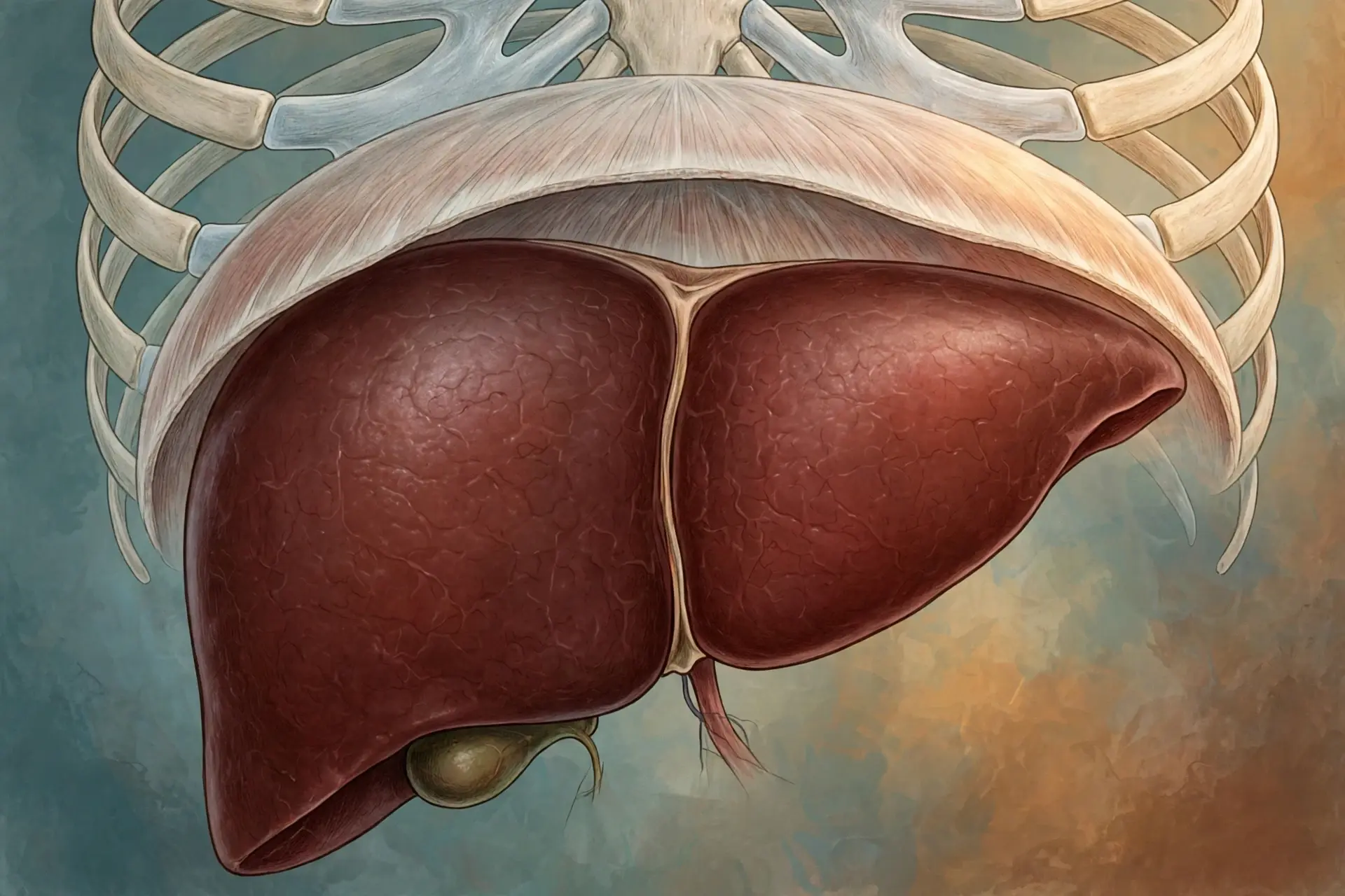
The liver sits in the upper right of the abdomen, tucked under the diaphragm and partly protected by the lower ribs. It is the largest internal organ in the body, weighing roughly 1.5 kilograms in an average adult — about three pounds — and making up around 2 to 2.5 percent of body weight. Its colour is a deep reddish-brown, its surface is smooth and glossy, and its shape is a wedge: a large right lobe and a smaller left lobe, separated on the upper surface by the falciform ligament, with two smaller lobes, the caudate and quadrate lobes, tucked on the underside. Press gently under your right ribs and you are pressing, more or less, on the front of it — though in a healthy person you usually cannot feel it at all, because it sits high and the ribs shield it.
2.1 Two blood supplies, one organ
What makes the liver unlike any other organ is that it has two blood supplies, and they do different jobs. The hepatic artery brings oxygen-rich blood from the heart, the way an artery supplies any organ. The portal vein brings something no other organ receives: blood that has already passed through the gut. Everything absorbed from a meal — sugars, amino acids, fats in their packaged form, vitamins, minerals, and also alcohol, medicines and anything else swallowed — travels first to the liver before it reaches the rest of the body. The portal vein carries roughly 75 to 80 percent of the blood entering the liver, and the hepatic artery carries the remaining 20 to 25 percent. The split in flow is not the same as the split in oxygen: the artery, though smaller in volume, supplies about half the liver's oxygen, because portal blood has already given up much of its oxygen to the gut. Total blood flow through the liver is roughly 1.2 to 1.5 litres per minute at rest — about a quarter of the heart's entire output, flowing through an organ that is only about two percent of body weight. Over a day, that adds up to something like 2,000 litres of blood passing through the liver, every drop of it inspected by liver cells on the way.
2.2 Why the portal vein matters
The portal arrangement is a design choice with a purpose. The gut absorbs nutrients in a rush after every meal, and blood sugar and amino acid levels would spike dangerously if that flood went straight to the rest of the body. Instead, the liver stands between the gut and the general circulation like a customs post. It takes the first look at everything absorbed, stores what can be stored, converts what needs converting, and releases the rest into the bloodstream at a controlled rate. That first pass is also why many medicines are partly dismantled before they ever reach their target: a drug swallowed by mouth must run the gauntlet of the liver before it can act elsewhere, a phenomenon pharmacologists call first-pass metabolism. It is why the same drug can need a different dose when given by mouth than when given by injection, and why liver disease can change how medicines behave. The portal vein is the reason the liver gets the first word on everything you swallow — and the reason it takes the first hit from everything harmful you swallow, too.
2.3 The exits: hepatic veins and bile ducts
Blood leaves the liver through the hepatic veins, which drain into the inferior vena cava and from there back to the heart. Bile leaves through a separate system of ducts that run in the opposite direction to the blood, carrying bile from the liver cells out toward the gut. The two exit routes matter because they carry different cargo: the veins carry processed nutrients, proteins and cleaned blood; the ducts carry bile and the waste products the liver has chosen to throw away through the gut. A blockage in one system causes a different problem from a blockage in the other, which is why doctors distinguish between liver cell damage and bile duct obstruction — the blood tests for each look different, as section 15 explains.
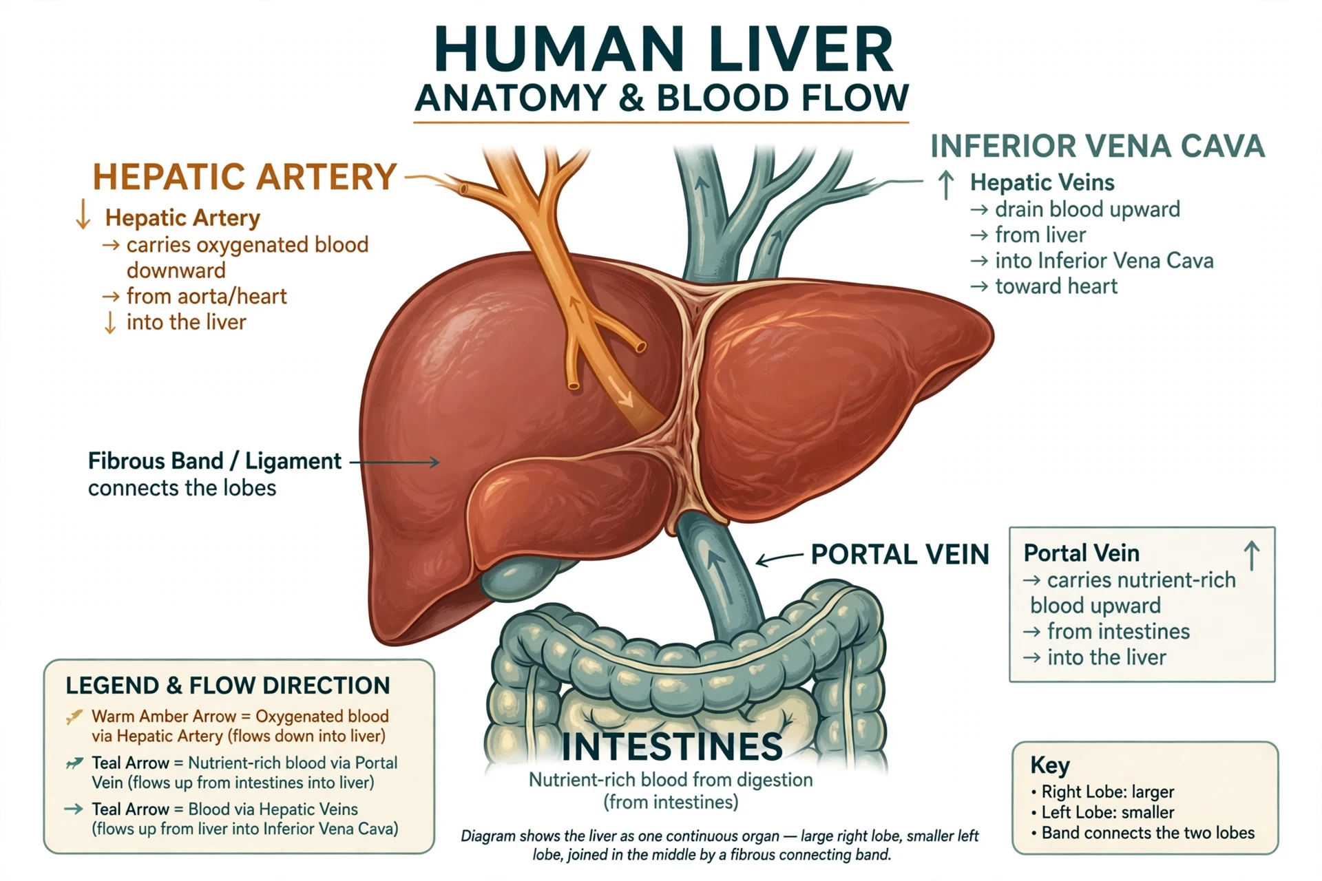
Vessel | Where the blood comes from | Share of flow into the liver | What it mainly delivers |
|---|---|---|---|
Portal vein | The gut, spleen and pancreas | About 75 to 80 percent | Nutrients from meals, plus medicines, alcohol and anything else absorbed |
Hepatic artery | The heart, via the coeliac trunk | About 20 to 25 percent | Oxygen-rich blood — about half the liver's oxygen supply |
Hepatic veins | The liver itself | Outflow | Processed blood back to the heart via the inferior vena cava |
Table 1: The liver's blood supply at a glance. The portal vein dominates by volume; the hepatic artery dominates by oxygen per litre.
3. The Lobule: The Liver's Repeating Unit
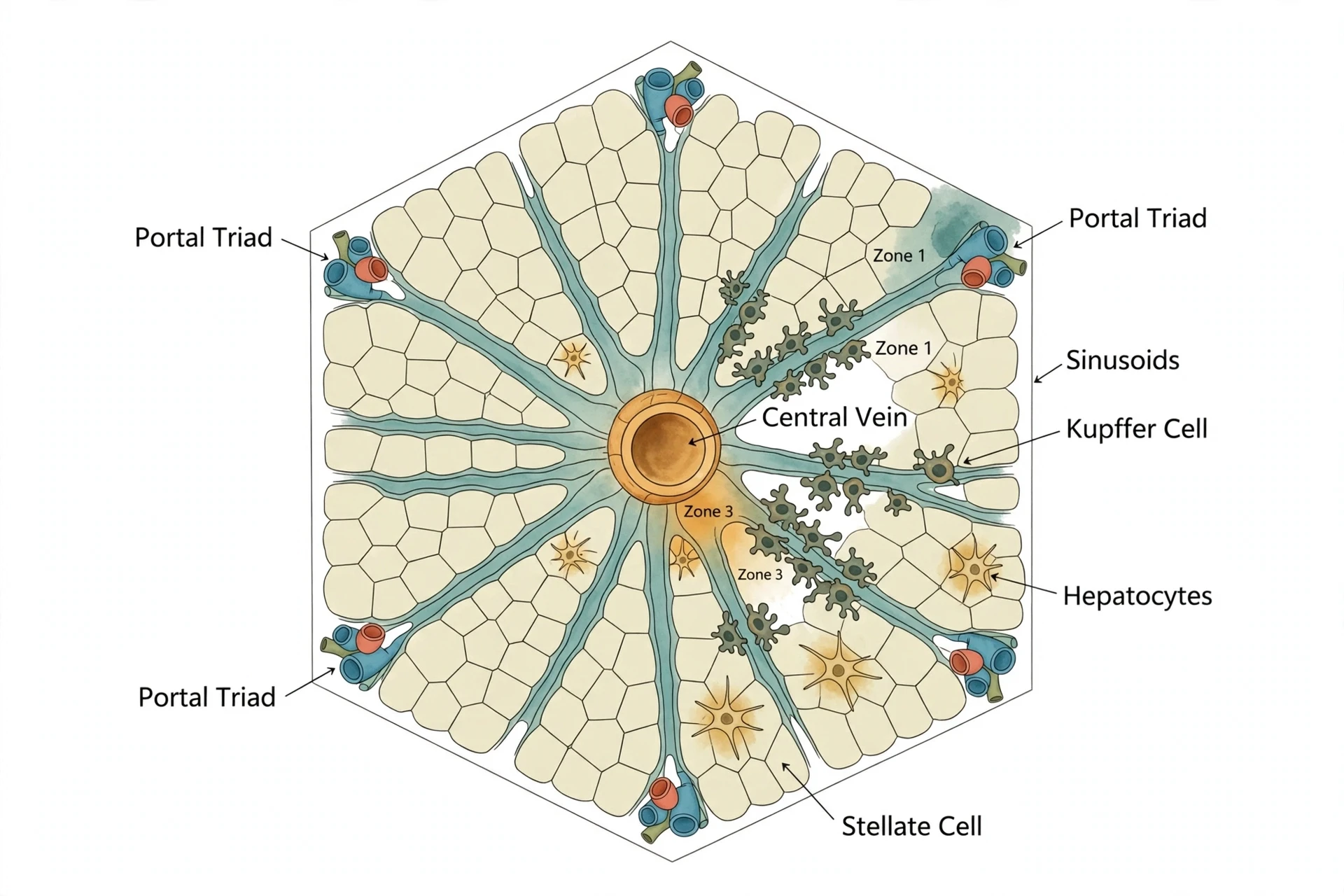
If you cut the liver open and look at it under a microscope, the smooth wedge from the outside resolves into a repeating pattern. The liver is built from about a million tiny hexagonal units called lobules, each roughly a millimetre across. Every lobule is a miniature copy of the whole organ's job: blood flows in at the corners, past rows of liver cells, and drains out through a vein in the centre, while bile flows the other way, outward from the centre toward the corners. That counterflow — blood in one direction, bile in the other — is the key to the whole design, and once you can picture a single lobule, you can picture how the liver works.
3.1 Hepatocytes: the workforce
The working cells of the liver are the hepatocytes. They make up about 80 percent of the liver's mass, and a single hepatocyte is a chemical generalist: it carries out hundreds of different reactions, and it can switch between them as conditions change. Hepatocytes are arranged in plates one or two cells thick, radiating out from the central vein like spokes from a hub. Between the plates run the sinusoids, wide, leaky capillaries through which blood flows slowly — slowly enough that hepatocytes have time to pull molecules out of it, and leaky enough that large molecules can pass through the gaps. The slow flow matters: blood crosses a lobule in seconds, but those seconds are the liver's working time, and the organ is built to make the most of them.
3.2 The portal triad and the central vein
At each corner of the hexagonal lobule sits a portal triad — three small vessels bundled together. One is a branch of the portal vein, bringing nutrient-rich blood from the gut. The second is a branch of the hepatic artery, bringing oxygen. The third is a bile duct, collecting bile. Blood from the first two mixes in the sinusoids and flows inward toward the central vein, which drains the lobule into the hepatic veins. Bile, meanwhile, is secreted by hepatocytes into tiny channels called canaliculi that run between the cells, and it flows outward, away from the central vein, toward the bile ducts at the corners. Blood and bile move in opposite directions through the same tiny unit, which keeps fresh bile from mixing with the blood it has just been cleaned from.
3.3 Zones, Kupffer cells and stellate cells
Not every part of a lobule does the same work. Hepatocytes near the portal triad — zone 1 — receive the freshest, most oxygen-rich blood, and they specialise in energy-hungry jobs such as making glucose and urea. Hepatocytes near the central vein — zone 3 — receive blood that has already been picked over, with less oxygen and more waste, and they specialise in detoxification, including the cytochrome P450 enzymes of section 6. That zoning explains a pattern doctors see again and again: damage that strikes zone 3, such as the injury from an overdose of paracetamol, hits the detoxification machinery hardest, while damage that strikes zone 1 looks different again. The lobule is not uniform; it is a gradient, and where an injury lands inside the gradient decides what goes wrong first.
Two other cell types share the lobule. Kupffer cells are the liver's resident macrophages, star-shaped immune cells that line the sinusoids and swallow bacteria, dead red blood cells and debris arriving from the gut — a first line of defence, since the portal blood carries gut microbes and their fragments straight to the liver. Hepatic stellate cells sit in the narrow gap between hepatocytes and sinusoids, the space of Disse, and in health they store vitamin A in fat droplets. When the liver is injured repeatedly, those same stellate cells change character and start laying down scar tissue, which is how fibrosis begins — a story that returns in section 14.
3.4 A note on numbers: how many lobules, how many cells
Textbooks commonly describe the liver as containing around a million lobules and on the order of hundreds of billions of hepatocytes. Those are round estimates rather than counted figures, and the research for this article could not verify a single authoritative count, so no precise total is given here. What matters is the principle: the liver does its work not in one large chamber but in a million small ones, all running the same process in parallel. That redundancy is part of why the liver tolerates injury so well — lose some lobules and the rest can carry the load — and part of why it can regenerate, as section 13 describes.
4. What Does the Liver Do? Six Jobs, One Organ
Ask what the liver does and you will hear a number: more than 500 functions. That figure comes from Johns Hopkins Medicine, which describes the liver as performing over 500 vital functions, and it is best read as a round estimate of the organ's range rather than a counted list. Cleveland Clinic once quoted a larger figure in a patient brochure, but that number is not supported by a citable source, so this article sticks with the Johns Hopkins wording. Whatever the exact count, the functions fall naturally into six jobs, and holding those six in mind makes the rest of the article easy to follow.
4.1 The six jobs
First, the liver processes nutrients. It takes the sugars, amino acids and fats arriving from the gut and decides what happens to each: burn now, store for later, or convert into something else. Second, it makes bile, the fluid that digests fat and carries waste out of the body. Third, it detoxifies — dismantling drugs, alcohol and other foreign chemicals so they can leave safely. Fourth, it makes proteins: albumin, clotting factors, transport proteins and more. Fifth, it stores: glycogen for quick sugar, vitamins and iron for the months ahead. Sixth, it regulates the blood itself, keeping glucose, cholesterol and amino acid levels within the range the rest of the body needs. Every section that follows is one of those six jobs, told in detail.
4.2 Why one organ does all of this
The six jobs are not six separate departments. They share the same cells, the same blood and the same chemistry, and they trade with each other constantly. The cholesterol the liver makes becomes the raw material for bile acids. The amino acids it processes become both the proteins it builds and the ammonia it must dispose of. The sugar it stores becomes the glucose it releases overnight. That sharing is efficient, and it is also why liver disease is so wide-ranging in its effects — and why understanding how the liver works means understanding all six jobs together, not one at a time. When hepatocytes fail, they do not fail at one job. They fail at all six at once, which is why advanced liver disease touches the blood, the brain, the kidneys and the gut, not just the liver itself.
Job | What the liver does | Section |
|---|---|---|
Process nutrients | Sorts sugars, amino acids and fats from each meal into fuel, building blocks or storage | 9, 10 |
Make bile | Produces roughly 600 millilitres to 1 litre of bile a day to digest fat and carry away waste | 5 |
Detoxify | Dismantles drugs, alcohol and foreign chemicals in two phases so they can leave the body | 6, 7 |
Make proteins | Builds albumin, clotting factors and transport proteins for the blood | 10 |
Store | Keeps glycogen, vitamins A, D, E, K and B12, iron and copper for later use | 9, 12 |
Regulate blood | Holds glucose, cholesterol and amino acids within the range the body needs | 9, 10 |
Table 2: The liver's six jobs at a glance. The sections that follow take them one at a time.
5. Bile: The Green Fluid That Digests Fat
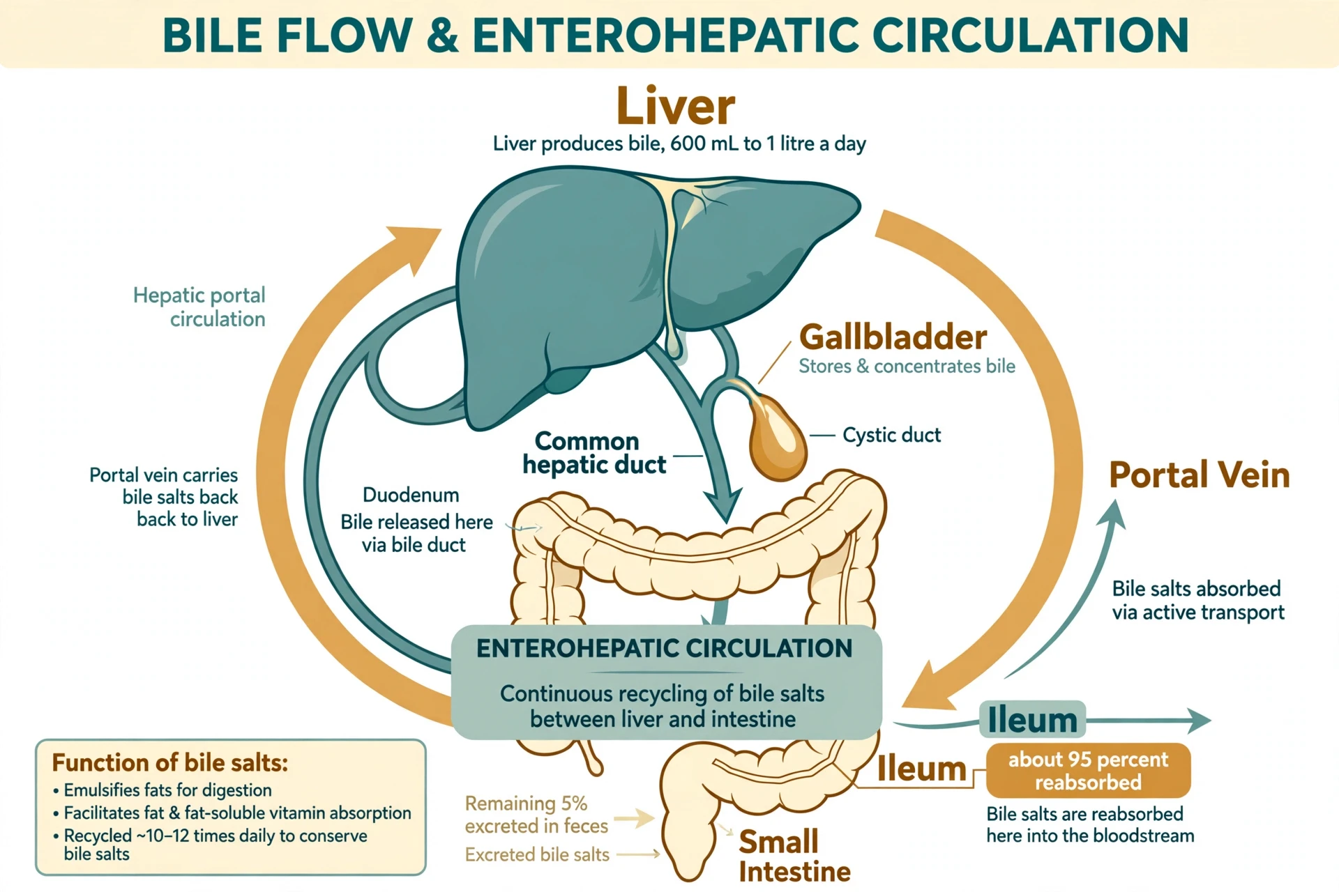
Bile is a greenish-yellow fluid, bitter and alkaline, and the liver makes roughly 600 millilitres to 1 litre of it every day. Different textbooks quote slightly different ranges — some say 400 to 800 millilitres, others up to 1.5 litres — because the amount varies with meals, fasting and how it is measured, so this article gives the range that the most commonly cited physiology texts agree on and treats it as approximate. Bile has two jobs, and both matter. It digests fat, and it carries waste out of the body. Without it, a meal's fat would pass through the gut largely undigested, and the waste products the liver has worked hard to package would have nowhere to go.
5.1 Bile salts: soap for fat
The active ingredients in bile are the bile salts, made in the liver from cholesterol. Fat and water do not mix, and the fat in a meal arrives in the gut as large globules that digestive enzymes cannot easily reach. Bile salts act like soap: they break those globules into tiny droplets, increasing the surface area so that the enzyme lipase can get to work. Without bile salts, fat digestion becomes inefficient, fat-soluble vitamins are poorly absorbed, and undigested fat passes onward, causing the pale, greasy stools that are a classic sign of bile duct blockage. Bile also carries bilirubin, the breakdown product of old red blood cells, which gives bile — and stool — its colour. That is why a blocked bile duct turns stools pale and the skin yellow at the same time: the pigment that should leave in the stool backs up into the blood instead.
5.2 The gallbladder: a storage tank, not a factory
Bile does not flow straight from the liver to the gut all day. Between meals it is diverted into the gallbladder, a small pear-shaped sac tucked under the liver, where water is removed and the bile is concentrated several-fold. When a fatty meal arrives, the gut releases the hormone cholecystokinin, the gallbladder contracts, and concentrated bile is squirted into the duodenum, the first part of the small intestine. People who have had their gallbladder removed can still digest fat, because the liver keeps making bile and it still reaches the gut — but it arrives as a steady trickle rather than a timed squirt, which is why some people notice that very fatty meals sit less comfortably afterwards. The gallbladder is a convenience, not a necessity: the factory is the liver.
5.3 Enterohepatic circulation: bile salts are recycled
One of the liver's cleverest economies is that bile salts are not thrown away after one use. After doing their work in the small intestine, about 95 percent of bile salts are reabsorbed in the last part of the ileum, returned to the liver through the portal vein, and secreted into bile again. The loop is called the enterohepatic circulation, and it runs several times a day — each bile salt molecule may be recycled a dozen or more times before it is finally lost in the stool. The liver only needs to make a small amount of new bile acids each day to replace what is lost. When the ileum is diseased or removed, that recycling breaks, the liver cannot keep up, and fat digestion suffers — a tidy example of how the gut and the liver depend on each other. The same loop explains why some medicines that bind bile salts in the gut lower cholesterol: they interrupt the recycling, the liver makes more bile acids from cholesterol to compensate, and blood cholesterol falls.
5.4 What bile carries away
Bile is also the liver's main waste route. Substances the liver has processed — conjugated bilirubin, excess cholesterol, some drugs and their metabolites, heavy metals — are secreted into bile and leave the body in the stool. That is the second half of detoxification: making a compound water-soluble is only useful if it can then leave, and for fat-soluble waste, bile is the exit. The colour of stool is mostly bilirubin and its breakdown products, and changes in stool colour are sometimes the first visible clue that bile flow has been interrupted. It is an unglamorous fact, but it is also a useful one: bile is the visible end of how the liver works, on show every day in a way most internal chemistry is not.
6. Detoxification: Phase I and Phase II
Detoxification is the liver's most famous job and its most misunderstood. In the liver, detoxification is not a vague cleansing. It is a specific, two-phase chemical process that turns fat-soluble foreign compounds — drugs, alcohol, pollutants, plant chemicals, the breakdown products of the body's own hormones — into water-soluble forms that can leave in urine or bile. The process has a logic to it: most foreign chemicals are fat-soluble, which means they can slip through cell membranes and hide in fat tissue, and the body cannot easily excrete them. The liver's job is to bolt water-loving chemical groups onto them until they dissolve in water and can be flushed out. That is what "detox" means, and it happens in hepatocytes, in two phases, thousands of times a day.
6.1 Phase I: the cytochrome P450 enzymes
Phase I is the alteration step. A family of enzymes called cytochrome P450 — CYP for short — sits in the smooth endoplasmic reticulum of hepatocytes and adds or exposes a reactive chemical handle on the foreign molecule, usually by oxidation. The best-known members are CYP3A4, CYP2D6 and CYP2C19, and CYP3A4 alone handles roughly half of all prescription drugs. Phase I has a catch: the altered molecule is often more reactive — and sometimes more toxic — than the one it started from. That is why Phase I cannot work alone. Its products are intermediates, not finished goods, and they must be passed immediately to Phase II. The classic example is paracetamol, known as acetaminophen in the United States. Most of a paracetamol dose is handled safely by Phase II directly, but a small fraction goes through a CYP enzyme, CYP2E1, and becomes a reactive compound called NAPQI. In a normal dose, the liver mops NAPQI up with glutathione and nothing bad happens. In an overdose, the glutathione runs out, NAPQI attacks liver cells, and the damage is concentrated in zone 3 of the lobule, where the CYP enzymes are densest. That is why paracetamol overdose is one of the commonest causes of acute liver failure — not because the drug is poisonous in itself, but because the liver's own first-phase chemistry creates a poison it must then catch.
6.2 Phase II: conjugation
Phase II is the finishing step, called conjugation. Enzymes attach a large, water-soluble molecule to the Phase I product — a sugar-like group in glucuronidation, a sulphate group in sulfation, the amino acid glycine, or glutathione, the liver's own antioxidant. The result is a compound that dissolves in water, cannot easily slip back into cells, and can be carried away in urine or bile. Glucuronidation handles the largest share of drugs; sulfation and glutathione conjugation cover the rest. Newborn babies are vulnerable precisely because this system is immature: their glucuronidation is slow, which is one reason newborn jaundice is common, as section 11 explains. Genetic differences matter too. Some people carry versions of CYP2D6 that work slowly or quickly, and the same dose of a drug can be too strong for one person and too weak for another. That variation is the whole field of pharmacogenetics, and it begins in the liver.
6.3 Phase III and the exits
Some accounts add a Phase III: transporter proteins that pump the conjugated products out of the hepatocyte, into the blood for the kidneys or into the bile for the gut. Whether you count it as a separate phase or as part of Phase II, the point is the same — detoxification is a pipeline, and a blockage anywhere in it causes trouble. If Phase I runs faster than Phase II can keep up, reactive intermediates accumulate. When the bile duct is obstructed, conjugated waste backs up into the blood. If the kidneys fail, water-soluble waste has nowhere to go. The liver does the chemistry, but it depends on the gut and the kidneys to finish the job — and that partnership is part of how the liver works as a whole.
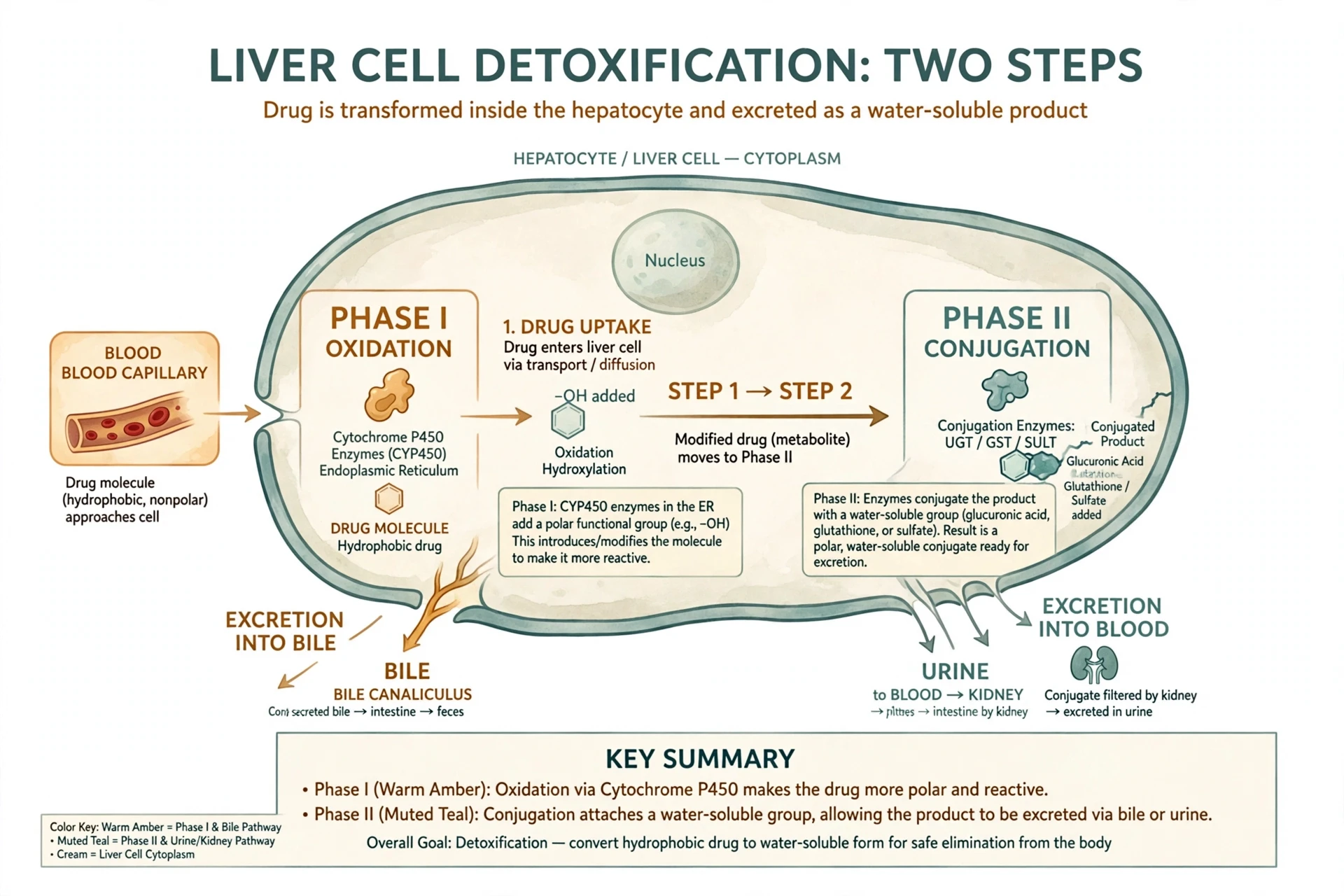
Phase I | Phase II | |
|---|---|---|
What happens | Functionalisation — usually oxidation by cytochrome P450 enzymes | Conjugation — a water-soluble group is attached |
Where | Smooth endoplasmic reticulum of hepatocytes | Cytosol of hepatocytes |
Key players | CYP3A4, CYP2D6, CYP2C19, CYP2E1 | Glucuronidation, sulfation, glutathione conjugation |
Result | A more reactive, more polar intermediate | A water-soluble compound ready for excretion |
Risk if it goes wrong | Reactive intermediates accumulate and damage cells | Waste cannot leave and backs up |
Table 3: The two phases of detoxification compared. Phase I alters; Phase II finishes.
6.4 What detox products cannot do
A word of caution belongs here, because the word "detox" has been borrowed by an industry. No tea, juice, supplement or cleanse has been shown to speed up the liver's Phase I or Phase II chemistry in a healthy person, and some herbal products have themselves caused liver injury — a cruel irony that hepatologists see regularly. The liver's detoxification system is switched on by the compounds it meets, not by a product, and it is limited by enzyme capacity and by glutathione supply, not by a lack of cleansing. Eating well, avoiding excess alcohol and not taking unnecessary medicines or supplements are the only evidence-based ways to keep the system working. Everything else is marketing dressed as physiology.
6.5 When medicines meet each other in the liver
Because so many drugs pass through the same small set of CYP enzymes, medicines can interfere with each other inside the liver. One drug can block an enzyme, so a second drug that depends on that enzyme builds up to toxic levels. Another can induce an enzyme — switch on more of it — so a second drug is cleared too quickly and stops working. Grapefruit juice is the classic food example: it blocks CYP3A4 in the gut wall — the enzyme in the intestinal lining that would otherwise break down part of the drug before it reaches the bloodstream — which is why some statins and blood-pressure medicines carry a warning about grapefruit. (Only very large, repeated amounts of grapefruit juice affect the liver's own CYP3A4.) St John's wort, a herbal remedy, does the opposite, inducing CYP3A4 and making some medicines — including some contraceptives and transplant drugs — less effective. Alcohol adds a third interaction: chronic drinking induces CYP2E1, which changes how the liver handles paracetamol and makes an overdose more dangerous. None of this is a reason to fear medicines. It is a reason to tell a clinician or pharmacist everything you take — prescription drugs, over-the-counter medicines, supplements and herbal products — because the liver is where they all meet, and only someone who knows the full list can spot the collisions.
7. Alcohol: The Liver's Hardest Customer
No substance tests the liver's processing plant quite like alcohol. Ethanol is small, it is absorbed quickly, it arrives through the portal vein in a concentrated wave, and the liver treats it as a priority — partly because it must. The liver cannot store alcohol the way it stores sugar or fat, so it has to dismantle it as it arrives, and while it does so, other work waits. That priority is why a drink with a meal changes how the meal is processed: fat burning pauses, sugar regulation shifts, and the liver's attention is monopolised until the alcohol is gone.
7.1 Three enzymes, one priority
The main route is a two-step relay. Alcohol dehydrogenase, ADH, converts ethanol into acetaldehyde. Aldehyde dehydrogenase, ALDH, then converts acetaldehyde into acetate, which the body can burn for energy or turn into fat. Acetaldehyde is the problem: it is far more toxic than ethanol itself, it damages proteins and DNA, and it is responsible for much of the flushing, nausea and headache that follow drinking. People whose ALDH works slowly — common in East Asian populations — accumulate acetaldehyde quickly and feel unwell after small amounts, which is a protective quirk of genetics rather than a weakness. A second route, the microsomal ethanol-oxidising system, uses CYP2E1 and handles roughly a tenth of ethanol at low intakes, but a larger share when drinking is heavy or chronic — and it generates reactive oxygen species as a by-product, adding oxidative damage to the bill. A third, minor route uses catalase. Together they clear alcohol at a roughly fixed rate, about one standard drink per hour in an average adult, and no amount of coffee, cold air or exercise speeds it up. The liver sets the pace, and the rest of the body waits.
7.2 From fat to fibrosis
When alcohol arrives faster than the liver can clear it, the chemistry shifts. Breaking down ethanol generates excess NADH, which pushes the liver toward making fat and away from burning it, and fat droplets begin to accumulate inside hepatocytes — the earliest stage of alcohol-related liver disease, and one that is reversible if drinking stops. Continued heavy drinking adds inflammation, then scar tissue laid down by activated stellate cells, then fibrosis, then cirrhosis, in which the liver's architecture is replaced by scar and its blood flow is strangled. The progression is not inevitable and it is not quick — it usually takes years of heavy drinking — but it is also not rare, and it is the reason alcohol remains one of the leading causes of liver disease worldwide. The same pathway, driven by metabolic factors rather than alcohol, produces the fatty liver disease described in section 14, and Sciory's dedicated article on fatty liver follows that story in detail.
7.3 Why the liver takes the first hit
Alcohol's damage is concentrated in the liver for the same reason medicines are: the portal vein delivers it there first, at its highest concentration, before it is diluted in the general circulation. The gut absorbs it, the liver meets it, and only then does the rest of the body. That first pass protects the brain and other organs from the full concentration, and the liver pays for the service. It is a pattern that repeats across this article: the liver's position between the gut and the rest of the body makes it both the body's guardian and its first casualty — and that is the clearest single picture of how the liver works.
8. The Urea Cycle: Turning Poison into Fertiliser
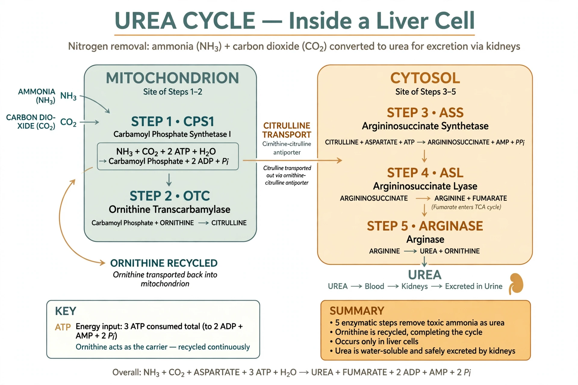
Every hour, your body breaks down protein — from food, and from its own tissues being renewed — and every breakdown releases ammonia. Ammonia is a problem out of all proportion to its size. It is highly toxic to the brain in particular, and even modest rises cause confusion, drowsiness and, at high levels, coma. The body has no way to store ammonia and no way to excrete it directly. The liver's answer is the urea cycle, a five-step chemical loop that converts ammonia into urea, a harmless compound that the kidneys can excrete in urine. The cycle runs in hepatocytes and nowhere else in its complete form, which makes the liver the only organ that can do this job — and makes liver failure dangerous in a very specific way, because ammonia then has nowhere to go.
8.1 Five steps, two compartments
The urea cycle is sometimes called the ornithine cycle or the Krebs-Henseleit cycle, after Hans Krebs and Kurt Henseleit, who described it in 1932 — the first metabolic cycle ever discovered, and the model for the citric acid cycle that Krebs would describe five years later. Its five steps are split between two compartments of the hepatocyte: the first two happen inside the mitochondria, the last three in the cytosol. Ammonia and carbon dioxide enter, are combined with the carrier molecule ornithine, and are passed through a series of intermediates — citrulline, argininosuccinate, arginine — until arginase splits arginine into urea and regenerates ornithine, ready to carry the next ammonia. Each turn of the cycle costs the equivalent of four high-energy phosphate bonds, which is a measure of how seriously the body takes ammonia: it pays a substantial energy price to keep it out of the blood. The urea leaves the liver in the blood, travels to the kidneys, and is excreted in urine.
8.2 When the cycle fails
Because the urea cycle exists only in the liver, its failure is a liver problem with brain symptoms. In advanced liver failure, ammonia rises in the blood, crosses into the brain, and causes hepatic encephalopathy — confusion, personality change, sleep disturbance and, in severe cases, coma. It is one of the most frightening complications of cirrhosis, and it is treated, in part, by reducing ammonia production in the gut, which is why the gut microbiome and the liver are so closely linked. The same principle explains why a high-protein meal can worsen confusion in someone with advanced liver disease while barely registering in a healthy person: the healthy liver simply runs the cycle faster. Inborn errors of the urea cycle are rarer but more dramatic: babies born without one of the five enzymes develop severe ammonia poisoning within days of birth, and they are medical emergencies. The cycle is a quiet, constant piece of housekeeping, and you only notice it when it stops.
8.3 The kidneys complete the job
Urea is harmless, but it still has to leave. That is the kidneys' work, and it is why the liver and the kidneys are a partnership rather than rivals. Sciory's article on how the kidneys work follows the other half of the story: filtration, reabsorption and the excretion of urea in urine. The liver converts the poison; the kidneys throw it away. Neither organ can do the other's job, and the blood tests that monitor them — urea and creatinine — reflect both at once, which is why doctors read them together. That division of labour is a good summary of how the liver works: chemistry here, excretion elsewhere.
9. Sugar: Glycogen, Glucose and the Overnight Shift
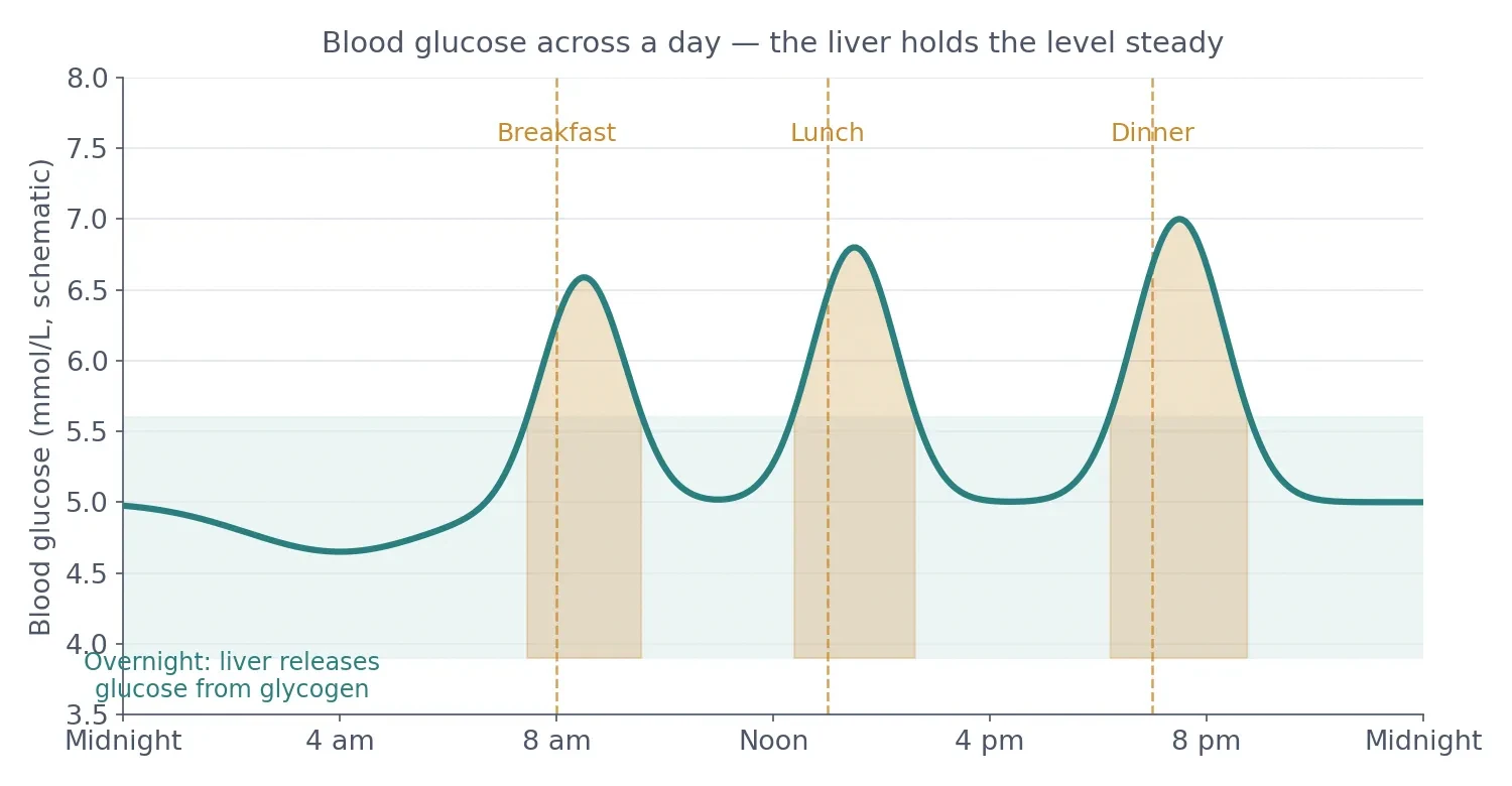
Of all the liver's jobs, the one that keeps you alive between meals is the regulation of blood glucose. Your brain runs almost entirely on glucose, and it cannot store any of it. It needs a steady supply, minute by minute, whether you have just eaten or have not eaten for twelve hours. The liver is the organ that guarantees that supply, and it does so by switching between two modes: after a meal it stores sugar, and between meals it releases it. The switch is thrown by two hormones, insulin and glucagon, and the liver is the main place where their orders are carried out.
9.1 Glycogen: the short-term store
After a meal, blood glucose rises, the pancreas releases insulin, and the liver takes up glucose and chains it into glycogen — a large, branched molecule that is essentially glucose in storage form. The liver holds roughly 100 grams of glycogen, about 6 to 10 percent of its weight. That sounds small, and it is: the muscles hold three or four times as much. But muscle glycogen is for the muscles alone — muscle cells lack the enzyme that frees glucose back into the blood. Liver glycogen is the only store that can be released for the rest of the body, which makes those 100 grams disproportionately important. At the rate the body uses glucose at rest, the liver's glycogen store lasts roughly a day of fasting before it runs low, and that window is the body's first line of defence against low blood sugar.
9.2 Glycogenolysis and gluconeogenesis
When blood glucose falls — between meals, during exercise, overnight — the pancreas releases glucagon, and the liver answers in two ways. First, glycogenolysis: the glycogen chains are cut apart and glucose is released into the blood. That is the fast response, and it works within minutes. Second, gluconeogenesis: the liver builds new glucose from scratch, from lactate, glycerol and amino acids. That is the slow response, and it takes over as glycogen runs down. The two processes overlap and hand over to each other seamlessly, which is why blood sugar stays within a narrow band through a night's sleep even though no food has arrived for hours. Claude Bernard, the nineteenth-century French physiologist, discovered the liver's sugar-making role in the late 1840s and isolated the stored substance in the 1850s, naming it glycogen — literally, "sugar former" — after showing that a liver removed from the body still released sugar into its veins. It was one of the first proofs that an organ could manufacture a substance rather than merely filtering blood, and it changed how physiology understood the liver.
9.3 What happens when the system is strained
The glucose system works beautifully until it is overloaded. In type 2 diabetes, the liver becomes resistant to insulin's signal to stop releasing glucose, so it keeps making sugar even when blood sugar is already high — a major reason fasting glucose rises in diabetes. In prolonged starvation, the liver's gluconeogenesis keeps the brain fed while the body burns fat, and the ketone bodies the liver makes from fat become an alternative fuel. In liver failure, the opposite happens: the liver cannot store or release glucose reliably, and low blood sugar becomes a danger. Sciory's articles on type 2 diabetes and on how exercise changes the body follow the insulin and muscle sides of the same story; the liver is the third partner, and how the liver works through the night is what keeps the other two fed.
- Protein and Fat: Building, Shipping and Storing
If sugar is the liver's daily balancing act, protein and fat are its factory output. The liver is the main site of plasma protein synthesis — almost every protein floating in the blood that is not made by immune cells is made by hepatocytes — and it is the body's central clearing house for cholesterol and fat. The amino acids arriving from the gut through the portal vein are the raw material, and the liver decides their fate: build them into proteins, burn them for energy, or convert them into glucose and urea.
10.1 Albumin and the proteins of the blood
The most abundant protein the liver makes is albumin, which accounts for more than half of all the protein in blood plasma. Albumin does two things: it carries things — hormones, fatty acids, medicines, bilirubin — that do not dissolve well in water, and it holds water in the bloodstream by osmosis, keeping fluid from leaking into the tissues. When the liver fails and albumin falls, fluid leaks out, and the belly and legs swell — the ascites and oedema that are among the most visible signs of advanced liver disease. The liver also makes most of the clotting factors — factors II, V, VII, IX and X, and fibrinogen — which is why liver failure causes bleeding as well as swelling: the blood simply cannot clot properly. It makes angiotensinogen, the starting point of the blood-pressure system, and a range of transport proteins, including transferrin for iron and the binding proteins for thyroid and steroid hormones. The blood tests that measure albumin and clotting, described in section 15, are therefore direct windows onto the liver's factory output.
10.2 Cholesterol: made, used and shipped
The liver is the body's main cholesterol factory. Cholesterol is often talked about as something that arrives in food, but the liver itself is the body's main cholesterol factory, building it from the building block acetyl-CoA. The liver uses cholesterol for three things: it builds it into cell membranes, it turns it into bile acids, and it ships it out to the rest of the body packaged in lipoproteins. The best-known of those packages are VLDL, which carries triglycerides out to the tissues, LDL, which delivers cholesterol to cells, and HDL, which collects cholesterol and brings it back to the liver. That return journey — reverse cholesterol transport — is the reason HDL is called "good" cholesterol: it is the shuttle that brings cholesterol home for disposal as bile acids. Statins, the most widely prescribed cholesterol drugs, work by blocking the liver's cholesterol-making enzyme, HMG-CoA reductase, which forces the liver to pull more LDL out of the blood — a neat example of a drug that works by talking to the liver's factory rather than to the blood itself.
10.3 Fat storage and the liver's own supply
The liver stores some fat, but it is not designed to hold much of it. Hepatocytes normally contain only small fat droplets. When fat accumulates — because too many calories arrive, because alcohol forces fat-making, or because insulin resistance drives fat into the liver — the droplets grow, the cells swell, and the organ begins the journey toward fatty liver disease described in section 14. The liver also makes the fat it needs from excess sugar and protein, a process called de novo lipogenesis, which is why a diet high in sugar can fill the liver with fat even when the diet itself is low in fat. It is a point worth remembering: the liver does not care whether the excess arrived as fat, sugar or alcohol. It converts what it cannot use into fat and stores it, and the storage has a limit.
Protein | What it does | What happens when the liver fails |
|---|---|---|
Albumin | Carries hormones, drugs and bilirubin; holds water in the blood | Fluid leaks into the belly and legs |
Clotting factors (II, V, VII, IX, X) and fibrinogen | Build blood clots | Bleeding and bruising; clotting tests lengthen |
Angiotensinogen | Starting point for blood-pressure control | Blood-pressure regulation weakens |
Transferrin and binding proteins | Carry iron, thyroid hormone and steroid hormones | Iron and hormone transport falter |
Complement proteins and acute-phase proteins | Part of the immune response and inflammation | Infection defence weakens |
Table 4: Some of the proteins the liver makes, and what goes wrong when it cannot make them.
10.4 The gut connection
None of this happens in isolation from the gut. The nutrients the liver processes arrive through the portal vein, and the bile the liver makes returns to the gut to digest the next meal. The two organs are a loop, and the microbes of the gut are part of it: they ferment fibre into short-chain fatty acids that travel to the liver, they modify bile acids, and their fragments arrive in portal blood where Kupffer cells meet them. Sciory's articles on how the digestive system works and on the gut microbiome describe the other half of that loop. The liver is the gut's downstream partner, and the health of one shows up in the other.
11. Bilirubin and Jaundice: When the Yellow Signal Appears
Red blood cells live for about 120 days, and when they die, their haemoglobin is dismantled. The iron is recycled, the protein is reused, and the haem ring is converted into bilirubin — a yellow pigment that is toxic if it accumulates and harmless once the liver has dealt with it. The liver's handling of bilirubin is a small, elegant story that explains one of the most visible signs in medicine: jaundice.
11.1 Unconjugated to conjugated
Bilirubin arrives at the liver in its unconjugated form, bound to albumin and insoluble in water. Hepatocytes take it up and conjugate it — attaching glucuronic acid, the same Phase II chemistry from section 6 — which makes it water-soluble. Conjugated bilirubin is then secreted into bile and leaves the body in the stool, where gut bacteria convert it into the pigments that give stool its brown colour. If any step in that chain breaks, bilirubin accumulates in the blood and deposits in the skin and the whites of the eyes, turning them yellow. That yellowing is jaundice, and its cause depends on where the chain broke.
11.2 Three places it can break
If too many red blood cells are destroyed at once, the liver cannot keep up, and unconjugated bilirubin rises — the pattern of haemolytic anaemia. If the liver cells themselves are damaged, they cannot take up and conjugate bilirubin properly, and both forms rise — the pattern of hepatitis. If the bile duct is blocked, conjugated bilirubin that the liver has already processed cannot leave, and it backs up into the blood — the pattern of obstruction, with pale stools and dark urine as companions. Doctors use the split between the two forms of bilirubin to tell these apart, which is why a bilirubin result is reported in two parts. Gilbert syndrome, a common and harmless genetic variation affecting a few percent of the population, slows conjugation slightly and causes mild, intermittent jaundice, usually noticed during illness or fasting — a reminder that small variations in the liver's chemistry are normal, not disease.
11.3 Newborn jaundice
Newborn babies are often jaundiced in the first days of life, and the reason is developmental: their livers are still switching on the conjugation enzyme, and they are breaking down a large number of fetal red blood cells at the same time. In most babies the jaundice is mild and fades as the enzyme matures. In a few, bilirubin rises high enough to need treatment with light, which changes bilirubin into a form the body can excrete without conjugation. It is the same chemistry as in adults, running on a system that has not yet reached full speed — a small, early lesson in how the liver works, and a good illustration of why Phase II conjugation matters as much as Phase I.
12. The Store Cupboard: Vitamins, Iron and Copper
Beyond sugar, the liver is the body's long-term store cupboard. It holds vitamins A, D, E and K, and vitamin B12 in quantities that last far longer than any other store in the body — vitamin B12 for months to years, vitamin A for months. It stores iron as ferritin, ready to be released for making red blood cells, and copper, which it packages into the protein ceruloplasmin. These stores are the reason a deficiency can take months or years to appear even after intake falls: the liver is quietly spending its savings. They are also the reason the liver is vulnerable to excess. Vitamin A is stored in stellate cells, and too much vitamin A — usually from supplements rather than food — can injure the liver. Iron overload, whether from the genetic condition haemochromatosis or from repeated transfusions, deposits iron in hepatocytes and damages them. The store cupboard is generous, but it is not infinite in either direction.
12.1 Vitamin D and the liver's quiet contribution
The liver also plays a step in vitamin D activation. Vitamin D made in the skin or taken in food is first hydroxylated in the liver to 25-hydroxyvitamin D, the form measured in blood tests, and then activated further in the kidneys. Liver disease can therefore lower vitamin D levels even when sun exposure and intake are adequate. It is one of many small connections between the liver and the rest of the body that only become visible when the liver falters — and one more reason how the liver works can only be understood alongside the rest of the body.
13. Regeneration: The Organ That Grows Back
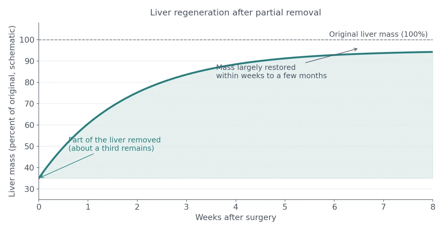
The liver's most famous trick is regeneration. Remove part of a healthy liver — as surgeons do to remove a tumour, or as living donors do when they give part of their liver for transplant — and the remaining tissue grows back. Not by growing a new lobe in the old shape, but by the remaining hepatocytes dividing and enlarging until the liver's mass is restored. In humans the process takes weeks to a few months; in rats, the classic experimental model, it is faster, and figures from rat studies should not be quoted as human timelines. The regrown liver is not a perfect copy of the original — its shape is different — but its mass and function return, which is what matters.
13.1 How regeneration works
Regeneration is not stem cells doing the work. It is ordinary hepatocytes, which in a healthy liver rarely divide, re-entering the cell cycle in large numbers after a signal. The triggers include the growth factors and cytokines released after injury, and the process is tightly controlled: it stops when the liver reaches its target mass, a control system that researchers still do not fully understand. The same machinery runs, in miniature, every day: hepatocytes are constantly renewed at a slow rate, and the liver's cells turn over across months. That slow renewal is part of why the liver tolerates single injuries well — a heavy night, a short illness — and only succumbs to repeated injury over years.
13.2 The limits of regrowth
Regeneration has limits, and they matter. It works when the remaining liver is healthy. It fails when the liver is scarred, because scar tissue replaces the hepatocytes that would have divided, and the architecture that guides regrowth is gone. That is why a person with cirrhosis cannot regenerate the way a healthy donor can, and why surgery that is safe in a healthy liver is dangerous in a scarred one. The Prometheus story — the Titan whose liver was eaten by an eagle each day and regrew each night — is often told as if the Greeks knew about regeneration. It is a myth, not a medical observation, and there is no evidence that its tellers knew the liver could regrow. The real story is more impressive than the myth: an organ that can rebuild itself from a fraction, provided it is healthy enough to do so.
13.3 Living-donor transplantation
Regeneration is what makes living-donor liver transplantation possible. A healthy donor gives a portion of their liver — usually the right lobe — and both the donor's remaining liver and the recipient's graft grow toward full size. It is one of the few transplants in which the donor's organ regrows, and it is also one of the reasons the liver's regenerative ability is so intensively studied: if researchers could switch that ability on in a diseased liver, or grow liver tissue in the laboratory, the shortage of donor organs would matter less. That work is ongoing, and it is one of the most active frontiers in understanding how the liver works well enough to rebuild it.
14. When the Liver Struggles: Fatty Liver, Hepatitis, Cirrhosis and Cancer
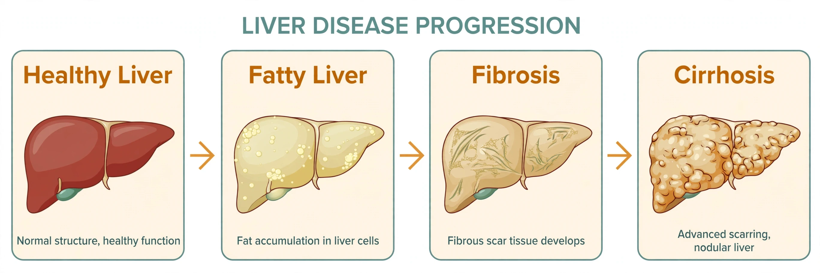
A healthy liver is silent and resilient. A struggling liver is often silent too, which is the danger: liver disease commonly causes no symptoms until it is advanced, and the first sign is often a blood test done for another reason. This section describes the main ways the liver gets into trouble, in the order they usually unfold — fat, inflammation, scarring, failure — and what the latest numbers say about how common each one is.
14.1 Fatty liver: the commonest liver disease
The commonest liver disease in the world today is metabolic dysfunction-associated steatotic liver disease, MASLD — the condition formerly called non-alcoholic fatty liver disease. Fat accumulates in hepatocytes because too many calories arrive, insulin resistance drives fat into the liver, and the liver cannot export it fast enough. The 2025 meta-analysis cited in section 1 put its global prevalence at 33.6 percent of adults — about one in three — and at 70.2 percent among people with type 2 diabetes. In most people, fatty liver causes no symptoms and stays as simple fat accumulation. In some, the fat triggers inflammation — a stage called metabolic dysfunction-associated steatohepatitis, or MASH — and in a smaller share that inflammation leads to fibrosis. The encouraging fact, and the reason early detection matters, is that the early stages are reversible: weight loss, exercise and better metabolic control can clear the fat and quiet the inflammation. The dedicated article on fatty liver follows the diet and lifestyle side of that story in detail.
14.2 Hepatitis: five viruses, one organ
Hepatitis means inflammation of the liver, and its best-known causes are five viruses, A through E. Hepatitis A and E spread through contaminated food and water and usually cause short illnesses that do not become chronic — although hepatitis E can become chronic in people with weakened immune systems, such as organ-transplant recipients. Hepatitis B and C spread through blood and body fluids and can become chronic, silently damaging the liver for decades; both are now preventable or curable — B by vaccination, C by antiviral drugs that cure the infection in a matter of weeks. Hepatitis D only infects people who already have hepatitis B. Together, viral hepatitis remains a major global killer, mostly through the cirrhosis and liver cancer that chronic infection causes. Other causes of hepatitis include alcohol, some medicines and supplements, and the immune system attacking the liver itself — autoimmune hepatitis — which is rarer but important because it is treatable when recognised.
14.3 Fibrosis, cirrhosis and portal hypertension
Whatever injures the liver repeatedly — fat, alcohol, viruses, immune attack — converges on the same pathway. Activated stellate cells lay down scar tissue, a process called fibrosis. Early fibrosis is reversible if the cause is removed. Advanced fibrosis is not, and when scar tissue replaces large areas of the organ and distorts its architecture, the result is cirrhosis. In cirrhosis, blood flow through the liver is obstructed, pressure rises in the portal vein — portal hypertension — and blood finds detours around the liver, through veins in the oesophagus and stomach that can bleed dangerously. The liver's synthetic work fails at the same time: albumin falls, clotting factors fall, bilirubin rises, ammonia rises. The Global Burden of Disease study published in 2025 estimated that cirrhosis killed 1.43 million people in 2021, and the US National Institute of Diabetes and Digestive and Kidney Diseases reported in 2025 that about one in 400 adults in the United States lives with cirrhosis. Those are large numbers for a condition that usually announces itself late.
14.4 Liver cancer
Most liver cancer is hepatocellular carcinoma, HCC, which arises from hepatocytes themselves and accounts for roughly three-quarters to 85 percent of primary liver cancers. It almost always develops in a liver that is already damaged — by cirrhosis, chronic hepatitis B or C, or increasingly by MASLD. The GLOBOCAN 2024 figures quoted in section 1 — 843,045 new cases and 732,489 deaths in 2024 — put liver cancer seventh among cancers by new cases and third by deaths worldwide. The numbers are sobering, and they are also the reason surveillance matters: people with cirrhosis are offered regular ultrasound scans, because a cancer caught small can sometimes be treated, and one caught late usually cannot. No single statistic in this section should be read as a prediction about any individual reader — these are population figures, and individual risk depends on the cause, the stage and the care received.
14.5 Transplantation and surveillance
When the liver fails beyond repair, the only treatment that replaces it is transplantation — and the liver is unusual among transplanted organs because a living person can donate part of one. The donor's liver regrows, the recipient's graft grows, and both end up with a working organ. Deceased-donor transplantation is more common, and organs are allocated by scoring systems built largely from the bilirubin, INR and creatinine numbers of section 15 (the current MELD 3.0 score also adds albumin and sex), which is why those three tests carry so much weight. Demand far exceeds supply, which is why prevention and early treatment matter so much: every case of fatty liver caught early, every hepatitis infection cured, every person who stops drinking before cirrhosis develops is one fewer person on a waiting list. People who already have cirrhosis are offered surveillance — regular ultrasound scans, usually every six months — because liver cancer caught small can sometimes be treated with surgery, ablation or transplant, while cancer caught late usually cannot. Surveillance is not a cure, but it is the best tool medicine has for a cancer that rarely causes symptoms until it is advanced.
15. How Liver Health Is Measured: Liver Function Tests
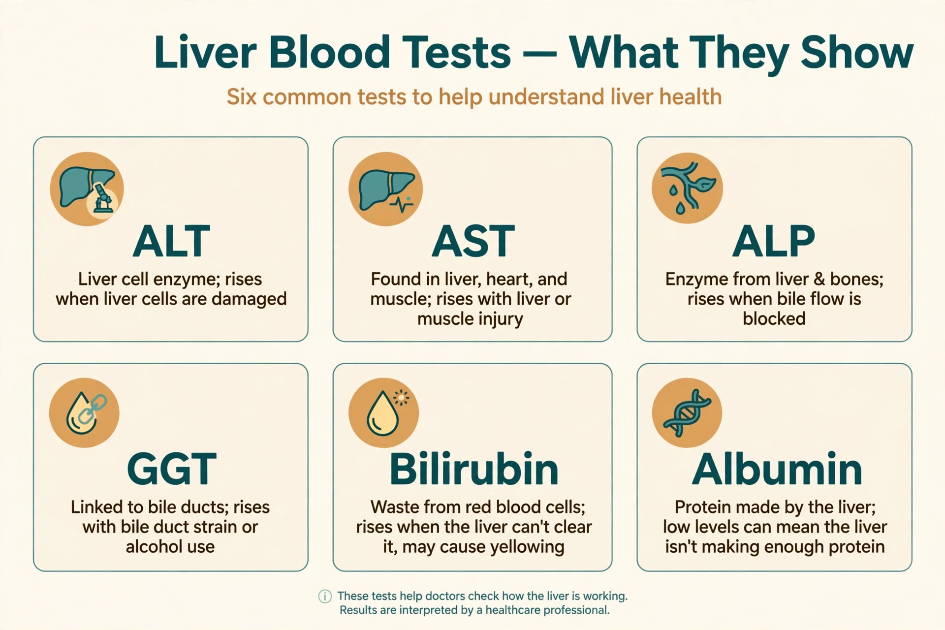
Liver function tests, usually called LFTs, are among the commonest blood tests in medicine — and the name is slightly misleading, because most of them do not measure function at all. They measure damage. The distinction matters, because a person can have abnormal LFTs and a liver that still works, or normal LFTs and a liver that is already scarred. The tests are clues, not verdicts, and they are read as a pattern, not one by one.
15.1 ALT and AST: the leakage enzymes
Alanine aminotransferase, ALT, and aspartate aminotransferase, AST, are enzymes that live inside hepatocytes. When liver cells are injured and their membranes leak, the enzymes spill into the blood and their levels rise. ALT is the more liver-specific of the two; AST is also found in muscle and heart, so a high AST with a normal ALT points away from the liver. A large rise suggests acute injury, such as viral hepatitis or a drug reaction. A small, persistent rise suggests ongoing low-grade damage, such as fatty liver. But the size of the rise does not measure how much liver is left — it measures how much is being injured right now — which is why a very damaged, burnt-out liver can have nearly normal ALT.
15.2 ALP and GGT: the bile duct enzymes
Alkaline phosphatase, ALP, and gamma-glutamyl transferase, GGT, rise when bile flow is obstructed or when the cells lining the bile ducts are irritated. ALP is also made by bone, so a high ALP alone can mean bone disease; a high ALP together with a high GGT points to the liver and bile ducts. GGT is sensitive but not specific — it rises with alcohol use and with many medicines — so it is used mainly to confirm that a raised ALP is hepatic in origin rather than as a test on its own. Together, ALT and ALP let doctors distinguish the two main patterns of liver injury: hepatocellular, in which ALT dominates, and cholestatic, in which ALP dominates.
15.3 Bilirubin, albumin and INR: the function tests
The tests that genuinely reflect function are the ones that measure what the liver makes or clears. Bilirubin measures clearance: a high level means the liver is not conjugating or excreting it properly. Albumin measures synthesis: a low level, in the absence of other causes, means the liver is not making enough protein. INR, the international normalised ratio, measures clotting: it lengthens when the liver cannot make enough clotting factors. These three are the ones that carry the most weight in assessing how sick a liver is, and they are components of the scoring systems used to prioritise liver transplants. Normal ranges for all of these tests vary between laboratories, so a result should always be read against the range printed on the report, not against a figure remembered from an article.
Test | What it measures | What a high or low result suggests |
|---|---|---|
ALT | Enzyme leaking from injured hepatocytes | High: liver cell injury — hepatitis, fatty liver, drug injury |
AST | Enzyme from liver, muscle and heart | High: liver or muscle injury; read with ALT |
ALP | Enzyme from bile ducts and bone | High: bile duct obstruction or bone disease |
GGT | Enzyme from bile duct cells | High: bile duct irritation, alcohol use, some medicines |
Bilirubin | Pigment the liver clears | High: jaundice — haemolysis, liver damage or obstruction |
Albumin | Protein the liver makes | Low: reduced synthesis in advanced liver disease |
INR | How quickly blood clots | Long: the liver is not making enough clotting factors |
Table 5: Liver function tests at a glance. ALT and AST signal injury; ALP and GGT signal bile duct trouble; bilirubin, albumin and INR reflect function.
15.4 What one abnormal result does not mean
A single abnormal LFT is not a diagnosis. Liver enzymes rise after a heavy meal, after vigorous exercise, during a viral illness, with many common medicines, and for no identifiable reason at all — and they often return to normal on repeat testing. Doctors therefore repeat the tests, look at the pattern, ask about alcohol, medicines, supplements and viral risks, and only then decide whether imaging or further tests are needed. If you have ever had a mildly raised ALT and been told to come back in a few weeks for a repeat, that is why. The liver's chemistry is sensitive, and a single reading is a snapshot, not a sentence — which is why how the liver works is judged by patterns, not by one number.
16. A Day in the Life of the Liver: How the Liver Works Around the Clock
The liver never stops, but it does not do the same thing all day. Its work follows the rhythm of meals, sleep and waking, and following one ordinary day through the organ is the fastest way to see how the liver works as a whole — not as a list of functions, but as a schedule.
16.1 Overnight: the long fast
At three in the morning, you have not eaten for eight hours. Your gut is empty, your blood sugar would be falling, and your brain still needs glucose. The liver is in release mode. Glycogenolysis is running, cutting glucose off the glycogen chains stored after dinner, and as those stores thin out, gluconeogenesis takes over, building new glucose from lactate and amino acids. Insulin is low, glucagon is high, and the liver is the only organ standing between your brain and an empty tank. At the same time, the liver is clearing the last of the evening's alcohol and medicines, finishing the detoxification work that began hours earlier, and packaging the day's bilirubin into bile. The bile, with no meal to meet, is diverted into the gallbladder, where it waits, concentrated, for breakfast.
16.2 Breakfast: the flood arrives
Breakfast changes everything within minutes. Glucose, amino acids and fats arrive through the portal vein in a rush, and the liver switches from release to storage. Insulin rises, glucagon falls, and hepatocytes begin chaining glucose into glycogen, rebuilding the store that the night consumed. Amino acids are sorted: some are built into albumin and other proteins, some are burned for energy, and the ammonia left over from the rest is fed into the urea cycle. Fats are packaged into VLDL particles and shipped out, or stored as small droplets inside the hepatocytes. The gallbladder contracts, bile flows into the duodenum, and fat digestion begins. If breakfast included coffee, the liver's CYP1A2 enzyme starts dismantling the caffeine — which is why a cup of coffee has a different effect on different people, depending on how fast their version of that enzyme works.
16.3 Midday and afternoon: steady processing
Through the middle of the day, the liver settles into its steady state: processing lunch, topping up glycogen, making bile, clearing medicines, and keeping blood glucose level between meals. This is also when the liver does much of its protein manufacturing, turning over albumin and clotting factors at a constant rate. The work is unglamorous and invisible, which is exactly the point. A healthy liver in the middle of an ordinary day is doing hundreds of small jobs at once, none of them dramatic, all of them necessary — and the only sign that any of it is happening is that nothing goes wrong. Blood sugar stays level, the blood clots when it should, bile arrives when fat does, and the brain never notices the organ that is feeding it.
16.4 Evening: the hardest shift
Evening is when the liver works hardest, because evening is when people eat the largest meal, drink alcohol and take medicines. A large dinner sends the biggest nutrient flood of the day through the portal vein. Alcohol, if there is any, is given priority over everything else: fat burning pauses, sugar regulation shifts, and the liver's detoxification machinery is monopolised until the ethanol is cleared. A headache tablet taken at bedtime is dismantled overnight, its safe metabolites leaving in urine while its reactive by-products are caught by glutathione. By midnight, the cycle is complete and the liver is back to its overnight role — releasing glucose, clearing waste, preparing for the morning. The organ has run a full day without a break, without a complaint and without being noticed. That is how the liver works, day after day, for a lifetime.
17. Looking After Your Liver: What the Evidence Supports
The liver asks for very little, and the evidence for what helps it is refreshingly ordinary. There is no secret food, no supplement and no ritual that the research supports. What the research supports is an unglamorous list:
Keep body weight in a healthy range.
Move regularly.
Drink little or no alcohol.
Avoid unnecessary medicines and supplements.
Get vaccinated against hepatitis A and B where that is recommended. Each of those has a mechanism in this article behind it. Weight and movement reduce the fat arriving at the liver and improve insulin sensitivity, which is the core driver of MASLD. Less alcohol means less acetaldehyde and less fat-making in hepatocytes. Fewer unnecessary medicines mean fewer compounds for Phase I and Phase II to process. Vaccination prevents the chronic infections that cause cirrhosis and cancer.
17.1 The heart, the hormones and the immune system
The liver does not work alone, and looking after it means looking after its partners. The heart supplies the liver's oxygen and receives its output, and the blood-pressure system that starts with the liver's angiotensinogen is a shared project between the two organs — Sciory's article on how the human heart works follows the cardiac side. The hormones that tell the liver when to store and when to release sugar come from the pancreas and the adrenal glands, and the endocrine system article follows those signals. The immune cells that guard the liver — the Kupffer cells and the complement proteins the liver itself makes — are part of the wider immune system described in the article on how the human immune system works. A liver problem is rarely only a liver problem, and a healthy liver is usually the product of a healthy system, not a single habit.
17.2 When to seek care
This article cannot tell any individual reader whether their liver is healthy, and it does not try. But the general information is worth stating plainly: liver disease is often silent, so the people most likely to benefit from a conversation with a clinician are those with risk factors:
Heavy alcohol use.
Type 2 diabetes.
Obesity.
A family history of liver disease.
A history of hepatitis.
Long-term use of medicines known to affect the liver. The conversation itself is simple: a history, an examination, blood tests, and, if needed, an ultrasound or a scan that measures liver stiffness. Early liver disease is often reversible. Late liver disease often is not. The gap between those two sentences is the reason the subject matters, and it is the reason this article exists.
18. Common Mistakes and Misconceptions
Most people assume the liver is a filter, like the filter in a coffee machine, that fills up with toxins and needs to be cleaned out. What the anatomy actually shows is different: the liver is a chemical factory that dismantles molecules and sends the pieces out, and it never fills up in the way a filter does. That single misunderstanding drives most of the others, so it is worth taking them one at a time.
18.1 "A detox tea will clean my liver"
No product has been shown to clean the liver, and the National Center for Complementary and Integrative Health states that detox diets and cleanses lack evidence for removing toxins. The liver cleans itself by chemistry, continuously, and a tea cannot add enzymes it does not have. Worse, some herbal and dietary supplements have caused liver injury themselves, including cases of acute liver failure. If a product promises to detox the liver, the promise itself is the warning sign.
18.2 "Liver pain means my liver hurts"
The liver itself has few pain nerves. Pain in the upper right abdomen usually comes from the capsule around the liver stretching, from the gallbladder, or from neighbouring structures — not from liver cells complaining. That is why liver disease is so often painless until it is advanced, and why pain is a poor guide to liver health. A normal-feeling abdomen says nothing about the liver inside it.
18.3 "The liver filters blood like the kidneys"
The kidneys filter blood: they pass it through a sieve and keep what is needed. The liver does something different: it changes molecules chemically. It does not filter; it transforms. Blood passes through the liver's sinusoids slowly so that hepatocytes can take molecules out and alter them, not so that the blood can be sieved. The distinction is not pedantic — it is the reason kidney failure and liver failure look and feel completely different.
18.4 "One heavy night damages the liver"
A single heavy night can injure the liver acutely — alcohol is toxic to hepatocytes, and a large dose can cause inflammation — but the liver recovers from single insults remarkably well. What it cannot recover from is repetition: years of heavy drinking, years of excess calories, years of viral infection. Liver disease is a disease of repeated injury and failed repair, not of single events. That is both reassuring and serious: reassuring, because one mistake is rarely the end of the story, and serious, because the pattern is what counts.
18.5 "Natural supplements are safe for the liver"
Natural does not mean harmless. Many plant compounds are handled by the same cytochrome P450 enzymes as prescription drugs, and some — including certain herbal weight-loss and bodybuilding products — have caused severe liver injury. The liver treats a herbal compound exactly as it treats a drug: it dismantles it, and if the dismantling creates a reactive intermediate, the damage is the same. Anyone taking supplements regularly, especially in combination with prescription medicines, should mention them to a clinician — not because supplements are always dangerous, but because the liver cannot tell a natural molecule from a synthetic one.
18.6 "A normal liver test means a healthy liver"
As section 15 explains, liver function tests measure injury and some aspects of function, but they do not measure scarring. A person with established fibrosis can have normal ALT, and a person with a high ALT can have a liver that recovers fully. Normal tests are reassuring, not conclusive, and they are one piece of a larger picture that includes risk factors, examination and imaging.
19. Frequently Asked Questions
The questions below are the ones readers ask most often about how the liver works, answered in brief.
19.1 How does the liver work?
How the liver works, in one sentence, is this: blood from the gut arrives through the portal vein, flows slowly past plates of liver cells in tiny hexagonal lobules, and those cells process everything in it — storing sugar as glycogen, building proteins, making bile, converting ammonia to urea, and dismantling drugs and alcohol in two chemical phases — before the cleaned blood leaves through the hepatic veins. The full answer is the article above: the anatomy of section 2, the lobule of section 3, the six jobs of section 4, and the chemistry of sections 5 to 10.
19.2 What does the liver do?
The liver does more than 500 jobs, according to Johns Hopkins Medicine, and they fall into six groups: processing nutrients from each meal, making bile, detoxifying drugs and alcohol, making proteins for the blood, storing glycogen, vitamins and iron, and regulating blood sugar, cholesterol and amino acids. No other single organ covers that range, which is why liver failure affects so many systems at once.
19.3 How does the liver detoxify?
The liver detoxifies in two phases. In Phase I, cytochrome P450 enzymes alter fat-soluble compounds, usually by oxidation. In Phase II, conjugation reactions attach a water-soluble group — glucuronic acid, sulphate, glycine or glutathione — making the compound soluble enough to leave in urine or bile. A third step, transport out of the cell, completes the pipeline. No tea, juice or cleanse has been shown to speed this up.
19.4 What is bile?
Bile is a greenish-yellow fluid the liver makes — roughly 600 millilitres to 1 litre a day — that digests fat and carries waste out of the body. Its bile salts act like soap, breaking fat globules into droplets so enzymes can digest them. Bile is stored and concentrated in the gallbladder between meals, and about 95 percent of its salts are reabsorbed in the gut and recycled to the liver.
19.5 Can the liver regenerate itself?
Yes. If part of a healthy liver is removed, the remaining hepatocytes divide and the liver grows back to its full mass over weeks to a few months in humans. It regrows mass and function, though not the original shape. Regeneration fails when the liver is scarred, because scar tissue replaces the cells that would divide — which is why cirrhosis is so serious and why living-donor transplantation works only with a healthy donor liver.
19.6 How much alcohol can the liver handle?
The liver works through alcohol at a steady pace — roughly a single standard drink each hour for a typical adult — and coffee, cold air and exercise do not hurry it. Drinking faster than that means alcohol and its toxic by-product acetaldehyde accumulate. Occasional moderate drinking is cleared without lasting harm in most healthy people, but repeated heavy drinking overwhelms the system, fills hepatocytes with fat, and over years can lead to inflammation, fibrosis and cirrhosis. The safe amount varies between people, and this article gives no individual limit.
19.7 What do liver function tests show?
Liver function tests show two things: injury and function. ALT and AST rise when liver cells are damaged; ALP and GGT rise when bile flow is obstructed; bilirubin, albumin and INR show how well the liver is clearing and making things. A single abnormal result is not a diagnosis — tests are repeated, read as a pattern, and interpreted alongside risk factors and imaging. Normal ranges vary between laboratories.
20. Sources
Johns Hopkins Medicine — Liver Health. https://www.hopkinsmedicine.org/health/conditions-and-diseases/liver-health
Cleveland Clinic — Liver: Anatomy, Function, Location and Conditions. https://my.clevelandclinic.org/health/body/21481-liver
OpenStax, Anatomy and Physiology 2e — 23.6 Accessory Organs in Digestion: The Liver, Pancreas, and Gallbladder. https://openstax.org/books/anatomy-and-physiology-2e/pages/23-6-accessory-organs-in-digestion-the-liver-pancreas-and-gallbladder
Johns Hopkins Medicine — Detoxing Your Liver: Fact Versus Fiction. https://www.hopkinsmedicine.org/health/expert-qa/detoxing-your-liver-fact-versus-fiction
National Center for Complementary and Integrative Health — "Detoxes" and "Cleanses": What You Need To Know. https://www.nccih.nih.gov/health/detoxes-and-cleanses-what-you-need-to-know
National Institute of Diabetes and Digestive and Kidney Diseases — Cirrhosis: Definition and Facts. https://www.niddk.nih.gov/health-information/liver-disease/cirrhosis/definition-facts
National Institute of Diabetes and Digestive and Kidney Diseases — Liver Disease. https://www.niddk.nih.gov/health-information/liver-disease
Mayo Clinic — Liver Function Tests. https://www.mayoclinic.org/tests-procedures/liver-function-tests/about/pac-20394595
MedlinePlus, US National Library of Medicine — Liver Diseases. https://medlineplus.gov/liverdiseases.html
NHS — Liver Disease. https://www.nhs.uk/conditions/liver-disease/
World Health Organization — Hepatitis B Fact Sheet. https://www.who.int/news-room/fact-sheets/detail/hepatitis-b
World Health Organization — Hepatitis C Fact Sheet. https://www.who.int/news-room/fact-sheets/detail/hepatitis-c
Ho, S. Y. et al. — Global Prevalence of Metabolic Dysfunction-Associated Steatotic Liver Disease, Clinical Gastroenterology and Hepatology, 2025. https://doi.org/10.1016/j.cgh.2025.02.006
Duo, Y. et al. — Global, regional, and national burden of cirrhosis and other chronic liver diseases, PLOS One, 2025. https://journals.plos.org/plosone/article?id=10.1371/journal.pone.0328493
Sung, H. et al. — Global Cancer Statistics 2024: GLOBOCAN Estimates of Incidence and Mortality Worldwide for 34 Cancers in 186 Countries, CA: A Cancer Journal for Clinicians, 2026. https://doi.org/10.3322/caac.70090
Krishna, M. — Microscopic Anatomy of the Liver, Clinical Liver Disease, 2013. https://pmc.ncbi.nlm.nih.gov/articles/PMC6448667/
Ceni, E. et al. — Alcohol-Related Liver Disease: From Pathogenesis to Treatment, Journal of Clinical Medicine, 2020. https://pmc.ncbi.nlm.nih.gov/articles/PMC7148483/
Li, Y. et al. — Hepatocellular Carcinoma: Recent Advances, Journal of Clinical Medicine, 2024. https://pmc.ncbi.nlm.nih.gov/articles/PMC11441870/
Thieme — Liver Blood Flow: Physiology, Measurement and Clinical Relevance, Seminars in Interventional Radiology, 2023. https://www.thieme-connect.de/products/ejournals/html/10.1055/s-0043-1767690
Colorado State University — Liver Regeneration. https://vivo.colostate.edu/hbooks/pathphys/digestion/liver/regen.html
US Department of Veterans Affairs — The Liver as a Factory. https://www.hepatitis.va.gov/basics/liver-as-factory.asp
Mayo Clinic — Fatty Liver Disease. https://www.mayoclinic.org/diseases-conditions/nonalcoholic-fatty-liver-disease/symptoms-causes/syc-20354567
Cleveland Clinic — Bile Ducts. https://my.clevelandclinic.org/health/body/24523-bile-duct
Cleveland Clinic — Gallbladder. https://my.clevelandclinic.org/health/body/21690-gallbladder
National Institute on Alcohol Abuse and Alcoholism — Alcohol's Effects on the Body: The Liver. https://www.niaaa.nih.gov/alcohols-effects-health/alcohols-effects-body
PubMed — Ho et al., Global Prevalence of MASLD, 2025. https://pubmed.ncbi.nlm.nih.gov/40204206/
StatPearls — Physiology, Urea Cycle. https://www.ncbi.nlm.nih.gov/books/NBK513323/
StatPearls — Physiology, Glycogen. https://www.ncbi.nlm.nih.gov/books/NBK537329/
OpenStax, Anatomy and Physiology 2e — 24.3 Lipid Metabolism. https://openstax.org/books/anatomy-and-physiology-2e/pages/24-3-lipid-metabolism
OpenStax, Anatomy and Physiology 2e — 24.4 Protein Metabolism. https://openstax.org/books/anatomy-and-physiology-2e/pages/24-4-protein-metabolism
OpenStax, Anatomy and Physiology 2e — 24.2 Carbohydrate Metabolism. https://openstax.org/books/anatomy-and-physiology-2e/pages/24-2-carbohydrate-metabolism
Centers for Disease Control and Prevention — Alcohol Use and Your Health. https://www.cdc.gov/alcohol/about-alcohol-use/index.html
European Association for the Study of the Liver — EASL Clinical Practice Guidelines: Management of MASLD. https://www.journal-of-hepatology.eu/article/S0168-8278(24)00329-5/fulltext
National Health Service — Jaundice. https://www.nhs.uk/conditions/jaundice/
MedlinePlus — Bilirubin Blood Test. https://medlineplus.gov/lab-tests/bilirubin-blood-test/
Mayo Clinic — Cirrhosis. https://www.mayoclinic.org/diseases-conditions/cirrhosis/symptoms-causes/syc-20351487
StatPearls — Physiology, Renin Angiotensin System. https://www.ncbi.nlm.nih.gov/books/NBK470410/
Fuhr, U. et al. — Grapefruit Juice and CYP3A4: Clinical Pharmacology and Therapeutics, 2023. https://doi.org/10.1002/cpt.2968