

Put your hand flat on the centre of your chest. Wait a second. There it is — a quiet, steady thump, thump, thump. You have felt it thousands of times without thinking about it, after climbing stairs, during a scary film, or in the silence before sleep. The first time I really listened to a heartbeat through a stethoscope, I was struck by how unglamorous and how extraordinary it sounded at once: no whirring, no drama, just a small muscle doing a job it has never once taken a day off from. That sound is the engine of the circulatory system, and this article explains how the human heart works, from the first beat to the farthest capillary in your body.
Learning how the human heart works means learning how a fist-sized pump keeps every cell in your body fed, cleaned, and alive. Your heart beats around 100,000 times a day and moves roughly 2,000 gallons of blood through a network of vessels that, laid end to end, would stretch more than 60,000 miles. It does all of this without a single conscious instruction from you. By the end of this article, you will know exactly how that happens — chamber by chamber, valve by valve, beat by beat.
1. What the Circulatory System Actually Is
1.1 A definition that sticks
Your circulatory system — also called the cardiovascular system — is your body's transport network. It has three parts: the heart, the blood vessels, and the blood itself. The heart is the pump. The vessels are the roads. The blood is the fleet of delivery trucks carrying oxygen, nutrients, hormones, and heat out to the tissues, and hauling carbon dioxide and other waste back for disposal. The Cleveland Clinic describes it plainly: your circulatory system moves blood and oxygen through your entire body, day and night, even while you sleep (Cleveland Clinic).
Think of it like a city's water system. A central pumping station pushes water through large mains, which branch into smaller pipes, which finally reach a tap in every single house. If the pumping station stops, every tap in the city runs dry within minutes. Your body works the same way — except the "city" is about 37 trillion cells, and the pumping station never closes.
1.2 The three jobs of the system
The circulatory system does three things, all at the same time:
Delivery. It carries oxygen from your lungs and nutrients from your digestive system to every tissue and organ.
Collection. It picks up carbon dioxide and other waste products and carries them to the lungs, kidneys, and liver for removal.
Balance. It moves heat around the body, carries hormones to their target organs, and helps keep the chemical environment of your blood steady.
I once watched a colleague try to explain circulation to a group of schoolchildren by filling a loop of clear tubing with red water and squeezing a rubber bulb in the middle. The children got it instantly: squeeze, and the water moves; stop squeezing, and everything stops. That rubber bulb was the heart, and it is still the best one-minute demonstration of circulation I have ever seen.
2. Where Your Heart Sits and What It Looks Like
Most people assume the heart sits on the far left of the chest. It does not. It sits in the middle of your chest, tucked between your lungs, just behind the breastbone, with its lower tip — the apex — pointing slightly to the left. That is why a heartbeat feels strongest on the left side, even though most of the organ is central.
Your heart is roughly the size of your own clenched fist, and it weighs about 8 to 12 ounces in an adult. Its shape is often compared to a pinecone: broad at the top, tapering toward the bottom. It sits inside a tough, double-layered sac called the pericardium, which holds it in place and lets it slide smoothly as it beats. A thin layer of fluid inside that sac works like oil in an engine, cutting down friction with every contraction.
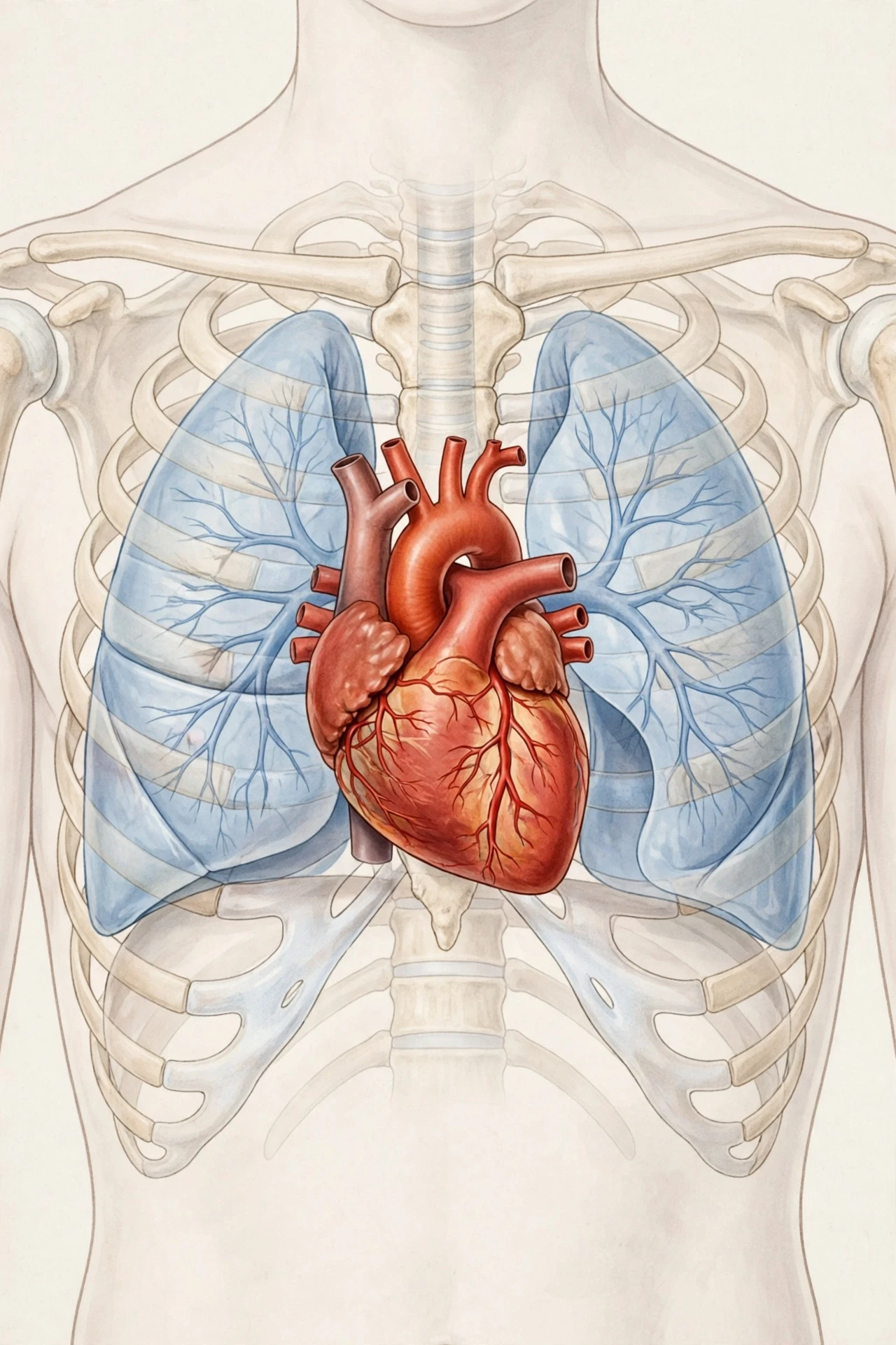
The heart is not a single hollow bag. It is a muscle — the hardest-working muscle you own — divided into chambers, wrapped in layers, and wired with its own electrical system. The outer layer is the epicardium, the thick middle layer of muscle is the myocardium, and the smooth inner lining is the endocardium. When people say someone has a "strong heart," they are mostly talking about the myocardium: the thicker and healthier that muscle, the more forcefully it can push blood out with each beat.
Your heart is about the size of your fist, yet it will beat roughly 3 billion times over a typical lifetime. No machine humans have built runs that long without a single service break.
3. Inside the Heart: Four Chambers, One Team
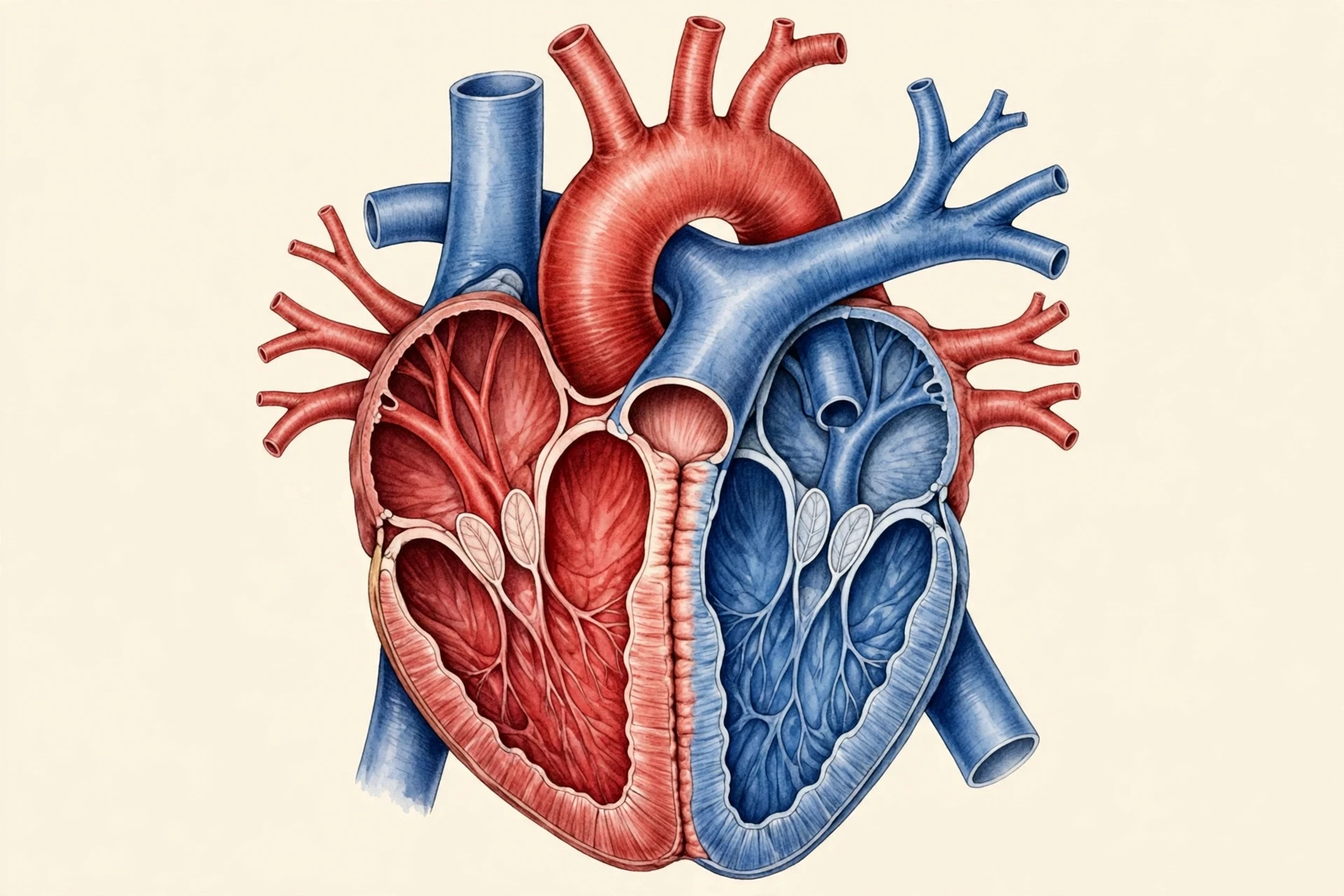
Cut the heart open — in a textbook diagram, at least — and you find four hollow rooms. A muscular wall called the septum divides the heart into a right side and a left side, and each side has an upper chamber and a lower chamber. The two sides never mix their blood. That separation is the whole point of the design.
3.1 The atria: the receiving rooms
The two upper chambers are the atria (singular: atrium). They are the receiving rooms. Blood returning from the body flows into the right atrium; blood returning from the lungs flows into the left atrium. The atria are relatively thin-walled because their job is gentle: they collect blood and give it a small push downward. Think of them as the lobby of a building — people arrive, pause briefly, and move on.
3.2 The ventricles: the pumping rooms
The two lower chambers are the ventricles, and they do the heavy lifting. The right ventricle pumps blood a short distance to the lungs. The left ventricle pumps blood all the way around the body — to your toes, your brain, your fingertips — so its wall is by far the thickest in the heart, roughly three times as thick as the right ventricle's wall. When a doctor talks about a "strong left ventricle," this is the muscle they mean.
The first time I held a plastic model of a human heart in a teaching lab, the left ventricle surprised me most. I expected the two sides to look alike. They do not. One side is a modest pump for a short trip; the other is a thick-walled engine built for a journey around the entire body. The anatomy tells the story before any textbook does.
3.3 Why two sides matter
The right side handles oxygen-poor blood, and the left side handles oxygen-rich blood. Keeping them apart matters because mixing would dilute the oxygen supply. The septum is the wall that keeps the two streams separate, and a hole in it — a condition doctors call a septal defect — is one of the most common heart problems found in newborns. The design is simple, but it has no room for error.
4. How Blood Flows Through the Heart, Step by Step
Blood moves through the heart in one direction only, in a sequence that repeats with every beat. Follow one drop of blood through a full circuit:
Oxygen-poor blood from the body arrives at the right atrium through two large veins, the superior and inferior vena cava.
The right atrium pushes it down into the right ventricle.
The right ventricle pumps it out through the pulmonary artery to the lungs.
In the lungs, the blood drops off carbon dioxide and picks up fresh oxygen.
Oxygen-rich blood returns through the pulmonary veins into the left atrium.
The left atrium pushes it down into the left ventricle.
The left ventricle pumps it out through the aorta, the largest artery in the body, to every tissue and organ.
After delivering oxygen and collecting waste, the blood returns through the veins to the right atrium — and the loop begins again.
Two details catch most people out. First, the pulmonary artery is the only artery in the body that carries oxygen-poor blood, and the pulmonary veins are the only veins that carry oxygen-rich blood. The usual rule — arteries carry oxygen-rich blood, veins carry oxygen-poor blood — holds everywhere except on the short trip to and from the lungs. Second, the whole circuit takes less than a minute: a drop of blood can travel from your heart to your toes and back in roughly sixty seconds.
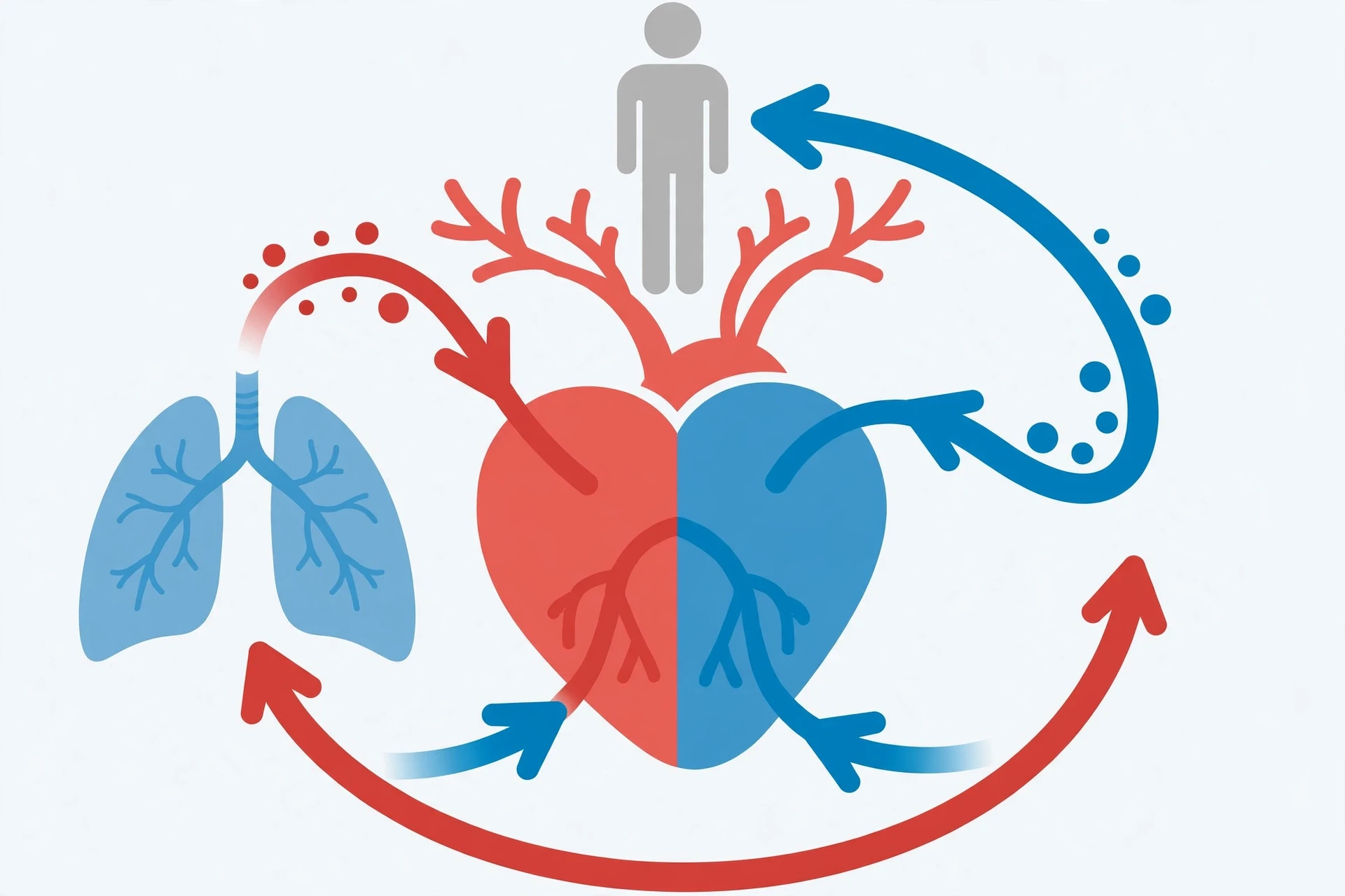
OpenStax Anatomy and Physiology traces this same pathway in detail and confirms the chamber-by-chamber route described here (OpenStax).
5. The Cardiac Cycle: How the Human Heart Works, Beat by Beat
A single heartbeat is not one squeeze. It is a coordinated two-part cycle called the cardiac cycle, and it has two phases.
Systole is the contraction phase. The ventricles squeeze, pressure rises inside them, and blood is forced out — to the lungs from the right side, to the body from the left. Diastole is the relaxation phase. The muscle loosens, pressure falls, and the chambers fill with blood again, ready for the next beat. At a resting heart rate of about 75 beats per minute, one full cycle lasts roughly 0.8 seconds.
Here is the part that surprised me when I first measured it on a heart-rate monitor during a run: the heart spends more of each cycle resting than squeezing. Diastole is the longer phase at rest, and it matters, because the heart muscle itself receives most of its own blood supply during diastole, when the muscle is relaxed and its own vessels are not being squeezed shut. A heart that never rested could not feed itself.
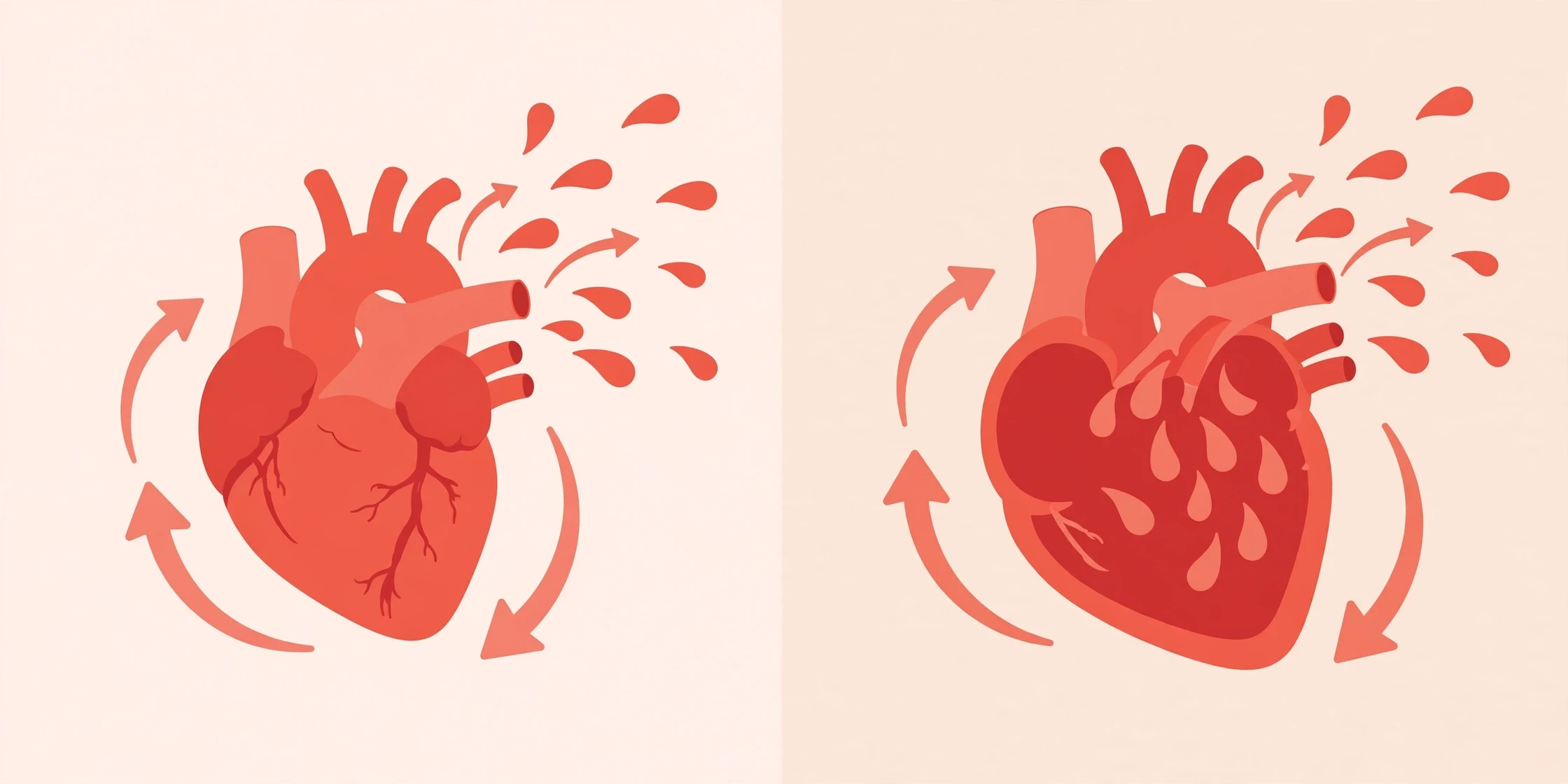
Your resting heart rate usually sits between 60 and 100 beats per minute. Athletes often rest lower, sometimes below 60, because a stronger heart can move the same amount of blood with fewer beats. During exercise, the rate climbs because working muscles demand more oxygen, and the heart answers by beating faster and harder. The U.S. National Heart, Lung, and Blood Institute explains how the heart adjusts its rate and force to match what the body needs (NHLBI).
6. The Valves: Four One-Way Doors
Blood must never flow backward through the heart. Four valves make sure of that, and each one is a set of thin, tough flaps that snap open and shut like doors that only swing one way.
Tricuspid valve — between the right atrium and right ventricle.
Pulmonary valve — between the right ventricle and the pulmonary artery.
Mitral valve — between the left atrium and left ventricle.
Aortic valve — between the left ventricle and the aorta.
When the ventricles contract, the atrioventricular valves (tricuspid and mitral) slam shut, and the semilunar valves (pulmonary and aortic) swing open. When the ventricles relax, the pattern reverses. That double snap is the "lub-dub" you hear through a stethoscope: the first sound is the atrioventricular valves closing, the second is the semilunar valves closing.
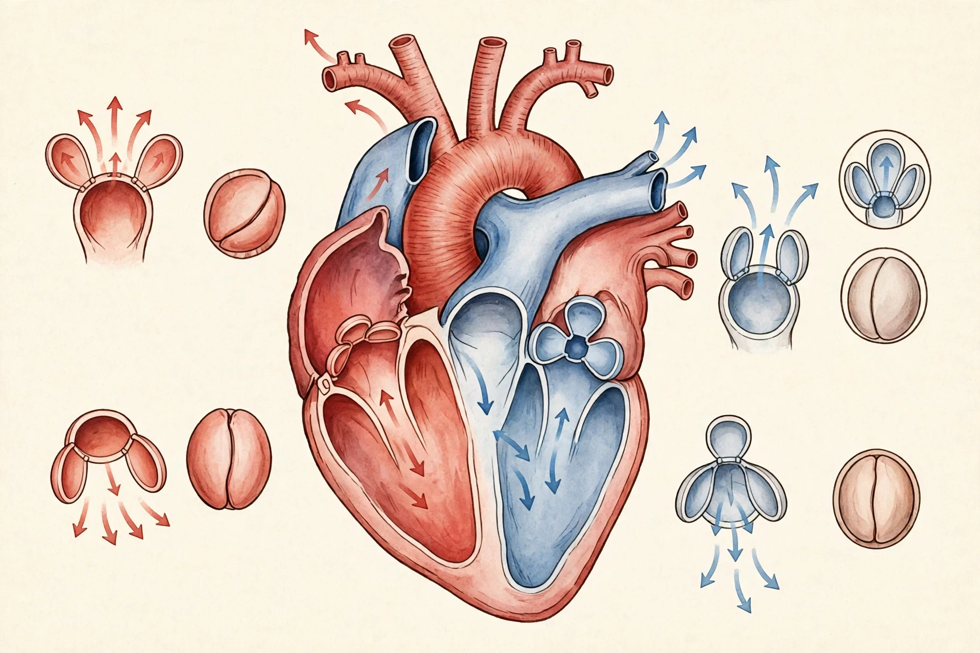
Valves can wear out. They can stiffen and narrow, forcing the heart to push harder, or they can leak, letting blood slosh backward. Either way the heart compensates for a while — it is good at that — and then, gradually, it cannot. This is one reason a heart murmur, the whooshing sound of turbulent blood, is worth a doctor's attention rather than a shrug.
7. The Electrical System: Your Heart's Own Pacemaker
Your heart does not wait for your brain to tell it to beat. It generates its own electrical signals, which is why a heart removed from the body can keep beating for a short time in the right conditions. The system works like a small relay race:
The sinoatrial (SA) node, a cluster of specialised cells high in the right atrium, fires an electrical impulse. This is the natural pacemaker, and it sets the pace — usually 60 to 100 impulses per minute at rest.
The signal spreads across both atria, making them contract and push blood into the ventricles.
The signal pauses briefly at the atrioventricular (AV) node, giving the ventricles time to fill.
It then races down the bundle of His and the Purkinje fibres, making the ventricles contract from the bottom up, squeezing blood out efficiently.
An electrocardiogram (ECG) is simply a recording of this electrical activity, drawn as a wavy line. Each bump on the trace matches one step in the relay. Doctors read that line the way a mechanic reads an engine's sound — a delay, a missing bump, or an extra spike can point to exactly where the wiring has gone wrong.
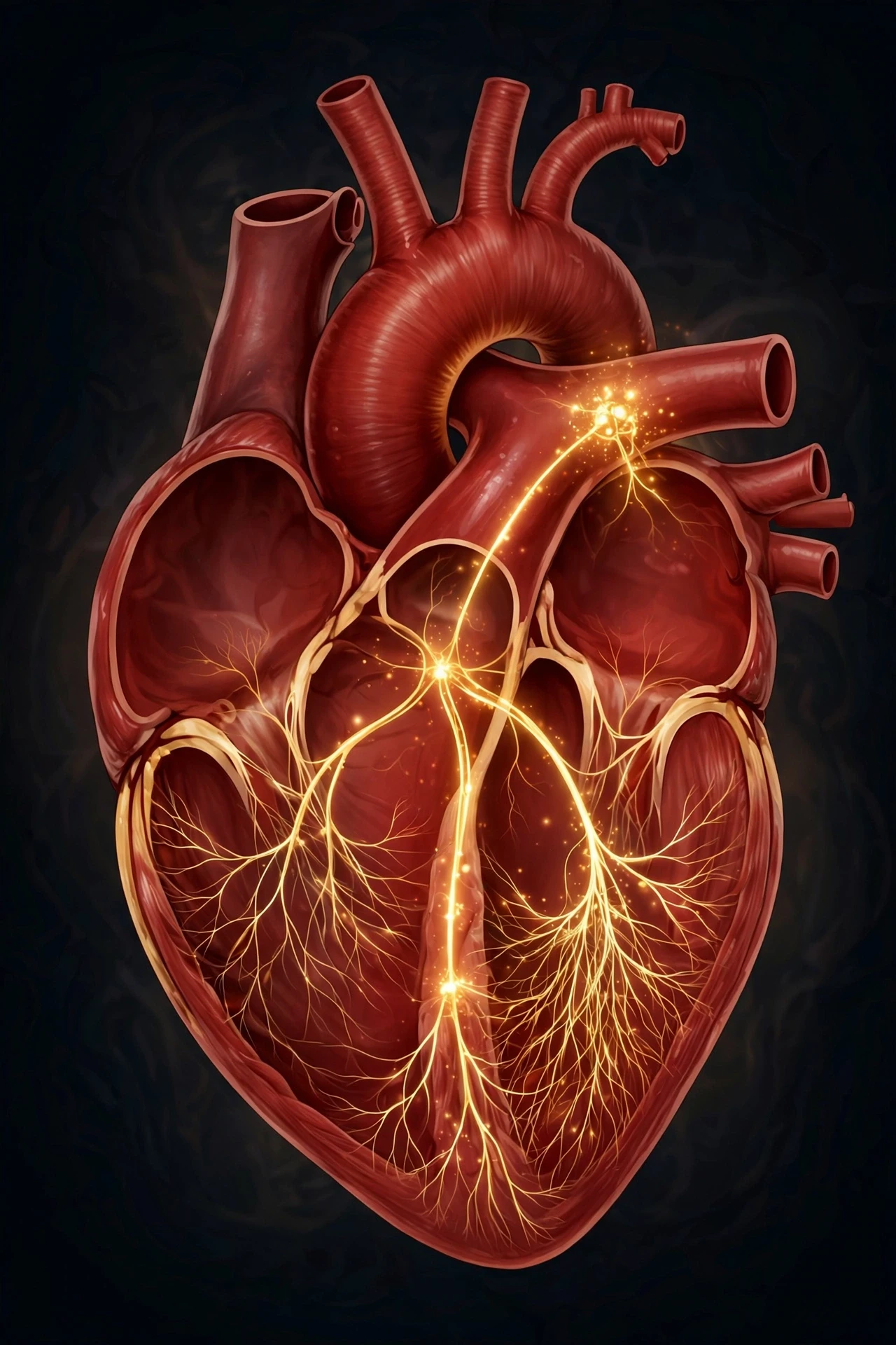
The electrical side of the heart is a good reminder that the body runs on signals as well as plumbing. The heart's wiring is, in its own way, an electrical circuit — a small, self-powered one. If you want to see how electrical signals behave in circuits more generally, our explainer on the fundamentals of electricity covers voltage, current, and resistance in plain language. The heart uses the same basic idea — a signal travelling along a path — inside living tissue.
Your brain can influence the pace through the autonomic nervous system — speeding the heart up when you are frightened or exercising, slowing it down when you rest — but it does not start the beat. The heart starts itself. To see how the brain and heart communicate, read our guide to human brain anatomy and functions.
8. The Blood Vessels: Highways, Streets, and Doorways
If the heart is the pump, the blood vessels are the road network — and like any road network, they come in three sizes, each built for a different job.
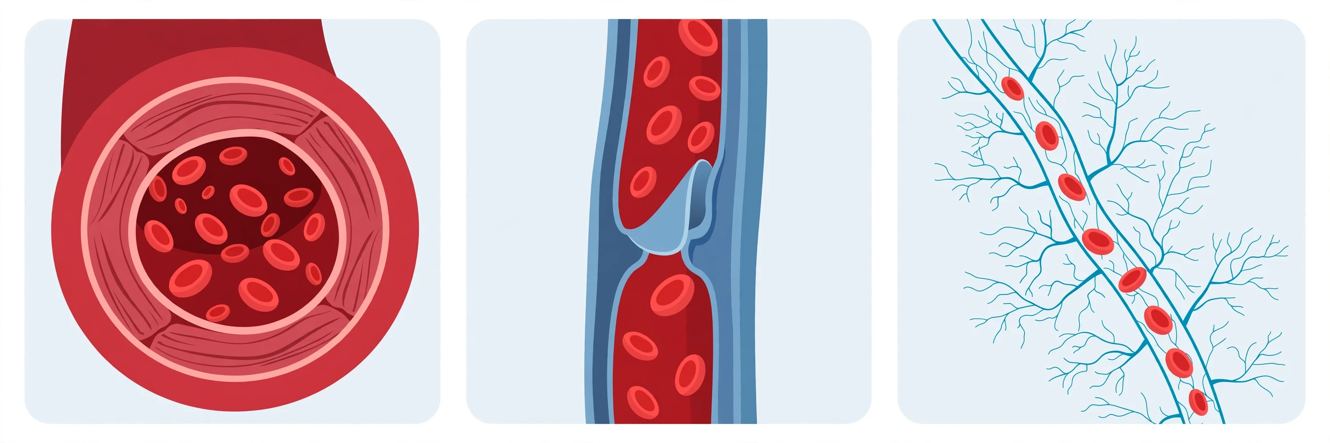
Feature | Arteries | Veins | Capillaries |
|---|---|---|---|
Direction of flow | Away from the heart | Toward the heart | Between arteries and veins |
Wall thickness | Thick, muscular, elastic | Thinner, less muscle | One cell thick |
Pressure inside | High | Low | Very low |
Valves | None (except at the heart) | Yes, to prevent backflow | None |
Main job | Deliver blood under pressure | Return blood to the heart | Exchange oxygen, nutrients, and waste with cells |
Typical diameter | Millimetres to centimetres | Millimetres to centimetres | About the width of a single red blood cell |
Arteries are the highways. Thick, muscular, elastic walls let them stretch with each surge of blood and then spring back, smoothing the flow between beats. The aorta, the largest artery, is about as wide as a garden hose. Arteries branch into smaller arterioles, which act like taps — tightening or loosening to direct more blood to the organs that need it most at any moment.
Veins are the return roads. Their walls are thinner and their pressure is much lower, so many veins — especially in the legs — have one-way valves that stop blood from pooling downward under gravity. The squeezing of your leg muscles as you walk helps push blood back up toward the heart, which is why long periods of sitting still can leave your ankles swollen.
Capillaries are the doorways, and they are where everything that matters actually happens. Their walls are a single cell thick, so thin that oxygen, nutrients, carbon dioxide, and waste can pass straight through them into and out of the surrounding tissue. A red blood cell must fold itself to squeeze through a capillary single file. There are so many capillaries that almost no cell in your body sits more than a fraction of a millimetre from one.
9. Two Loops at Once: Pulmonary and Systemic Circulation
The heart runs two circuits at the same time, and understanding this is the key to understanding the whole system.
Pulmonary circulation is the short loop: right ventricle to lungs and back to the left atrium. Its only job is gas exchange — dropping off carbon dioxide and picking up oxygen. Systemic circulation is the long loop: left ventricle to the entire body and back to the right atrium. Its job is everything else — feeding organs, warming tissues, and collecting waste.
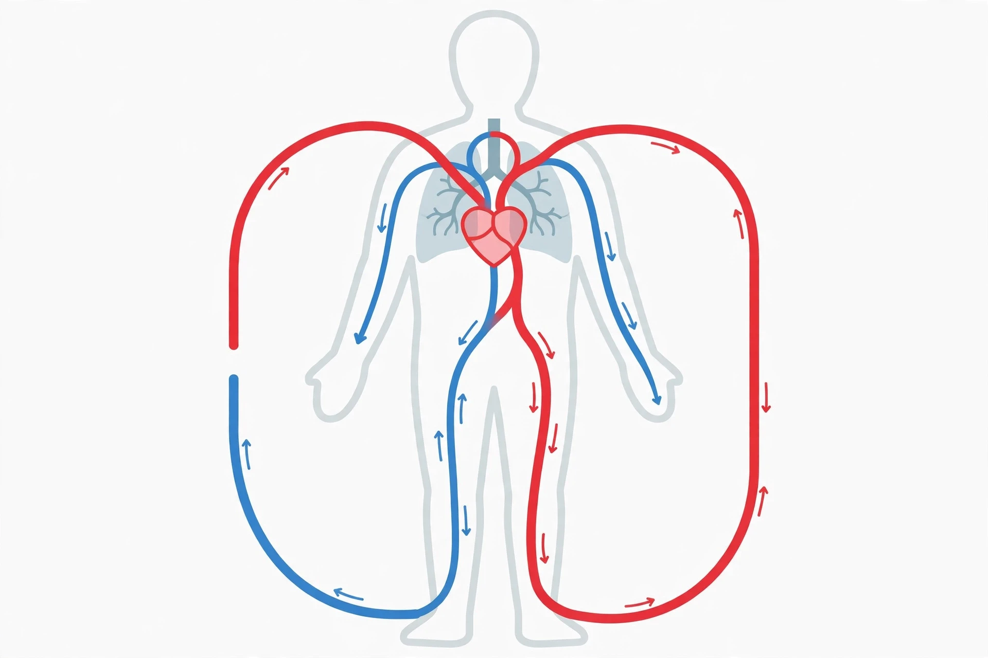
There is also a third, smaller loop that is easy to forget: the coronary circulation. The heart muscle itself needs oxygen, and it gets it from the coronary arteries, which branch off the aorta and wrap around the heart's surface like a crown. When one of these arteries narrows or blocks, part of the heart muscle is starved of oxygen — that is a heart attack. The heart, for all its power, depends on its own small supply line like any other organ.
10. What the Blood Actually Carries
Blood is more than red liquid. Roughly half of it is plasma, a pale yellow fluid that carries proteins, hormones, and nutrients. The other half is cells:
Red blood cells carry oxygen, bound to a protein called haemoglobin, and bring carbon dioxide back for disposal. They have no nucleus, which leaves more room for haemoglobin — a neat piece of biological engineering.
White blood cells defend the body against infection. They are part of the immune system, which works hand in hand with circulation: white blood cells ride the bloodstream to reach infections anywhere in the body. Our article on how the human immune system works explains that partnership in detail.
Platelets are tiny cell fragments that rush to a cut and clump together to form a clot, sealing the leak before too much blood escapes.
Each cell in your body depends on this delivery service. A muscle cell needs oxygen to burn fuel, a skin cell needs nutrients to repair itself, and every cell needs its waste removed. The exchange happens in the capillaries, one cell at a time — which is why cell biology and circulation are inseparable. For the cellular side of the story, see our explainer on cell biology: organelles and functions.
11. How Your Heart Talks to the Rest of Your Body
The heart is not an island. It listens constantly.
The brain sends signals through the autonomic nervous system that raise or lower the heart rate. Stress hormones such as adrenaline can double your heart rate in seconds — the familiar pounding before a big moment.
The lungs set the oxygen level of the blood. When oxygen runs low, breathing deepens and the heart beats faster to circulate what oxygen there is.
The kidneys help set blood pressure by controlling how much water and salt the body keeps. Keep too much fluid, and blood volume rises, and so does pressure.
Hormones travel in the blood itself, so the circulatory system is both the messenger and the message. Thyroid hormone, for example, can raise the heart rate when levels run high.
I once watched my own resting heart rate fall by several beats per minute over a few months of regular brisk walking — no medicine, no special diet, just a daily walk. It was a small, measurable reminder that the heart is not a fixed machine. It adapts to how you treat it, in both directions.
12. Common Misconceptions About the Heart
"The heart is on the left side of your chest." It sits in the centre, behind the breastbone, with only its tip pointing left.
"Your heart beats the same speed all day." It changes constantly — with exercise, emotion, temperature, sleep, and even posture.
"Heart disease only affects old people." The damage that leads to heart disease often builds quietly for decades, starting in young adulthood. Habits in your twenties and thirties shape risk in your sixties.
"A heart attack always looks like the movies — clutching the chest and collapsing." Many heart attacks begin with mild discomfort, breathlessness, or nausea, and some people — particularly women — experience symptoms that do not match the dramatic picture at all.
"If your heart is beating, everything is fine." A heart can beat at a normal rate while its valves leak, its arteries narrow, or its muscle weakens. Rate is only one number.
The British Heart Foundation's guide to how a healthy heart works is a clear, plain-language companion to this article and covers the same anatomy from a patient-facing angle (British Heart Foundation).
13. When Things Go Wrong
Most heart and circulation problems develop slowly, and most share the same few roots: high blood pressure, high cholesterol, smoking, diabetes, inactivity, and a diet heavy in salt and saturated fat. High blood pressure is especially quiet — it usually causes no symptoms at all while it strains artery walls year after year, which is why it is often called the silent killer. If you want a practical plan for it, read our guide to high blood pressure management.
Cholesterol matters because it builds up inside artery walls as plaque, narrowing the vessels and making them stiffer. Food plays a real role here, and our guide to foods that lower cholesterol covers what the evidence supports eating more — and less — of.
A blocked or burst blood vessel in the brain causes a stroke, which is a circulation problem with brain consequences. The warning signs are worth knowing by heart: face drooping, arm weakness, speech difficulty — time to call emergency services. Our article on the warning signs of stroke explains each sign and what to do in the first minutes.
A heart attack or stroke is not the moment the damage starts. It is the moment years of quiet strain finally show themselves. That is why prevention is measured in decades, not days.
None of this is a substitute for medical advice. If you have chest pain, breathlessness, or any symptom that worries you, see a clinician — this article teaches you how the system works so you can have a better conversation with them, not so you can replace them.
14. Keeping Your Heart Strong
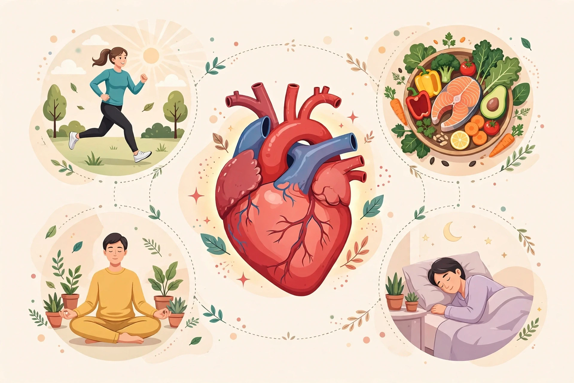
The habits that protect your heart are not exotic. They are the same ones doctors have recommended for decades, because they work:
Move every day. Brisk walking, cycling, swimming, or dancing — about 150 minutes of moderate activity a week is the widely recommended target for adults.
Eat for your arteries. Plenty of vegetables, fruit, whole grains, beans, nuts, and oily fish; less salt, less processed meat, and fewer sugary drinks.
Do not smoke. Smoking damages the lining of blood vessels and makes blood more likely to clot. Quitting is the single most powerful step a smoker can take for their heart.
Keep blood pressure and cholesterol in check. Both can be measured in a few minutes at a clinic or pharmacy, and both can be managed.
Sleep and stress matter too. Chronic poor sleep and long-term stress raise blood pressure and heart rate, and they make healthy habits harder to keep.
MedlinePlus, the U.S. National Library of Medicine's patient resource, offers plain-language overviews of heart conditions, tests, and treatments for readers who want to go further (MedlinePlus).
15. What Researchers Are Learning Now
Heart science is moving quickly, and three directions stand out.
First, researchers are learning how much the heart can repair itself. For a long time, the textbook view was that heart muscle, once damaged, never comes back. That picture is softening: scientists have found that heart muscle cells renew themselves slowly throughout life, and research teams are studying how to encourage that renewal after a heart attack.
Second, wearable sensors have turned the heart into one of the most measured organs in the body. Wrist devices now track heart rate, rhythm, and blood oxygen around the clock, and researchers are studying whether patterns in that data can flag problems — such as an irregular rhythm — earlier than a clinic visit would.
Third, the link between the heart and the rest of the body keeps widening. Researchers are mapping how gut bacteria, sleep patterns, air pollution, and even loneliness relate to cardiovascular risk. The heart turns out to be less of an isolated pump and more of a meeting point where diet, mood, environment, and genes all converge. Expect the advice to keep evolving as that picture sharpens — and treat any single dramatic headline with the caution it deserves.
Conclusion
Your heart is a pump, a muscle, an electrical circuit, and a tireless worker all at once. It sits in the centre of your chest, divides its labour between four chambers, guards its flow with four one-way valves, sets its own pace with its own wiring, and pushes blood through two loops — one to the lungs, one to the whole body — about 100,000 times a day. It asks for little in return: movement, decent food, rest, and a bit of attention to blood pressure and cholesterol.
The next time you feel your pulse, remember what is behind that small thump. A system of highways and doorways, of red and blue streams that never mix, of a muscle that has been beating since before you were born and will keep beating, without a single day off, for as long as you live. Understanding how it works is the first step toward looking after it — and it is a system well worth understanding.
Frequently Asked Questions
How does the human heart work?
Your heart works as a muscular pump with four chambers. The right side collects oxygen-poor blood from the body and sends it to the lungs to pick up oxygen. The left side collects that oxygen-rich blood and pumps it out through the aorta to every tissue in your body. Four valves keep the blood flowing in one direction, and an electrical system centred on the SA node sets the rhythm of each beat.
How many times does the human heart beat per day?
Around 100,000 times per day at a typical resting rate. That adds up to roughly 3 billion beats over a typical lifetime. The rate rises with exercise, stress, fever, or excitement, and falls during sleep and rest.
What is the difference between the circulatory system and the cardiovascular system?
They are two names for the same system. "Cardiovascular" refers to the heart (cardio) and the blood vessels (vascular), while "circulatory" describes what the system does — circulate blood. Doctors and textbooks use the terms interchangeably.
Why is the left ventricle thicker than the right ventricle?
The left ventricle pumps blood all the way around the body, so it must generate much more pressure than the right ventricle, which only pumps blood the short distance to the lungs. Its thicker muscle wall provides that extra force.
What is a normal resting heart rate?
For most adults, a resting heart rate between 60 and 100 beats per minute is considered normal. Well-trained athletes often rest below 60. A rate that is consistently above 100 at rest, or a pulse that feels irregular, is worth discussing with a clinician.
Can the heart repair itself after damage?
Heart muscle renews itself very slowly throughout life, but a large area damaged by a heart attack does not fully regrow on its own. Researchers are actively studying ways to encourage repair, including cell-based therapies, but these remain experimental rather than routine treatment.
Sources
Cleveland Clinic — "How Your Circulatory System Works": https://my.clevelandclinic.org/health/body/circulatory-and-cardiovascular-system
National Heart, Lung, and Blood Institute (NHLBI) — "How the Heart Works": https://www.nhlbi.nih.gov/health/heart
MedlinePlus (U.S. National Library of Medicine) — "Heart Diseases": https://medlineplus.gov/heartdiseases.html
OpenStax, Anatomy and Physiology — "19.1 Heart Anatomy": https://openstax.org/books/anatomy-and-physiology/pages/19-1-heart-anatomy
British Heart Foundation — "How Your Heart Works": https://www.bhf.org.uk/informationsupport/how-a-healthy-heart-works