
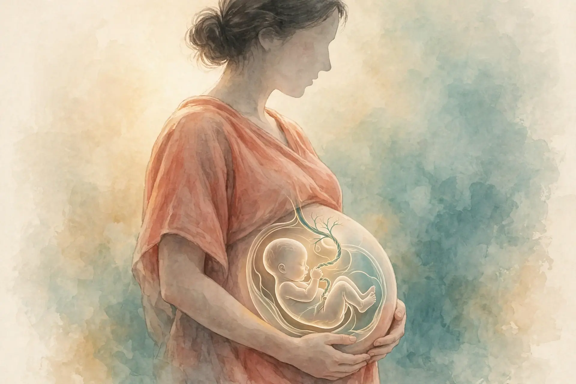
1. How Pregnancy Works: A Quiet Beginning
1.1 The scene: a missed period, a small stick, a changed calendar
She had not been watching the calendar closely. Work had been busy, sleep had been short, and the date of her last period was one of those small facts that slips out of mind until it matters. Then one morning she counted back and realised she was late. The small stick from the pharmacy sat on the bathroom shelf for a day before she used it, because using it would make the question real.
A few minutes later two lines appeared, and the room seemed to tilt slightly. Nothing in her body felt dramatically different. She felt, if anything, ordinary — a little tired, a little warm, her breasts a little tender. Yet something enormous had already happened, and it had happened quietly, days earlier, in a place she could not feel and on a schedule she had not known she was keeping.
That is where this story starts, and it is also where most misunderstandings about pregnancy start. The test turned positive only a few days after implantation, when the trophoblast had secreted enough human chorionic gonadotropin, or hCG, for a home urine test to detect it. But the calendar of the pregnancy had started earlier still — about two weeks before conception, on the first day of her last menstrual period. She was already, by the counting doctors use, several weeks pregnant before she knew anything at all. That gap between what happened and when it was noticed is not a quirk of one person's story. It is built into how pregnancy works, and it explains why the numbers in this article sometimes look surprising at first glance.
I have watched that surprise play out many times in conversations with friends and students. Someone says, "I am six weeks pregnant," and then adds, "but conception was only four weeks ago — how can both be true?" Both can be true because pregnancy is counted from the last period, not from conception. Once that single fact clicks into place, the rest of the story — the heartbeat that seems to arrive early, the trimesters, the due date — falls into order. Without it, every week number feels slightly wrong.
1.2 The answer in 150 words: how does pregnancy work?
How does pregnancy work? In short, a sperm fertilises an egg, usually in a fallopian tube, and the single fertilised cell divides again and again as it travels toward the uterus. About a week after fertilisation it embeds in the uterine lining, in a process called implantation, and the outer layer of that tiny ball of cells begins to build the placenta. The placenta connects the developing fetus to the wall of the uterus through the umbilical cord, exchanging oxygen and nutrients for waste and carbon dioxide, while also acting as an endocrine gland. Hormones — above all oestrogen, progesterone and hCG — hold the pregnancy in place, stop ovulation, quiet the uterus and prepare the body for birth. Over about 38 weeks from conception, or 40 weeks counted from the last period, one cell becomes an embryo, then a fetus, then a newborn. Labour, driven by a feedback loop involving oxytocin, opens the cervix, delivers the baby, and then delivers the placenta. That, in one paragraph, is how pregnancy works. The rest of this article is the same story told slowly, week by week, so that each step earns its place.
The numbers behind that short answer are worth holding onto. Pregnancy is conventionally dated from the first day of the last menstrual period, so gestational age runs about two weeks ahead of fertilisation age. A full-term pregnancy lasts approximately 270 days from conception, and the due date is set at 284 days from the last menstrual period, assuming conception on day 14 of the cycle. The 40 weeks are usually discussed as three trimesters, each approximately 13 weeks long. Those three figures — 270 days, 284 days, three trimesters of about 13 weeks — are the skeleton of everything that follows.
Two headline figures set the scale of the subject before the detail begins. The World Health Organization reported, in figures published on 7 April 2025 covering the data year 2023, that about 260,000 women died during and following pregnancy and childbirth in 2023, a maternal death almost every two minutes, with a global maternal mortality ratio of 197 per 100,000 live births, down about 40 percent since 2000. That figure is a reminder that pregnancy, for all its ordinariness, is a major physiological undertaking and a global health story, not only a personal one. In the United States, the CDC National Center for Health Statistics final data for 2024, released on 9 June 2026, recorded that 10.41 percent of live births were preterm, before 37 completed weeks — about one baby in ten. Keep both figures in mind as you read: pregnancy is common, it is usually straightforward, and it is also a process in which the body does a remarkable amount of work under real strain.
This article is an educational explainer. It describes mechanisms — how fertilisation happens, how the placenta works, how hormones steer the process, how labour unfolds. It does not diagnose, it does not advise any individual reader, and it does not replace a clinician. Anything personal — a symptom, a test result, a decision about care — belongs in a conversation with the reader's own clinician, not with an article.
2. The Two Clocks: Gestational Age vs Fertilisation Age
Before any week number in this article can be trusted, the reader needs to know which clock it is read from. Pregnancy is counted on two clocks at once, and most of the confusion in popular writing about pregnancy comes from mixing them up without saying so. One clock starts at the last menstrual period. The other starts at conception. One naming point needs stating plainly: this article uses the clinical convention, in which a developing human is called a fetus from week 9 of gestation. Embryology texts that count from fertilisation place the end of the embryonic period at week 8 after fertilisation, about week 10 of gestation — the same boundary, read on the other clock. They differ by about two weeks, and that difference matters at almost every milestone. How pregnancy works cannot be understood without first learning to read both clocks, because the same event can be described as happening in week 5 on one clock and week 3 on the other, and both descriptions are correct.
2.1 Why pregnancy is counted from the last period, not from conception
The convention sounds backwards. Why count a pregnancy from a day when the person was not yet pregnant? The answer is practical. The first day of the last menstrual period is a date most people can recall or find in a calendar, and it is a visible event. Conception, by contrast, is invisible. Fertilisation happens inside a fallopian tube, without a sensation that marks the moment, and the exact day of ovulation varies from cycle to cycle. A doctor who needs a date to plan scans, screening and a due date cannot wait for a date nobody can observe.
So medicine settled, long ago, on the observable date. Gestational age is counted from the first day of the last menstrual period, and fertilisation age is counted from conception, about two weeks later. The two-week gap is an average, built on the assumption that conception happens on about day 14 of the cycle. In a real cycle that gap can be a little shorter or longer, which is one reason a dating ultrasound, typically done at 11 to 14 weeks, is used to check the dates. The convention is a tool, not a claim that pregnancy begins at menstruation. Nobody believes the embryo existed on the first day of the last period. The clock simply starts there because that is where the calendar can be read.
There is a second, quieter reason the convention survives. It gives every pregnancy a shared language. When a midwife says "week 12," a sonographer says "week 12," and a textbook says "week 12," they all mean the same thing: 12 weeks from the last period. If each of them counted from conception instead, they would each be guessing at a different invisible day. The LMP convention trades a small fiction — counting two weeks that came before conception — for a large clarity: everyone is reading the same clock. That clarity is what lets the rest of this article state a week number and mean something definite by it.
2.2 Converting between the two clocks
The conversion itself is simple, and it is worth practising until it feels automatic. Add about two weeks to fertilisation age to get gestational age. Subtract about two weeks from gestational age to get fertilisation age. A heart that starts its first movements in the fourth week after fertilisation is doing so in about the sixth week of pregnancy counted from the last period. An embryo that is three weeks old after fertilisation is five weeks along by the gestational count. A fetus at 20 weeks gestational age has been developing for about 18 weeks since conception.
Table 1 below sets out the conversion for the milestones that appear most often in this article. Read it once now, and return to it whenever a week number in a later section feels unfamiliar. The table uses gestational age as its first column, because that is the age a clinician, a scan report and this article will usually give, and it shows the fertilisation age beside it so the biology stays visible.
Gestational age (from last period) | Fertilisation age (from conception) | What it means |
|---|---|---|
Week 4 | About 2 weeks | Implantation complete; hCG rising; a home test can turn positive a few days after implantation |
Week 5 | About 3 weeks | Gastrulation; the three germ layers form; the neural tube begins to close |
Week 6 | About 4 weeks | Neural tube normally closed; first heart movements begin in the fourth week after fertilisation |
Week 7 | About 5 weeks | Cardiac activity usually visible on transvaginal ultrasound at 6 to 7 weeks |
Week 9 | About 7 weeks | The developing human stops being called an embryo and starts being called a fetus (week 9 of gestation; the clinical convention used in this article) |
Week 10 | About 8 weeks | All organ systems structured in rudimentary form (the embryonic period ends at week 8 after fertilisation) |
Week 12 | About 10 weeks | End of the first trimester; fetus about 9 cm crown to rump |
Week 20 | About 18 weeks | Quickening may be felt; anatomy scan typically at about 18 to 22 weeks |
Week 24 | About 22 weeks | Viability generally considered between 23 and 24 weeks in high-resource settings |
Week 28 | About 26 weeks | Start of the third trimester; rapid weight gain and surfactant production under way |
Week 40 | About 38 weeks | Due date; full-term pregnancy is about 38 weeks from conception |
A worked example helps. Suppose a person conceived on 1 March. Her last period began about 15 February, two weeks earlier. On 1 April, she is about four weeks past conception and about six weeks pregnant by the gestational count. A scan at that point might report "6 weeks," and a textbook describing the embryo at "4 weeks after fertilisation" is describing the same moment. Neither is wrong. They are reading different clocks, and the table above is the translation between them.
The habit to build is a small one: whenever a week number matters — the heartbeat, the neural tube, quickening, viability — ask which clock it is on, and state both. This article does that at every milestone. The payoff is that the apparent contradictions in other sources dissolve. A page that says the heart starts beating in week 5 and a page that says it starts in week 6 are often describing the same day, one in gestational age and one in fertilisation age, without labelling the clock.
2.3 Why 40 weeks, not 38 — and what a due date really means
Pregnancy typically lasts about 38 weeks from conception, or 40 weeks from the first day of the last period. The two figures describe one duration, measured from two starting lines. The full-term figure from conception is approximately 270 days, about 38.5 weeks, and the due date is set at 284 days, about 40.5 weeks, from the last menstrual period, on the assumption that conception happened on day 14 of the cycle. The half-week in each figure is a reminder that these are averages over many pregnancies, not appointments stamped on a calendar.
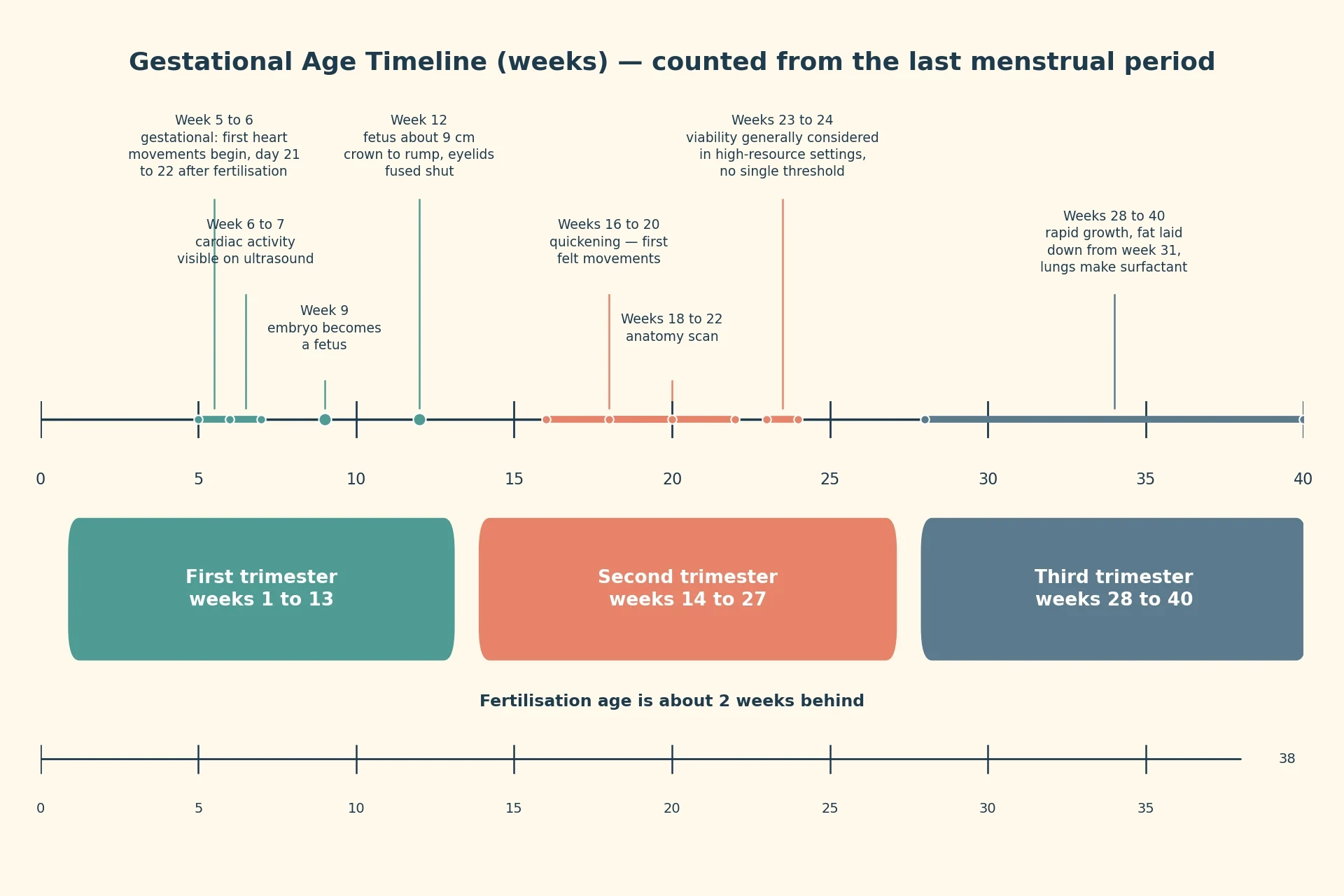
Those 40 weeks are usually grouped into three trimesters of about 13 weeks each. The boundaries are conventional rather than biological — nothing dramatic changes in the body at the stroke of week 14 or week 28 — but they are useful, because the character of pregnancy changes across them. The first trimester builds the body plan. The second grows it and makes it move. The third adds weight, matures the lungs and prepares for birth.
First trimester: weeks 1 to 13. Fertilisation, implantation, the embryo building every organ system, then the change to a fetus at week 9 of gestation.
Second trimester: weeks 14 to 27. Growth, quickening, the anatomy scan, and the approach of viability.
Third trimester: weeks 28 to 40. Rapid weight gain, surfactant production, fat laid down from week 31, and preparation for labour.
A due date, then, is an estimate built on an average. It marks 40 weeks from the last period, or about 38 weeks from conception, in a cycle in which conception happened on day 14. Real cycles vary, real conceptions happen on different days, and real babies arrive on their own schedule. Only a minority arrive on the due date itself. Treat the date as the centre of a window, not as a deadline, and the rest of the timeline makes more sense. The due date tells the clinician when to schedule checks and when to start watching more closely; it does not tell the body when to begin labour.
3. Fertilisation and Implantation: From One Cell to a Blastocyst
Fertilisation and implantation are the quietest part of how pregnancy works, and also the most time-sensitive. The whole sequence — from an egg leaving the ovary to a ball of about 100 cells embedding in the uterine lining — takes roughly two weeks, and most of it happens before a pregnancy test could possibly turn positive. It is a journey measured in hours and days, not weeks, and the timing windows on each side of it are strikingly uneven: sperm can wait for days, while an egg, once released, has about a day.
3.1 The fertile window: sperm, egg and capacitation
The fertile window is lopsided. An egg is viable for about 18 to 24 hours after ovulation, and sperm remain viable in the female reproductive tract for about 48 hours, although this can be quite variable. A second account puts the sperm's survival more generously: sperm can survive in the uterine tubes for 3 to 5 days, so fertilisation remains possible if intercourse occurs a few days before ovulation, while an egg can only survive independently for about 24 hours after ovulation. The two accounts are not in conflict so much as they describe different places and different conditions — viability in the tract as a whole, and survival in the tubes in particular — and both point the same way. The window opens days before ovulation and closes about a day after it.
Capacitation adds another layer of timing. Capacitation, the process in the female reproductive tract that prepares sperm to fertilise an egg, typically takes 5 to 7 hours. A sperm that arrives at the egg is not yet ready to fertilise it; it must first spend hours being chemically prepared by the environment of the reproductive tract. That preparation changes the sperm's surface and its swimming behaviour, and only then can it penetrate the egg's outer layers. The fertile window is therefore not only about survival. It is about readiness, and readiness takes time.
The egg: about 18 to 24 hours. Viable for roughly a day after ovulation; after that, the chance of fertilisation falls away quickly.
Sperm in the tract: about 48 hours, and 3 to 5 days in the uterine tubes. Sperm can wait for ovulation, which is why the fertile window stretches several days before ovulation.
Capacitation: 5 to 7 hours. The preparation sperm must undergo in the female reproductive tract before they can fertilise an egg.
Put together, these three windows explain why timing matters and why it is forgiving on one side and strict on the other. Intercourse a few days before ovulation can still lead to fertilisation, because sperm can survive and be capacitated in time. Intercourse more than a day after ovulation usually cannot, because the egg has gone. The biology is not mysterious once the clocks are laid out; it is a race in which one runner waits and the other hurries.
3.2 Fertilisation in the fallopian tube
Fertilisation usually takes place in a fallopian tube that links an ovary to the uterus. The egg, released from the ovary at ovulation, is drawn into the tube, and sperm that have travelled up through the uterus meet it there. One sperm enters the egg, the egg's surface changes to block the others, and the two sets of genetic material come together in a single cell called a zygote. That single cell carries a complete set of instructions for building a human body, and it also carries the first decision the embryo will make: whether the combination of chromosomes will lead to a male or female pattern of development, a decision whose visible consequences do not appear until weeks 9 to 12, when bipotential gonads develop into testes or ovaries.
There is something worth pausing on here. A common picture of fertilisation shows a sperm racing to an egg as if the egg were passive. The reality is more collaborative. The egg's layers select and prepare, the tract capacitates the sperm, and the tube itself moves the egg along with gentle contractions and the beating of tiny hair-like cilia. Fertilisation is not a single collision. It is the end of a coordinated journey through a living tube, and the tube's health matters as much as the cells travelling through it. That is one reason an embryo implanting outside the uterus — most often in the uterine tube itself — is a medical emergency rather than a variation; the tube is a passage, not a home.
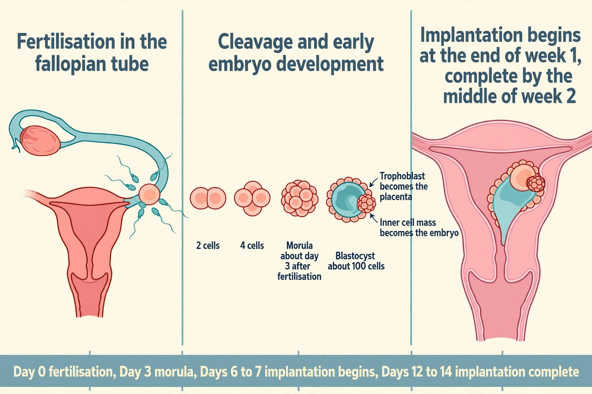
3.3 Cleavage, morula and blastocyst: the first week
After fertilisation, the zygote undergoes rapid mitotic divisions called cleavage, which increase cell number without increasing the total volume of the conceptus. The first division makes two cells, the next makes four, then eight, and so on, while the whole structure stays roughly the same size as the original egg. It is division without growth, a kind of careful unpacking: the same amount of material, cut into smaller and smaller pieces, each piece carrying a full copy of the genetic instructions.
About 3 days after fertilisation, a 16-cell conceptus reaches the uterus and is called a morula — the name comes from the Latin for mulberry, because the cluster of cells looks like a small dark berry. The morula continues to divide as it is carried along the tube, and by the time it has been in the uterus for a few days it has become a blastocyst, a ball of about 100 cells around a fluid-filled cavity, with an inner cell mass that becomes the embryo and an outer trophoblast layer that becomes the chorionic sac and the fetal part of the placenta.
That division of labour inside the blastocyst is the first great fork in the road. The inner cell mass, a small cluster tucked to one side of the cavity, is the part that will become the embryo itself — and, later, the fetus and the baby. The trophoblast, the outer shell, is the part that will do the work of connecting to the mother: it will invade the uterine lining, build the placenta, and secrete the hormone that announces the pregnancy. One ball of cells, two destinies, already separated before implantation has even begun. When people ask how pregnancy works, this is one of the answers that surprises them most: the organ that will feed the baby for nine months is built by the embryo itself, not by the mother.
3.4 Implantation: embedding in the uterine lining
Implantation begins at the end of the first week, when the blastocyst contacts and embeds in the uterine lining, and implantation is complete by the middle of the second week. The blastocyst does not simply sit on the surface of the endometrium. Its trophoblast cells burrow into the lining, breaking down the surface and sinking the blastocyst into the tissue beneath, where it can tap into the mother's blood supply. The uterine lining, thickened under the influence of progesterone in preparation for just this event, receives it.
The timing matters for the pregnancy test that will come later. The trophoblast secretes human chorionic gonadotropin, and just a few days after implantation the trophoblast has secreted enough hCG for an at-home urine pregnancy test to give a positive result. That is why a test taken too early can be negative even when implantation has happened: the hormone has to accumulate to a detectable level, and that takes a few days after the blastocyst has embedded. The missed period, the positive test and implantation are three events on one short timeline, each a few days after the last.
I remember the first time I saw a time-lapse film of a blastocyst hatching and settling onto a surface in a laboratory dish. It did not look like an invasion, which is the word textbooks often use. It looked deliberate and unhurried — the ball of cells turning, pausing, then pressing itself into place as if it knew where it belonged. The film was silent and lasted only a minute, but it changed how I thought about implantation. It is not a collision. It is a negotiation between two tissues, and when it works, it works quietly.
3.5 Why implantation often does not happen
A significant percentage — 50 to 75 percent in the OpenStax account — of blastocysts fail to implant and are shed with the endometrium during menstruation, which is one reason pregnancy typically requires several ovulation cycles to achieve. That figure is startling the first time it is read, and it deserves to be read plainly. More than half of all blastocysts, in this account, never implant at all. They are lost without the person ever knowing a fertilisation happened, because there was no missed period beyond an ordinary one, no test taken, no symptom to notice.
This is not a failure of the body in the ordinary sense. It is a feature of a system that is selective at its earliest stage. Many of the blastocysts that fail to implant carry chromosomal errors that would make further development impossible, and the uterine lining itself plays a part in deciding which blastocysts to accept. The high failure rate is also why fertility is best thought of in cycles, not in single attempts: a couple trying to conceive may need several ovulation cycles, and that is within the normal range of how pregnancy works, not evidence that something is wrong. The fact is stated here without alarm and without advice, because it is a fact about the biology, not a judgement about any reader.
4. The Embryo Builds Itself: Weeks 3 to 8 After Fertilisation
The first two weeks of prenatal development are the pre-embryonic stage, a developing human is called an embryo during weeks 3 to 8 after fertilisation, and a fetus from week 9 of gestation until birth. Those labels are not mere naming conventions. They mark a real change in what the developing human is doing. In the embryonic period, the body plan is drawn. In the fetal period, the plan is filled in and grown. Weeks 3 to 8 after fertilisation — about weeks 5 to 10 of gestational age — are the weeks when every organ system is laid down in rudimentary form, and that is why they matter out of proportion to their length.
4.1 Gastrulation: three layers, every organ
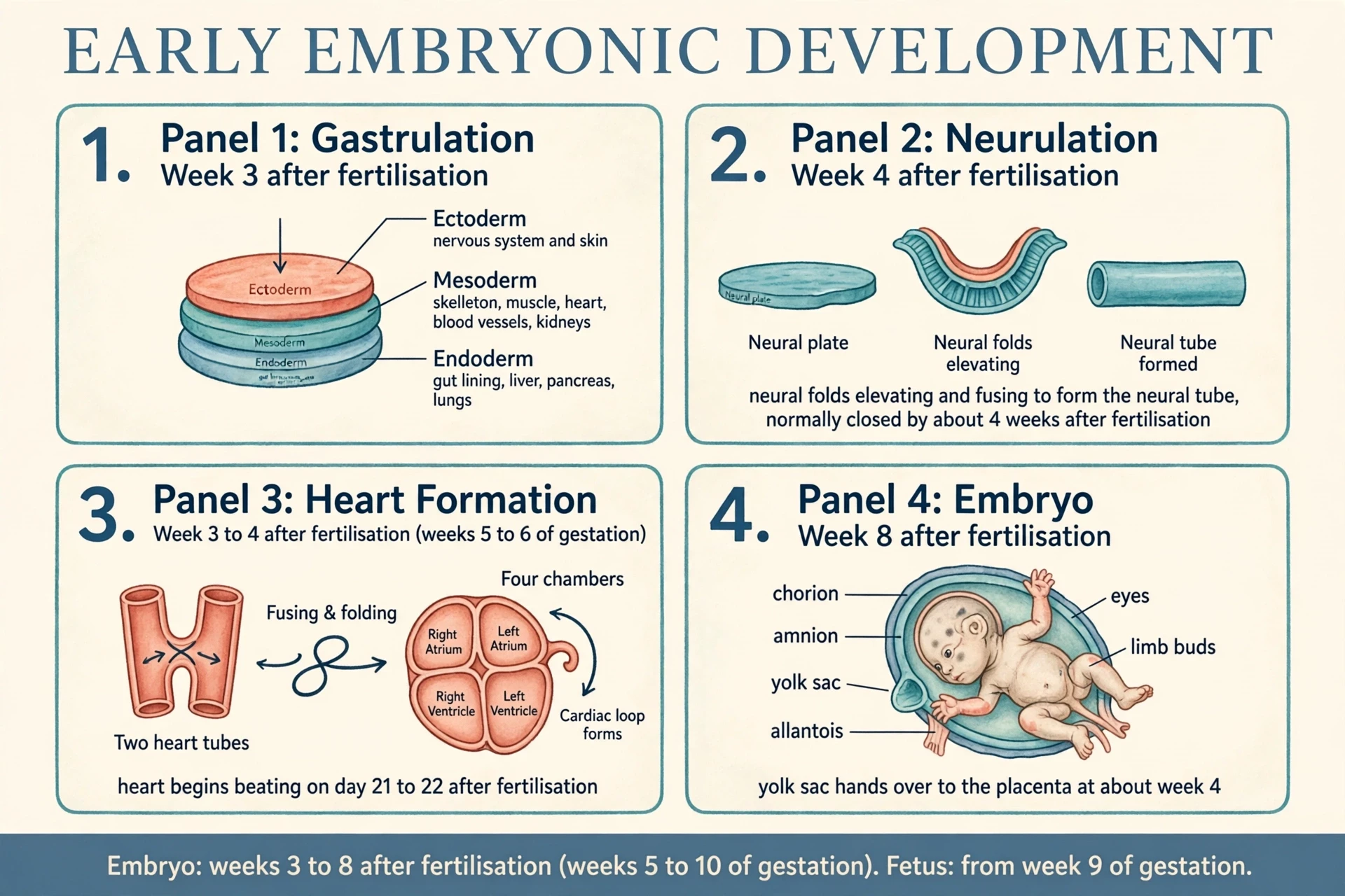
Gastrulation in the third week after fertilisation turns the two-layered disc into a three-layered disc with ectoderm, mesoderm and endoderm, which give rise respectively to the nervous system and skin, the skeleton, muscles, heart, blood vessels and kidneys, and the lining of the gut, liver, pancreas and lungs. Picture a flat disc of cells, two layers thick, folding and rearranging itself into three layers, each with a different destiny. It is one of the most consequential rearrangements in all of biology, and it happens in a structure smaller than a grain of rice.
The three layers are worth learning by name, because they explain the body's organisation for the rest of life.
Ectoderm — the outer layer. Gives rise to the nervous system and the skin. The brain, the spinal cord and the nerves all begin here, as does the surface that will face the world.
Mesoderm — the middle layer. Gives rise to the skeleton, muscles, heart, blood vessels and kidneys. The body's scaffolding, its pump, its pipes and its filters all begin in this layer.
Endoderm — the inner layer. Gives rise to the lining of the gut, liver, pancreas and lungs. The tubes and glands that process food and air begin here.
The link to epigenetics is the natural next thought. Every cell in the three layers carries the same genome, yet an ectoderm cell becomes a neuron and a mesoderm cell becomes a muscle fibre. The difference is not in the genes they carry but in which genes are switched on and off, and in what order. Gastrulation is the moment when that switching begins in earnest, and the three layers are the first visible result of it. The embryo does not grow new genes for each organ. It re-reads the same genome three different ways.
4.2 The neural tube closes
The neural tube is normally completely closed by about 4 weeks after fertilisation, with the rostral neuropore closing at about 24 days and the caudal neuropore at about 26 days. That is about week 6 of gestational age — before many people know they are pregnant, and before the first scan. The neural tube is the hollow cylinder that will become the brain and the spinal cord. It forms when the ectoderm along the embryo's back folds up and fuses along its length, like a sheet of paper rolling into a tube and sealing along the seam. The seam closes from the middle outward, and the two ends — the rostral neuropore at the head end, closing at about 24 days, and the caudal neuropore at the tail end, closing at about 26 days — are the last to seal.
The timing is the point. Because the tube closes so early, the period when it is most vulnerable is also early, often before a pregnancy is recognised. That is why the neural tube is discussed in every account of early pregnancy, and why the story of its closure is told here before the story of the heart. The tube's closure is not a detail. It is the founding event of the nervous system — the structure that will later carry every sensation, every movement and every thought.
I once spent an afternoon looking at models of neurulation, the folding process itself, and what stayed with me was how physical it is. The cells do not simply decide to become a tube. They change shape, they pull on each other, the sheet bends, and the edges meet. It is engineering done by cells, and when the edges fail to meet, the consequences are structural and permanent. That physicality — the tube as a thing that must close, on a deadline, by folding — is easier to hold in mind than any abstract description, and it is the right way to think about the embryo's second month.
4.3 The heart forms and starts to beat
The heart first forms as two parallel tubes derived from mesoderm, which fuse and then fold and differentiate into the four chambers of a mature heart. Two tubes become one, the single tube loops and folds on itself, and the folded tube divides into chambers. It is a piece of origami done in living tissue, and it happens while the heart is already beginning to work.
Here is the confusion this article exists to resolve. The heart begins its first pumping motions during the fourth week after fertilisation, which is roughly the sixth week of pregnancy counted from the last menstrual period. The baby's heart starts beating around day 21 to 22 after fertilisation, which is about 5 weeks and 1 to 2 days of gestational age. Those two statements describe the same event from two sides: the first contractions of the forming heart muscle, and the first movements that can be called a beat. A page that says "week 5" and a page that says "week 6" are usually both right, reading different clocks and different definitions of the first beat. State both clocks, and the contradiction vanishes.
By the end of the embryonic period, all organ systems are structured in rudimentary form, although the organs themselves are either nonfunctional or only semi-functional. — OpenStax Anatomy and Physiology 2e
That single line is worth carrying through the rest of the article. By the end of week 8 after fertilisation — about week 10 gestational — the embryo has a heart with chambers, a neural tube closed into brain and cord, limb buds, a gut tube, and the beginnings of kidneys, liver and lungs. None of those organs works properly yet. The heart moves blood, but the circulation is not the circulation of a newborn. The kidneys make urine, but the placenta still does the filtering. The lungs exist as buds, but they do not breathe air. Structure first, function later: that is the rule of the embryonic period, and it explains why the fetal period that follows is mostly about growth rather than invention.
4.4 The membranes and the yolk sac hand over to the placenta
The four extra-embryonic membranes are the amnion, the yolk sac, the allantois and the chorion. Each has a job, and each job is temporary. The amnion is the sac that will fill with fluid and surround the embryo. The chorion is the outer membrane that will become the fetal part of the placenta. The allantois contributes to the umbilical cord and the early blood supply. The yolk sac, despite its name, does not feed the human embryo the way a bird's yolk feeds a chick; it supplies nutrients and primitive blood circulation in weeks 2 and 3, and when the placenta takes over nourishing the embryo at about week 4, the yolk sac's main function becomes serving as the source of blood cells and germ cells.
The handover at about week 4 after fertilisation — about week 6 gestational — is the embryo's first change of supplier. For the first two or three weeks, the yolk sac and the uterine lining do the feeding. Then the placenta, built by the trophoblast, takes over, and the yolk sac shrinks into a supporting role. The embryo does not notice the change, in any sense that matters, but the change is what makes the rest of pregnancy possible. A yolk sac can feed an embryo a few millimetres long. Only a placenta can feed a fetus that will grow to about 51 cm and 2.5 to 4 kg.
5. What Happens in Each Trimester: Pregnancy Week by Week
This is the core of how pregnancy works, told pregnancy week by week. What happens in each trimester is not three separate stories but one story in three movements. First, the body plan is built. Then it grows, moves and is checked. Finally it gains weight, its lungs mature, and it gets ready for birth. Every milestone below is dated on both clocks, so that the week-5 versus week-6 confusion of other pages cannot recur here.
5.1 First trimester — Weeks 1 to 13
The first trimester runs from week 1 to week 13 of gestational age, which is about the first 11 weeks after fertilisation. It begins before conception, on the first day of the last period, and it ends with a fetus that is recognisably human in miniature. The change of name at week 9 of gestation — embryo until then, fetus from the ninth week of gestation until birth — marks the end of the period when organs are invented and the start of the period when they are grown.
The heart's story runs through the whole trimester. The heart's first contractions come around 5 weeks and 1 to 2 days gestational — day 21 to 22 after fertilisation — with the first pumping motions in the fourth week after fertilisation, about week 6 gestational. An ultrasound can typically pick up embryonic cardiac activity by 6 to 7 weeks of pregnancy, and a handheld Doppler can usually pick it up at 10 to 12 weeks. The American College of Obstetricians and Gynecologists uses the term "embryonic cardiac activity" before around 10 weeks of pregnancy and "fetal cardiac activity" after that point, rather than "heartbeat", because the early heart has no chambers or valves yet. A normal fetal heart rate is typically 110 to 160 beats per minute, once the early peak of about 170 beats per minute around weeks 9 to 10 has passed. The heart rate is fast because the heart is small and the body it serves is growing at a pace no adult body ever matches.
By the end of week 12, the fetus measures approximately 9 cm (3.5 in) from crown to rump, its fingers and toes are developing nails, and its eyes are well developed but the eyelids are fused shut. The eyes are a good image for the trimester as a whole: well developed, but closed. The structures are there; the functions are waiting. Sexual differentiation, which does not begin until the fetal period, happens during weeks 9 to 12, when bipotential gonads develop into testes in males or ovaries in females — a reminder that the decision made at fertilisation takes weeks to become anatomy.
The week-by-week milestones of the first trimester, each with both clocks, run as follows.
Weeks 1 to 4 gestational (up to about 2 weeks after fertilisation). The last period, ovulation, fertilisation in the fallopian tube, cleavage, the morula reaching the uterus about 3 days after fertilisation, the blastocyst forming, and implantation beginning at the end of the first week and completing by the middle of the second week. hCG rises enough for a home test a few days after implantation.
Weeks 5 to 8 gestational (about 3 to 6 weeks after fertilisation). Gastrulation in the third week after fertilisation; the neural tube normally closed by about 4 weeks after fertilisation; the heart forming from two tubes and beginning to pump in the fourth week after fertilisation, about week 6 gestational; the yolk sac handing over to the placenta at about week 4 after fertilisation.
Weeks 9 to 13 gestational (about 7 to 11 weeks after fertilisation). The embryo becomes a fetus at week 9 of gestation; sexual differentiation in weeks 9 to 12; heart rate topping out near 170 beats per minute around weeks 9 to 10; cardiac activity visible on ultrasound at 6 to 7 weeks and audible on a Doppler at 10 to 12 weeks; by week 12, about 9 cm crown to rump, nails developing, eyes well developed with eyelids fused shut.
The gut is forming through all of this, and the link is worth making explicit. The lining of the gut comes from endoderm, one of the three layers laid down in gastrulation, and the digestive system that will one day process food begins as a simple tube in the embryo. Meconium — fetal faeces consisting of ingested amniotic fluid, cellular debris, mucus and bile — begins to accumulate in the intestines during weeks 13 to 16, at the boundary between the first and second trimesters. The fetus is already swallowing amniotic fluid and producing urine, practising the movements it will need after birth, weeks before it needs them for nutrition.
5.2 Second trimester — Weeks 14 to 27
The second trimester, weeks 14 to 27 gestational, is often described as the most comfortable part of pregnancy, and the biology gives a reason for that description. The body plan is built, the placenta has taken over as the endocrine organ, nausea typically subsides by about week 12, and the work of the trimester is growth and movement rather than invention. It is also the trimester when the pregnant person first feels the fetus move, and when the anatomy of the fetus is checked in detail.
During weeks 13 to 16, the eyes move closer together, blinking motions begin, the lips show sucking motions, the scalp begins to grow hair, and the kidneys are well formed. The face is becoming a face, and the movements are becoming coordinated — blinking, sucking, the small rehearsals of life outside the uterus. During approximately weeks 16 to 20, the pregnant person may begin to feel quickening, or fetal movements, as limb movements become more powerful. Quickening is often described as a flutter, and it is easy to miss at first; the movements are small, the fetus is still small, and the sensation is unlike anything the body has felt before. It is the first direct evidence, felt rather than seen on a scan, that the fetus is moving on its own.
Sebaceous glands coat the skin with a waxy protective substance called vernix caseosa, and a silky hair called lanugo covers the skin during weeks 17 to 20. Vernix protects the skin from months of soaking in amniotic fluid, and lanugo helps hold the vernix in place. Both are shed or absorbed before birth, but for the middle weeks of pregnancy they are part of the fetus's working equipment. Fingerprints, in the timeline used for this article, arrive only as an approximate range — by around week 10 to 17 — because one timeline places them at week 10 while other embryology sources describe ridge formation across weeks 10 to 17. No single exact week is given here, because none is settled.
The anatomy scan follows at about 18 to 22 weeks, checking the brain, heart, spine, kidneys, limbs and placenta — the structures built in the embryonic period, now large enough to see clearly. The dating scan comes first, at 11 to 14 weeks. The two scans have different jobs: the first fixes the dates, the second checks the anatomy.
The week-by-week milestones of the second trimester run as follows.
Weeks 14 to 16 gestational (about 12 to 14 weeks after fertilisation). Eyes moving closer together, blinking beginning, sucking motions of the lips, scalp hair beginning, kidneys well formed; meconium beginning to accumulate in the intestines in weeks 13 to 16.
Weeks 17 to 20 gestational (about 15 to 18 weeks after fertilisation). Vernix caseosa coating the skin, lanugo covering the skin; quickening may begin to be felt in weeks 16 to 20; fingerprints forming across the approximate range of weeks 10 to 17.
Weeks 21 to 27 gestational (about 19 to 25 weeks after fertilisation). Rapid weight gain beginning in developmental weeks 21 to 30; the lungs beginning to produce surfactant; the anatomy scan at about 18 to 22 weeks; viability generally considered between 23 and 24 weeks in modern high-resource settings, with no single threshold in all settings.
5.3 Third trimester — Weeks 28 to 40: growth, lungs and getting ready
The third trimester, weeks 28 to 40 gestational, is the trimester of weight and readiness. Developmental weeks 21 to 30 are characterised by rapid weight gain, and the lungs begin producing surfactant, a substance that reduces surface tension and assists proper lung expansion after birth. Surfactant is the difference between lungs that open easily at the first breath and lungs that struggle to stay open. Inadequate surfactant production in premature newborns may result in respiratory distress syndrome, which is why lung maturity, not size alone, decides how ready a premature baby is for the world.
The fetus continues to lay down subcutaneous fat from week 31 until birth, the skin changes from red and wrinkled to soft and pink, lanugo is shed, and the nails grow to the tips of the fingers and toes. Fat is not decoration. It is insulation and an energy store, and a baby born without enough of it loses heat quickly. The skin's change from red and wrinkled to soft and pink is the visible sign of that fat arriving underneath it. The nails reaching the fingertips are a small detail that clinicians still check, because they mark a fetus that has had time to finish growing.
Immediately before birth, the average crown-to-rump length is 35.5 to 40.5 cm (14 to 16 in), the fetus weighs approximately 2.5 to 4 kg (5.5 to 8.8 lbs), and the average length at birth is approximately 51 cm (20 in). The range in weight — 2.5 to 4 kg — is wide, and it should be read as a range, not as a target. Babies are born at many sizes, and size at birth is shaped by genetics, by the placenta's function, by the length of the pregnancy and by the mother's health. The averages describe a population, not a prescription for any one baby.
The week-by-week milestones of the third trimester run as follows.
Weeks 28 to 31 gestational (about 26 to 29 weeks after fertilisation). Rapid weight gain continuing; surfactant production under way in the lungs; subcutaneous fat beginning to be laid down from week 31.
Weeks 32 to 36 gestational (about 30 to 34 weeks after fertilisation). Fat laid down steadily; skin becoming softer and pinker; lanugo shed; the fetus growing longer and heavier each week.
Weeks 37 to 40 gestational (about 35 to 38 weeks after fertilisation). Nails grown to the tips of the fingers and toes; average crown-to-rump length 35.5 to 40.5 cm and weight approximately 2.5 to 4 kg immediately before birth; average length at birth approximately 51 cm; labour approaching as progesterone plateaus and drops.
5.4 Reading the timeline: what changes fastest, and when
Step back from the week-by-week detail and the pattern is simple: structure first, growth later. The first trimester invents the organs. The second trimester grows them and makes them move. The third trimester fattens them, matures the lungs and prepares them to work outside the uterus. Nothing in the third trimester is as radical as gastrulation in the third week after fertilisation, and nothing in the first trimester is as heavy as the weight gained in the last ten weeks. The story changes its character as it goes, and that change is the story.
Table 2 collects the three trimesters in one place, so the pattern can be seen at a glance. The "key check" column is the practical thread: each trimester has a check that matches what is happening in it — the dating scan when dates matter, the anatomy scan when structures can be seen, and the screening for gestational diabetes at 24 to 28 weeks when the third trimester is approaching.
Trimester | Weeks (gestational) | The fetus | The mother | Key check |
|---|---|---|---|---|
First | Weeks 1 to 13 | Embryo to fetus at week 9 of gestation; all organ systems structured in rudimentary form by week 10 of gestation; about 9 cm crown to rump by week 12 | Nausea common in the first weeks to months, typically subsiding by about week 12; hCG rising; corpus luteum leading hormone production | Dating ultrasound, typically 11 to 14 weeks |
Second | Weeks 14 to 27 | Quickening at about weeks 16 to 20; vernix and lanugo in weeks 17 to 20; rapid weight gain from weeks 21 to 30 | Placenta takes over as the endocrine organ in weeks 12 to 17; blood volume expanding; minute volume rising in the second half | Anatomy scan, typically about 18 to 22 weeks |
Third | Weeks 28 to 40 | Surfactant production; fat laid down from week 31; lanugo shed; nails to the fingertips; about 2.5 to 4 kg and about 51 cm at birth | Uterus grown from fist-sized to full-term; bladder compressed; progesterone plateauing then dropping before labour | Screening for gestational diabetes at 24 to 28 weeks; monitoring as the due date approaches |
A reader who remembers only one sentence from this section should remember this: the embryo builds the body in eight weeks, and the fetus spends the next thirty weeks growing it, moving it and getting it ready to breathe. How pregnancy works, at the level of the timeline, is a short period of invention followed by a long period of preparation.
6. What Is the Placenta? The Organ That Is Three Organs at Once
What is the placenta? Ask that question of a textbook and the answer is short: the organ that connects the fetus to the uterus. Ask it of the biology and the answer is longer and more interesting. The placenta is three organs at once — an exchange organ, a partial barrier and an endocrine gland — and it is the only organ in the human body that is built for a single pregnancy and then discarded. Few pages explain it as an endocrine organ in plain language, and fewer still explain immune tolerance, blood-volume expansion and the oxytocin feedback loop as one connected system. This section explains both, because the placenta is where the threads of how pregnancy works meet.
6.1 How the placenta forms — the first fetal organ
The placenta joins the developing fetus to the wall of the mother's uterus during pregnancy, grows in the wall of the uterus, and is attached to the fetus by the umbilical cord. It is formed by cells that originate from the fetus and is therefore the first of the fetal organs to develop. That last fact is the one to hold onto: the placenta is a fetal organ, built by the embryo, not a maternal organ lent to the fetus. Its roots are in the trophoblast, the outer layer of the blastocyst, which invades the uterine lining at implantation and grows into the disc-shaped organ that will serve the fetus for the rest of the pregnancy.
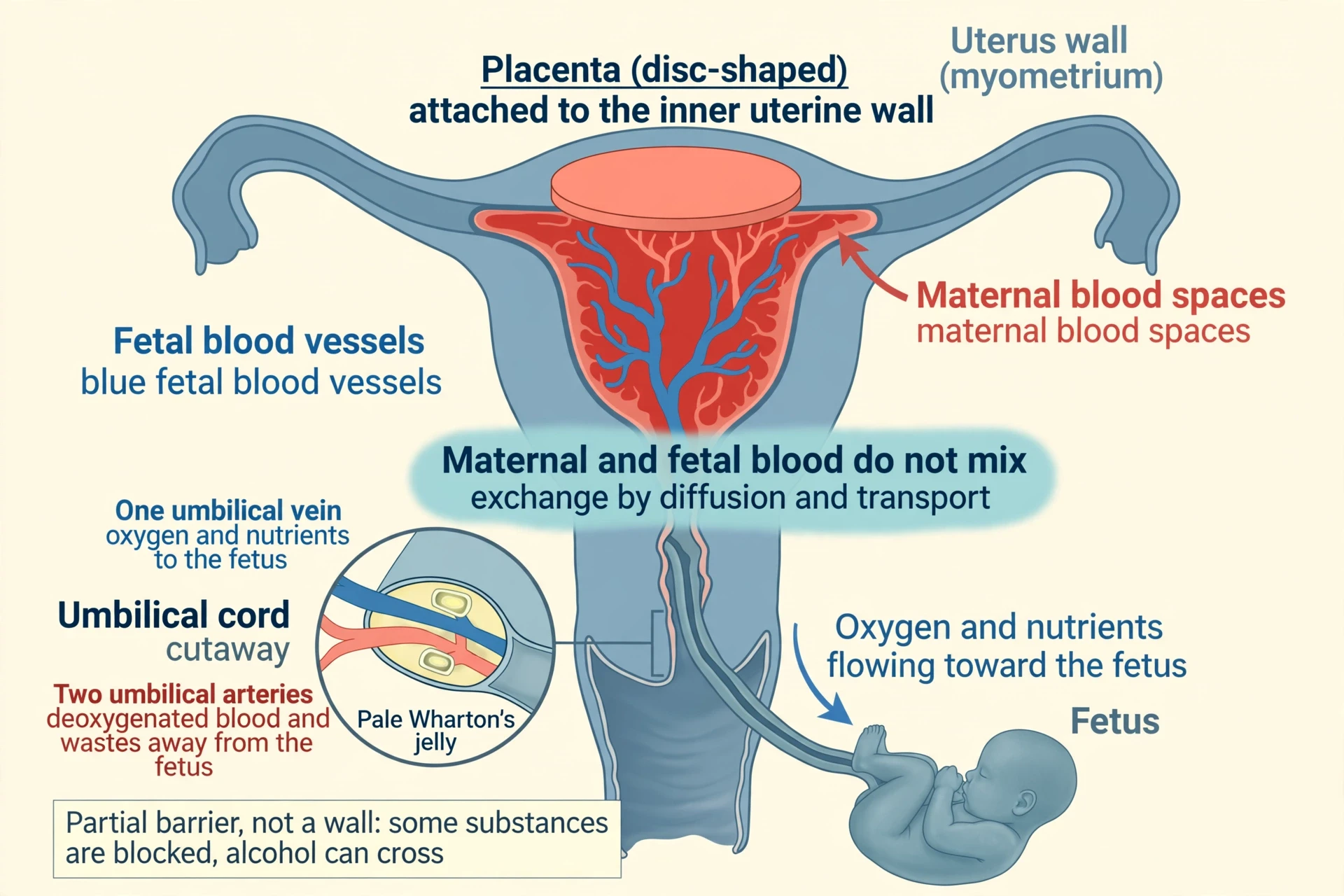
The mature placenta is composed of tissues derived from the embryo as well as maternal tissues of the endometrium, and it connects to the conceptus via the umbilical cord, which carries deoxygenated blood and wastes from the fetus through two umbilical arteries and nutrients and oxygen to the fetus through a single umbilical vein. The maternal contribution is the endometrium, the lining of the uterus, remodelled to receive the placenta. The fetal contribution is the chorion and its villi, the branching structures that reach into the mother's blood spaces. The two tissues interlock without merging, and that interlocking — close, but separate — is the physical basis of everything the placenta does.
6.2 Exchange without mixing: oxygen, nutrients and waste
The mother's blood supply does not actually mix with the fetus's blood supply; exchange occurs by diffusion of gases and transport of nutrients between the two blood supplies. That sentence corrects one of the most common misconceptions about pregnancy. The two bloods flow close to each other, separated by thin membranes in the placental villi, and substances cross those membranes without the bloods themselves ever meeting. Oxygen diffuses from the mother's blood into the fetal blood. Carbon dioxide diffuses the other way. Nutrients are transported across, some by diffusion and some by active transport that spends energy to move them against a gradient. Waste products follow the reverse route, back to the mother's blood for disposal.
The placenta transfers oxygen and nutrients from mother to fetus and waste products and carbon dioxide back from fetus to mother, and antibodies can also pass from mother to fetus, providing protection that can last for several months after birth. The antibody transfer is a quiet gift that continues after birth: the newborn arrives with some of the mother's immunity already in its blood, a borrowed defence while its own immune system learns its work. The umbilical cord is the pipeline for all of this exchange, carrying deoxygenated blood and wastes away from the fetus through two arteries and oxygen and nutrients toward the fetus through one vein — the opposite of the adult arrangement, in which arteries carry oxygenated blood away from the heart, and a detail worth remembering because it shows how thoroughly the fetal circulation is adapted to its job.
6.3 A partial barrier, not a wall
The placenta acts as a partial barrier, preventing some harmful substances in the mother's blood from damaging the fetus, but it cannot exclude all of them — for example, alcohol can cross the placental barrier. The word "partial" does the work in that sentence. The placenta filters, but it does not seal. Large molecules and many harmful substances are kept out, or kept out in part, while small molecules — including some drugs and alcohol — cross freely. A barrier that lets alcohol through is not a wall. It is a sieve with a particular mesh, and what passes through it depends on size, solubility and the transport systems in the placental membranes.
The antibody crossing described above is the other side of the same coin. The placenta does not simply block. It selects. Antibodies cross because the placenta carries them across deliberately, giving the fetus protection that can last for several months after birth. The same selectivity explains why the vaccines a mother has received, or the immunity she has built through past infection, can reach the fetus as protection. The mechanism is not magic. It is transport — the placenta moving a useful molecule across a membrane on purpose, while other molecules cross by accident or are held back.
Table 3 sets out the three jobs of the placenta side by side, so the "three organs at once" idea can be seen in one place.
Exchange organ | Partial barrier | Endocrine gland |
|---|---|---|
Oxygen and nutrients pass from mother to fetus; carbon dioxide and waste pass back | Some harmful substances are kept out; others, including alcohol, can cross | Oestrogen and progesterone, plus hCG, human placental lactogen, placental growth hormone, relaxin and kisspeptin |
Maternal and fetal blood do not mix; exchange is by diffusion and transport | Antibodies are carried across deliberately, giving protection for several months after birth | Takes over as the endocrine organ of pregnancy in weeks 12 to 17 |
Umbilical cord: two arteries carry deoxygenated blood and wastes away; one vein carries oxygen and nutrients in | Protection is partial, not complete — the barrier filters, it does not seal | Hormones hold the pregnancy, stop ovulation, quiet the uterus and prepare for birth |
6.4 The placenta as an endocrine gland
The placenta is an endocrine organ producing oestrogen and progesterone, plus protein hormones including human chorionic gonadotropin, human placental lactogen, placental growth hormone, relaxin and kisspeptin. That list is long, and it is worth reading slowly, because each name is a job. Oestrogen and progesterone are the steroid hormones that hold the pregnancy and prepare the body. hCG is the signal that kept the corpus luteum alive in the first weeks. Human placental lactogen and placental growth hormone adjust the mother's metabolism to favour the fetus. Relaxin loosens ligaments and softens the cervix. Kisspeptin helps regulate the placenta's own invasion of the uterine wall. Together they make the placenta a gland as surely as any gland in the adult body — a temporary one, built for nine months and then delivered as the afterbirth, but a gland all the same.
The endocrine system is usually taught as a set of permanent glands — thyroid, adrenals, ovaries, testes, pancreas — each with its own hormones and its own feedback loops. Pregnancy adds a temporary member to that set. The placenta joins the system in the first weeks, takes over its leadership in the second trimester, and leaves the system at birth. Thinking of it that way makes the hormone story in the next section easier to follow: the hormones of pregnancy are not a list of names to memorise, but the output of a gland that grows, takes over, and then hands the body back.
6.5 The umbilical cord, the amnion and amniotic fluid
The umbilical cord is surrounded by the amnion, and the spaces within the cord around the blood vessels are filled with Wharton's jelly, a mucous connective tissue. Wharton's jelly is the cord's padding. It cushions the two arteries and the single vein, keeping them from kinking or compressing as the fetus moves, and it gives the cord its thick, ropy feel. A cord without that cushioning would be a fragile tube; with it, it is a protected pipeline that can twist and bend without closing.
The amnion fills with amniotic fluid and eventually grows to surround the embryo; early in development the fluid is almost entirely a filtrate of maternal plasma, but from about the eighth week the fetal kidneys add urine to it. The fluid's source changes as the fetus grows, and so does its job. Floating within the amniotic fluid, the embryo and later the fetus is protected from trauma and rapid temperature changes, can move freely, and can prepare for swallowing and breathing outside the uterus. The fluid is a cushion, a thermostat, a gymnasium and a rehearsal room, all at once. The fetus circulates amniotic fluid by swallowing it and producing urine, and meconium begins to accumulate in the intestines during weeks 13 to 16. Swallowing and urinating in a loop, the fetus keeps the fluid turning over, practising the movements of eating and breathing long before either is needed for survival.
7. Hormonal Control: How Pregnancy Works from the Inside
If the placenta is the organ that feeds the fetus, hormones are the signals that tell the whole body what to do about it. How pregnancy works from the inside is a story of one gland handing over to another, of a signal that keeps a pregnancy alive in its first weeks, and of a hormone that rises thirty-fold and a hormone that falls so that labour can begin. Virtually all of the effects of pregnancy can be attributed in some way to hormones, particularly oestrogens, progesterone and hCG. That is a strong claim, and it is worth testing against each change described in this article: the missed period, the nausea, the growing uterus, the quieting of contractions, the start of labour itself. In each case, a hormone is somewhere in the chain of cause.
7.1 hCG: the signal that keeps pregnancy going
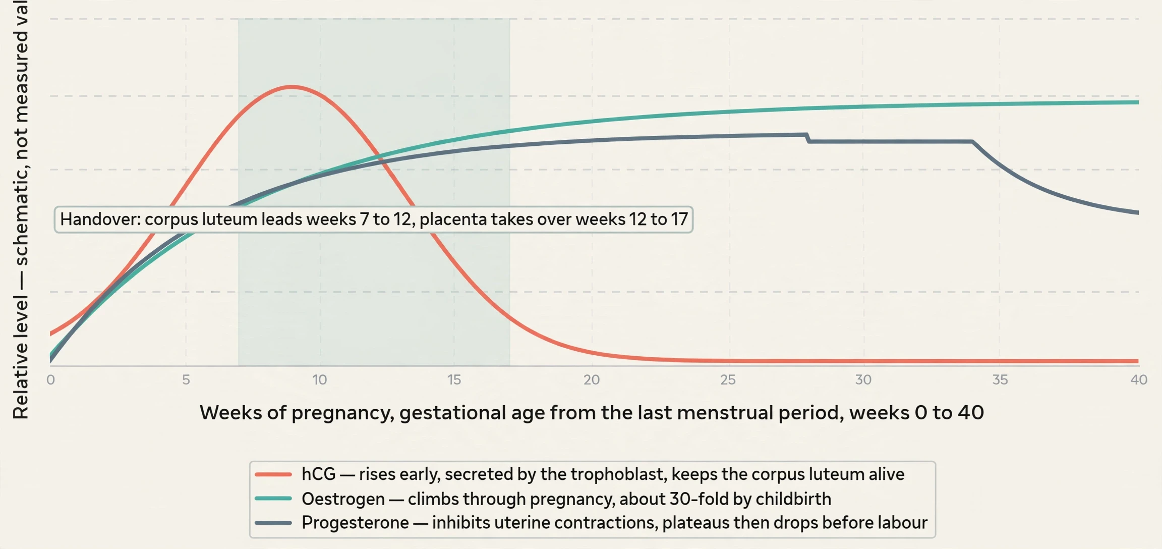
Human chorionic gonadotropin (hCG) is secreted by the trophoblast, and it directs the corpus luteum to survive, enlarge and continue producing progesterone and oestrogen to suppress menstruation. The corpus luteum is the remnant of the follicle that released the egg, and in a non-pregnant cycle it degenerates after about two weeks, progesterone falls, and menstruation follows. hCG interrupts that sequence. It tells the corpus luteum to stay alive and keep making progesterone and oestrogen, and those hormones keep the uterine lining in place and suppress the next period. The missed period that starts the story in the first section is, at the level of hormones, hCG doing its job.
Within a few days of implantation, the trophoblast has made enough hCG for a home urine test to turn positive. The test is not detecting the embryo. It is detecting the embryo's signal — the hormone the trophoblast makes to keep its own support system alive. That is why the test turns positive only after implantation, and why it can be negative a day or two earlier even when implantation has happened. The hormone has to accumulate, and accumulation takes days. The small stick on the bathroom shelf is, in effect, a chemical detector for a message the embryo sent to the ovary.
7.2 The handover: corpus luteum to placenta (Weeks 7–17)
During weeks 7 to 12 from the LMP, pregnancy hormones are primarily generated by the corpus luteum, and as the placenta develops and the corpus luteum degenerates during weeks 12 to 17, the placenta gradually takes over as the endocrine organ of pregnancy. This is the central changing of the guard in how pregnancy works, and it deserves its full weight in the story. For the first seven to twelve weeks, a small remnant in the ovary makes the hormones that hold the pregnancy. Then, over five weeks, the placenta — a new organ, built by the embryo, growing in the wall of the uterus — takes over the same job, and the corpus luteum quietly degenerates.
The handover is gradual, not sudden. The placenta does not switch on in a day, and the corpus luteum does not switch off in a day. They overlap, the placenta making more and the corpus luteum making less, until the placenta is doing the work alone. That overlap is a safety feature: if the handover were abrupt, any delay in the placenta's growth would leave the pregnancy without its hormones. As it is, the two sources share the job through the transition, and by week 17 the placenta is the endocrine organ of pregnancy in its own right. The timing of the handover also explains why the first trimester and the second feel different: the body is being run by a different gland.
7.3 Oestrogen and progesterone: growth, calm and preparation
Oestrogen levels climb throughout pregnancy, increasing 30-fold by childbirth, and oestrogens suppress FSH and LH production, effectively preventing ovulation. Thirty-fold is a large rise, and it is worth pausing on what it does. Oestrogen grows the uterus, increases blood flow to it, prepares the breasts for feeding, and — by suppressing follicle-stimulating hormone and luteinising hormone — stops the ovaries from releasing another egg. A pregnant person does not ovulate, not because the ovaries are tired, but because oestrogen is actively telling the brain not to start another cycle. The suppression is part of the design: one pregnancy at a time, with the body's resources devoted to it.
Progesterone inhibits uterine contractions, protecting the fetus from preterm birth, and its levels plateau and then drop in late gestation, allowing contractions to intensify and progress to true labour. Progesterone is the calm hormone of pregnancy. It quiets the muscle of the uterus, the myometrium, so that the organ that will one day contract powerfully to deliver a baby spends nine months not contracting. It also thickens the lining, supports the placenta and prepares the breasts. Without progesterone's calming effect, the uterus would contract too early and too often. With it, the pregnancy is held.
hCG — the early signal. Secreted by the trophoblast; keeps the corpus luteum alive; rises in the first weeks so a home test can turn positive a few days after implantation.
Oestrogen — the growth hormone of pregnancy. Climbs 30-fold by childbirth; grows the uterus and breasts; suppresses FSH and LH, preventing ovulation.
Progesterone — the calm hormone. Inhibits uterine contractions and protects against preterm birth; plateaus and then drops in late gestation so labour can begin.
The placenta's other hormones — the supporting cast. Human placental lactogen, placental growth hormone, relaxin and kisspeptin, each adjusting metabolism, softening tissues or regulating the placenta itself.
7.4 How hormones end pregnancy: the shift toward labour
Progesterone's fall is the hinge on which labour turns. As its levels level off and then fall in late gestation, contractions can intensify and progress to true labour. The uterus, held quiet for months by progesterone, becomes sensitive again as progesterone falls and oestrogen, now thirty times its starting level, makes the muscle more responsive to oxytocin. The balance shifts: less calm, more readiness. The cervix softens under the influence of relaxin and other signals. The fetus, now about 2.5 to 4 kg and about 51 cm long, presses on the cervix. And the feedback loop that will drive labour — contractions stretching the cervix, stretching boosting oxytocin, oxytocin driving stronger contractions — is ready to run. Hormones do not only start pregnancy and hold it. They end it, too, by withdrawing the calm and letting the contraction begin. The next section describes what the body does under those hormones; the section after that describes the labour they make possible.
8. The Mother's Body Changes: Blood, Breath, Bladder and Immunity
The pregnant person's body is not a passive container for the fetus. It is a system adapting on purpose, and the adaptations are large. Blood volume rises by a substantial fraction. Breathing deepens by half. The uterus grows from the size of a fist to an organ that holds a baby. The bladder is compressed. And the immune system, which should by its ordinary rules reject a fetus carrying foreign genes, learns to tolerate it instead. How pregnancy works, seen from the mother's side, is a story of a body remodelling itself for two.
8.1 Blood volume and the heart work harder
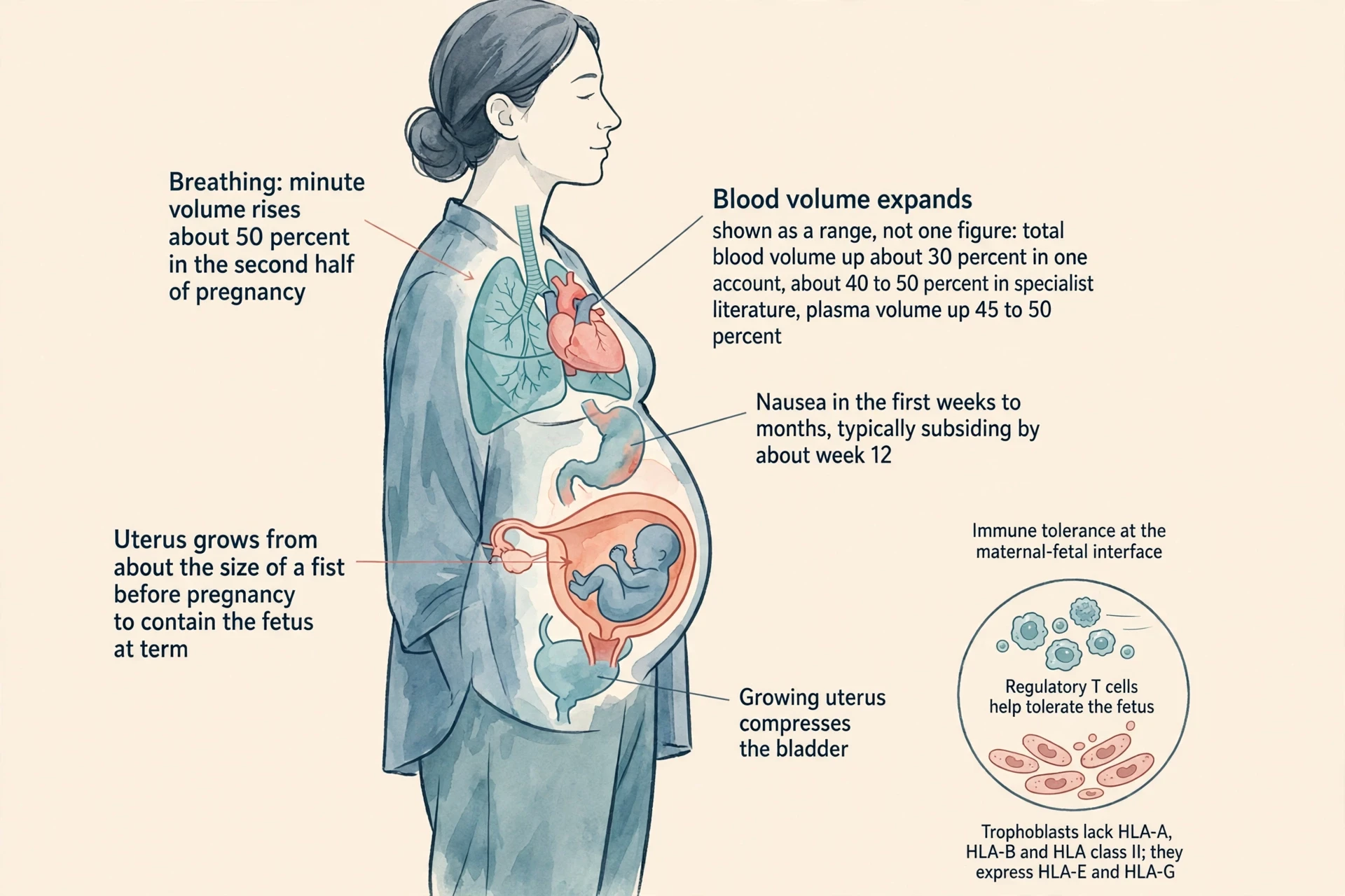
Blood volume increases substantially during pregnancy, and the figures for that increase need to be given as a range, with the measurement distinction explained, because the sources do not give a single settled number. In the OpenStax account, by childbirth blood volume exceeds its preconception volume by 30 percent, or approximately 1 to 2 litres. In specialist literature, plasma volume rises by 45 to 50 percent and total blood volume by about 40 to 50 percent. The difference is plasma versus total blood volume: plasma is the liquid part of blood, and it expands more than the total, because red blood cells are added more slowly than plasma. Both accounts are describing the same expansion through different measures, and neither should be presented as the single answer. The honest statement is the range, with the distinction named: total blood volume up by about 30 percent in one account and about 40 to 50 percent in specialist literature, plasma volume up 45 to 50 percent, an increase of roughly 1 to 2 litres by childbirth in the OpenStax account.
Why does the body make so much extra blood? The placenta needs it, the uterus needs it, the fetus needs it, and the body needs a reserve for the blood lost at birth. The heart works harder to move the larger volume, and the extra plasma dilutes the red cells slightly, which is why the blood of pregnancy looks thinner on a test even though there is more of it in total. The expansion is not a side effect. It is a planned increase, built over months, and it is one of the largest changes in the mother's body.
8.2 Breathing for two: minute volume rises
During the second half of pregnancy, respiratory minute volume increases by 50 percent to compensate for the oxygen demands of the fetus and the increased maternal metabolic rate. Minute volume is the amount of air moved in and out of the lungs in a minute, and a 50 percent rise is a large change. It happens mainly because each breath gets deeper, not because breathing gets much faster, and it happens while the growing uterus is pushing the diaphragm upward and leaving less room for the lungs to expand. The body compensates for a smaller space by using the space it has more fully.
The oxygen the extra breathing brings in is shared. The fetus's oxygen comes through the placenta, from the mother's blood, and the mother's own tissues need more oxygen too, because her metabolic rate has risen to support the pregnancy. The 50 percent rise in minute volume serves both demands at once. The same breathing and circulation systems that adapt to exercise — a faster heart, deeper breathing, more blood moved per minute — adapt here under a different demand: not a run, but nine months of carrying a growing passenger and a new organ. The parallel is instructive. The body has a limited set of adaptations, and it deploys them for pregnancy as it deploys them for exertion, with different triggers and a much longer duration.
8.3 Nausea, the bladder and the gut in early pregnancy
Nausea and vomiting are common during the first few weeks to months of pregnancy, thought to be caused by increased circulation of oestrogen, progesterone and hCG, and nausea typically subsides by about week 12. The timing matches the hormone story: hCG rises steeply in the first weeks, oestrogen and progesterone climb, and nausea follows the same curve, easing as hCG falls after its early peak and as the placenta takes over hormone production. The cause is described as "thought to be" in the source, and that hedging is kept here, because nausea has more than one contributor and the hormone link, while strong, is not the whole explanation. The reassurance in the fact is the timing: typically subsiding by about week 12, as the first trimester ends.
The growing uterus compresses the urinary bladder, leading to frequent urination, and the maternal urinary system processes both maternal and fetal wastes, further increasing total urine volume. The bladder, a flexible bag that sits just in front of the uterus, has less room to expand as the uterus grows, so it fills sooner and empties more often. At the same time, the kidneys are filtering more blood — the expanded blood volume, plus the waste the fetus sends back through the placenta — and making more urine. Frequent urination in pregnancy is therefore two effects at once: less space to store urine, and more urine being made. The gut microbiome is mentioned here only as background — the gut is a system that also shifts during pregnancy, and readers who want to understand it can read about it separately. No claim is made here that pregnancy changes the microbiome in a particular way, because no such claim was in the verified research for this article.
8.4 The immune puzzle: tolerating a semi-allogeneic fetus
The fetus is, genetically, half foreign. It carries genes from the father that the mother's immune system has never seen, and by the ordinary rules of immunology a tissue carrying foreign genes should be rejected. A transplanted kidney from a stranger is rejected unless the immune system is suppressed. A fetus carrying a stranger's genes is not rejected, in a normal pregnancy, for nine months. That is the immune puzzle of pregnancy, and it is the most distinctive depth in the research for this article.
The semi-allogeneic fetus needs to be tolerated by the mother's immune system, and regulatory T cells, known as Tregs, play a prominent role in that tolerance; low numbers of Tregs are associated with pregnancy failure and complications such as pre-eclampsia. Tregs are a specialised type of immune cell whose job is to calm the immune response and prevent it from attacking the body's own tissues — and, in pregnancy, from attacking the fetus. They accumulate at the boundary between mother and fetus, and their numbers rise during pregnancy. When they are too few, the tolerance weakens, and pregnancy failure and pre-eclampsia become more likely. The association is not proof that low Tregs cause those outcomes on their own, but it is a strong clue about where the tolerance is maintained.
Trophoblasts help evade maternal immune recognition in part by lacking the polymorphic HLA-A, HLA-B and HLA class II molecules on their cell surface and by expressing non-classical HLA-E and HLA-G. HLA molecules are the identity tags that immune cells read to decide whether a cell belongs to the body or not. The trophoblast, the fetal tissue that faces the mother's immune system, hides the most variable of those tags — HLA-A, HLA-B and HLA class II — and displays instead the quieter, less variable tags HLA-E and HLA-G, which signal "do not attack" rather than "inspect me closely." It is a form of camouflage at the molecular level: the fetus does not announce itself loudly, and what it does show is a signal for tolerance.
The immune system normally uses regulatory T cells and HLA molecules to tell self from non-self and to prevent autoimmunity. Pregnancy borrows that machinery and turns it to a new purpose: tolerating a fetus that is half self and half other. I find this the most elegant part of how pregnancy works. The body does not switch its immune system off — that would leave mother and fetus defenceless against infection. It retunes it, locally and precisely, at the boundary where mother and fetus meet, so that defence continues everywhere else while tolerance holds at the placenta. A system that can do both at once, in the same body, is a system worth admiring.
8.5 The uterus grows: from fist-sized to full-term
During the second and third trimesters, the pre-pregnancy uterus — about the size of a fist — grows dramatically to contain the fetus. The uterus is a muscle, and like any muscle it can grow, but no other muscle in the body grows by this much, this fast, and then returns to its former size. Its wall thickens, its blood vessels multiply, its cavity expands from about 10 millilitres to several litres, and it does all of this while remaining quiet, held calm by progesterone, until the time comes to contract. The fist-to-full-term comparison is the simplest way to hold the scale in mind: an organ the size of a clenched hand becomes an organ large enough to hold a baby, a placenta, a cord and a sac of fluid, and then, after birth, shrinks back over weeks. The growth is not stretching alone. The muscle cells themselves grow larger and more numerous, and the organ is rebuilt from within. When labour finally begins, that rebuilt muscle is the engine that will deliver the baby, and its size is part of why it can do the work.
9. Stages of Labour: How Birth Actually Happens
The stages of labour are the last chapter of how pregnancy works, and they are also the chapter where the hormone story becomes a mechanical story. A positive feedback loop drives labour, and the three stages that follow from it — dilation, expulsion, afterbirth — are the working-out of that loop in the body. The description here is mechanical and factual. Labour is hard work, and the people who have been through it know that better than any article can say. What an article can do is explain the mechanism clearly, so that the work has a shape in the reader's mind.
9.1 What starts labour: the oxytocin positive-feedback loop
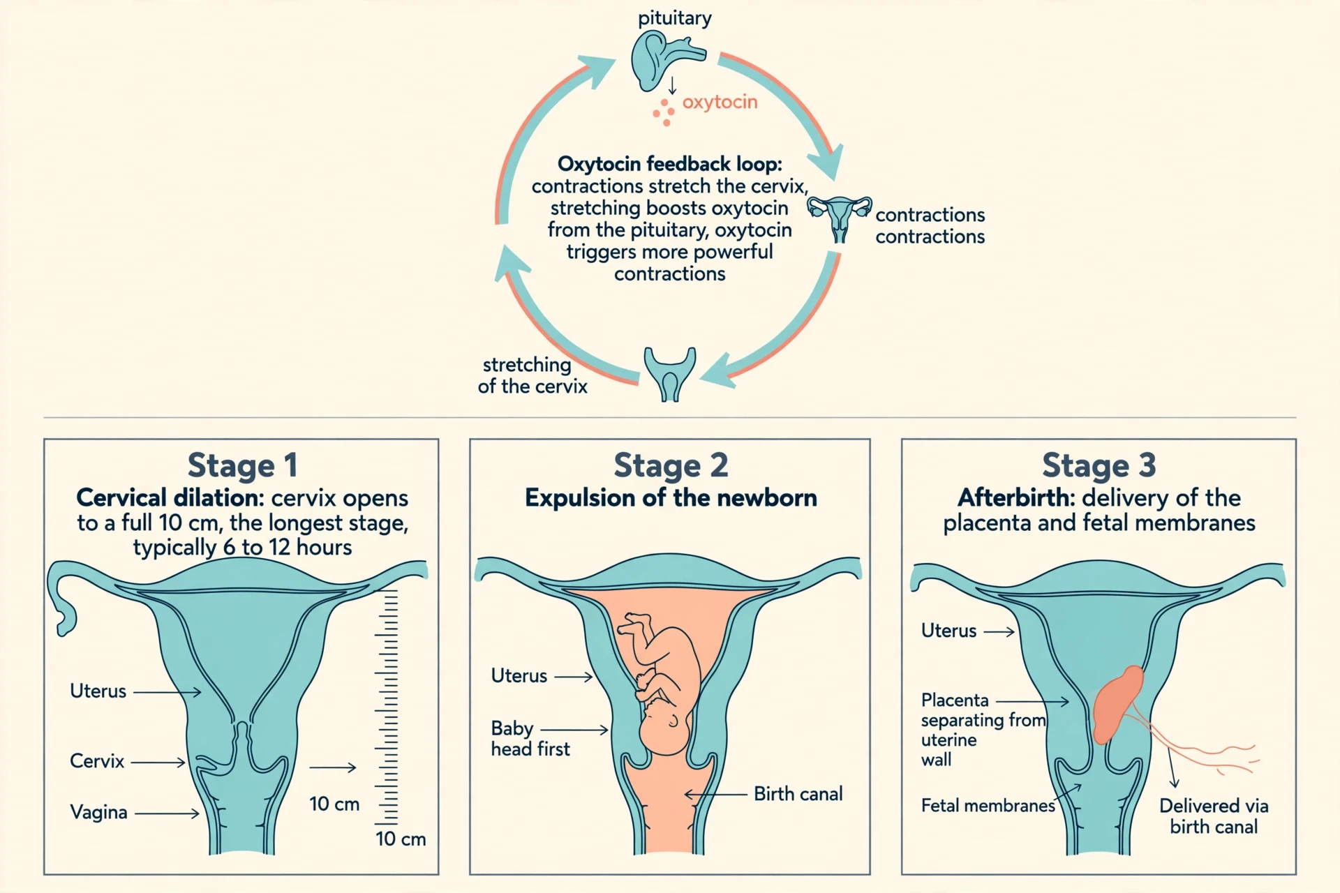
Labour is driven by a positive feedback loop: uterine contractions stretch the cervix, cervical stretching induces reflexive uterine contractions and boosts oxytocin secretion from the pituitary, which triggers more powerful contractions. A positive feedback loop is a loop that amplifies itself. Each contraction stretches the cervix a little more. The stretch signals the brain to release more oxytocin from the pituitary gland. Oxytocin makes the uterus contract more powerfully. The stronger contraction stretches the cervix further. Round and round it goes, each turn stronger than the last, until the cervix is fully open and the baby can pass through.
Positive feedback is rare in the body, because most systems use negative feedback to hold themselves steady. Labour is one of the few places where the body wants a runaway process, and it builds one deliberately. Progesterone's fall removes the brake, oestrogen's rise makes the uterus more sensitive to oxytocin, and the stretching of the cervix provides the trigger that keeps the loop turning. The loop does not need to be started from outside. Once it is running, it sustains itself.
True labor progresses in a positive feedback loop in which uterine contractions stretch the cervix, causing it to dilate and efface, or become thinner. Cervical stretching induces reflexive uterine contractions that dilate and efface the cervix further. In addition, cervical dilation boosts oxytocin secretion from the pituitary, which in turn triggers more powerful uterine contractions. — OpenStax Anatomy and Physiology 2e
9.2 Stage one: cervical dilation
Childbirth can be divided into three stages: cervical dilation, expulsion of the newborn, and afterbirth, the delivery of the placenta and associated fetal membranes. Stage one is the dilation stage, and it is the longest. For vaginal birth, the cervix must dilate fully to 10 cm in diameter, and the dilation stage is the longest stage of labour, typically taking 6 to 12 hours. Ten centimetres is about the width of a newborn's head, and the cervix — a firm, closed ring of tissue at the bottom of the uterus — must open from closed to that width, and also thin out, in a process called effacement, before the baby can pass.
Six to 12 hours is a typical duration, not a rule. First labours are often longer, later labours often shorter, and the range around the typical figure is wide. The work of stage one is done by the contractions of the feedback loop, each one pulling the cervix a little more open, each one a little stronger than the last as oxytocin rises. The person in labour feels the contractions as waves, building and easing, and the cervix, which cannot be seen from outside, is doing its slow, measurable work in response.
9.3 Stage two: expulsion of the newborn
Stage two is the expulsion of the newborn. Once the cervix is fully dilated to 10 cm, the baby can move down through the birth canal, and the contractions, now joined by the person's own pushing efforts, move the baby through the pelvis and out into the world. The baby turns as it descends, navigating the shape of the pelvis, and the head, the widest part, emerges first in the usual presentation. The stage ends when the baby is born, and the first breath follows — sometimes with a cry, sometimes quietly, as the lungs, filled with fluid until now, fill with air for the first time.
Stage two typically takes up to 2 hours, but it can last longer or be completed in minutes. What matters is the mechanism: the cervix is open, the uterus is contracting under the oxytocin loop, and the baby moves down the canal that the contractions and the pelvis together have made passable.
9.4 Stage three: the afterbirth — delivery of the placenta
Stage three is the afterbirth — the delivery of the placenta and associated fetal membranes. After the baby is born, the uterus continues to contract, smaller now, and those contractions shear the placenta away from the wall of the uterus. The placenta, the cord and the membranes follow the baby out, usually within minutes, and the uterus then contracts down hard to close the blood vessels that fed the placenta. That final contraction is as important as the delivery itself: it is what stops the bleeding from the place where the placenta was attached.
The afterbirth is the end of the placenta's life. An organ built by the embryo, the first fetal organ to develop, grown in the wall of the uterus for nine months, exchanged oxygen and nutrients for waste, made hormones, filtered what it could and let antibodies through — it is delivered, examined, and its job is done. The baby no longer needs it, because the baby can now breathe, eat and keep warm for itself, with help.
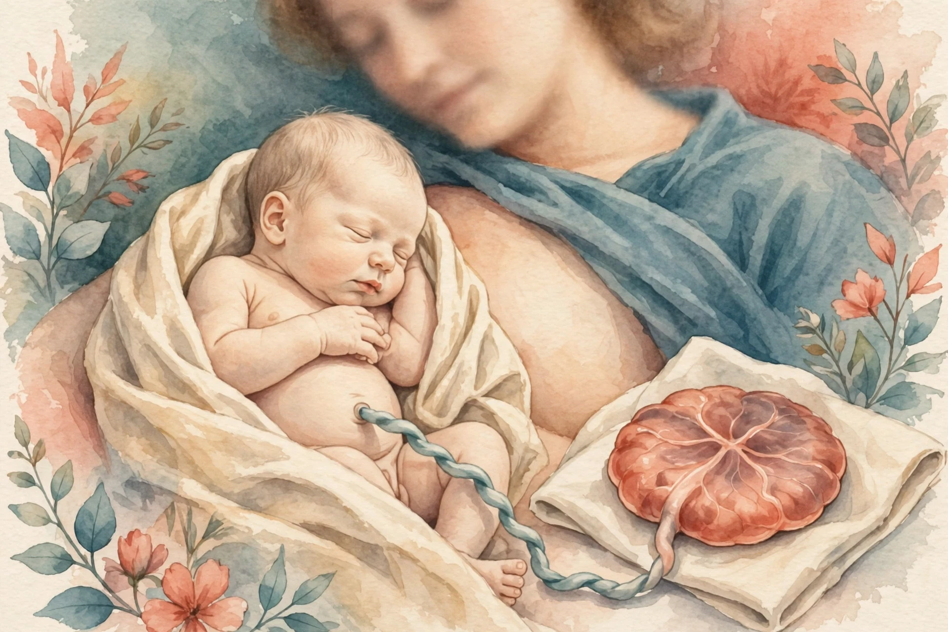
9.5 The first minutes after birth: what the numbers at birth mean
The baby arrives measuring about 35.5 to 40.5 cm crown to rump, weighing approximately 2.5 to 4 kg, and about 51 cm long. Those are the numbers the baby arrives with. In the first minutes after birth, the numbers stop being measurements of a fetus and start being measurements of a newborn, and the body has to make a series of rapid changes to match. The lungs, which have been making surfactant since developmental weeks 21 to 30, expand with air for the first time, and the surfactant keeps them open between breaths. The cord, which carried oxygen and nutrients through one vein and waste through two arteries, is no longer needed, because the baby's own lungs and gut can now do that work. The circulation rearranges itself, closing the shortcuts that let blood bypass the lungs in the womb. When a premature newborn has not made enough surfactant, respiratory distress syndrome may follow, and that is why a baby born before the lungs are ready needs help to breathe.
Table 4 collects the three stages in one place. Stage one typically takes 6 to 12 hours, and stage two typically takes up to 2 hours. Stage three is described by what happens, not by how long it takes.
Stage | What happens | Typical character |
|---|---|---|
Stage one — cervical dilation | The cervix opens and thins until it is fully dilated to 10 cm in diameter | The longest stage, typically 6 to 12 hours; driven by the oxytocin positive-feedback loop |
Stage two — expulsion of the newborn | The baby moves down the birth canal and is born; the lungs fill with air for the first time | Contractions plus the person's own efforts; usually up to 2 hours, though it may run longer or finish in minutes |
Stage three — afterbirth | The placenta and associated fetal membranes are delivered; the uterus contracts down to close the placental blood vessels | Follows the birth of the baby, usually within minutes; the uterus contracts down to close the placental blood vessels |
10. When Things Go Differently: Complications, Factually and Without Alarm
Pregnancy usually proceeds without serious complication, and the figures in this section should be read in that light. They are rates in a population, not predictions for any individual reader. A rate of 20 percent means that, across many pregnancies, about one in five ends in miscarriage; it does not mean that any particular pregnancy has a one-in-five chance in the way a coin has a one-in-two chance. Risk in pregnancy is shaped by age, health, history and care, and a population rate is a poor guide to an individual outcome. This section gives the verified rates with their dates and settings, defines the terms plainly, and points the reader to a clinician for anything personal. No alarm, no individual recommendations.
10.1 Miscarriage: how common it is, in plain terms
Approximately 20 percent of pregnancies miscarry, and early pregnancy loss accounts for over 50,000 admissions in the UK annually. Those figures come from NICE Guideline NG126, the UK guideline on ectopic pregnancy and miscarriage, and they describe a common event. Miscarriage is the loss of a pregnancy before the fetus can survive outside the uterus, and most miscarriages happen in the first trimester, often because of chromosomal errors in the embryo that make further development impossible. The 20 percent figure includes pregnancies that are lost very early, some before the person knew they were pregnant, and the figure is a reminder that the high failure rate of implantation described in the third section has a counterpart later in pregnancy: not every pregnancy that implants continues to birth.
That admissions figure is a measure of care, not only of loss. It means tens of thousands of people each year need hospital care for a miscarriage — for bleeding, for pain, for a procedure to complete the loss, or for observation. Miscarriage is common, it is often painful in every sense, and it is a normal subject for medical care and for conversation. Anyone experiencing bleeding or pain in pregnancy should seek care from their own clinician promptly, rather than waiting to see whether it resolves. This article does not advise on symptoms; it states that care exists and that a clinician is the person to contact.
10.2 Ectopic pregnancy: implantation outside the uterus
In 1 to 2 percent of cases, the embryo implants outside the uterus, an ectopic pregnancy, and in the vast majority of ectopic pregnancies it implants in the uterine tube. An ectopic pregnancy is a pregnancy that has implanted in the wrong place. The uterus is the only organ built to hold a growing fetus; the tube is a passage a few millimetres wide, and a blastocyst growing there cannot survive and can rupture the tube, causing internal bleeding. The rate of ectopic pregnancy is 11 per 1,000 pregnancies in the UK, with a maternal mortality of 0.2 per 1,000 estimated ectopic pregnancies, according to NICE Guideline NG126. The two figures together tell a story: ectopic pregnancy is uncommon, about one in a hundred pregnancies, and when it is recognised and treated, death from it is rare in the UK, about two deaths per ten thousand ectopic pregnancies. The key word is "recognised". Ectopic pregnancy is a reason to seek care early for pain or bleeding in pregnancy, and a clinician is the person to make that assessment.
10.3 Gestational diabetes and pre-eclampsia: ranges, not single figures
Gestational diabetes and pre-eclampsia are among the most common medical complications in pregnancy, with prevalence rates of 10 to 25 percent and 3 to 9.4 percent respectively in the cited scoping review. Both figures are ranges, and both must be presented as ranges, because the range is the fact. Gestational diabetes is diabetes that appears during pregnancy, when the hormones of pregnancy make the mother's body less sensitive to insulin and blood sugar rises. Pre-eclampsia is a condition of high blood pressure and organ effects, most often involving the kidneys, that appears after 20 weeks of pregnancy. The wide gestational-diabetes range, 10 to 25 percent globally, reflects different diagnostic criteria: high-income-country figures using American Diabetes Association criteria are 6 to 9 percent, while broader global criteria capture more cases. Pre-eclampsia's range, 3 to 9.4 percent, reflects different populations and definitions in the same way.
The lesson of the ranges is not that the figures are unreliable. It is that a prevalence depends on where the line is drawn and who is counted. A reader who sees a single figure for gestational diabetes — "14 percent" or "7 percent" — without a criteria set or a population attached to it is seeing a number stripped of its meaning. This article gives the range and the reason for it, and leaves the diagnosis and management of either condition to the reader's own clinician, who can apply the criteria used in their own health system.
10.4 Preterm birth and why lung maturity matters
In the United States, 10.41 percent of live births were preterm in 2024, before 37 completed weeks, according to the CDC National Center for Health Statistics' final 2024 data, released on 9 June 2026 — about one baby in ten, unchanged from 2023. Preterm birth is birth before 37 completed weeks of gestational age, and it is the most common reason a newborn needs intensive care. The connection to the rest of this article is surfactant. The lungs begin producing surfactant during developmental weeks 21 to 30, the same weeks when rapid weight gain begins, and a premature baby whose lungs have not made enough surfactant may develop respiratory distress syndrome. A baby born at 30 weeks has lungs that are still learning to make surfactant; a baby born at 38 weeks has lungs that have had weeks to practise. The 10.41 percent figure is therefore not only a statistic about timing. It is a statistic about how many babies are born before their lungs are fully ready, and how much neonatal care exists to support them until they are.
Viability belongs in this discussion, because it is often confused with preterm birth. In modern high-resource settings, medical viability is generally considered to be between 23 and 24 weeks of gestational age, and there is no single gestational-age threshold that defines viability in all clinical settings. In United States academic medical centres between 2013 and 2018, the percentage of newborns who survived long enough to leave hospital was 30 percent at 22 weeks, 55 percent at 23 weeks, 70 percent at 24 weeks and 80 percent at 25 weeks. Those are cohort figures from a specific place and time — US academic centres, 2013 to 2018 — and survival varies by hospital, by whether full intensive care is given, and by birth weight and sex. They are given here with their dates and setting, never as a universal prediction, because a single universal figure for viability does not exist.
10.5 What this section is not: no personal advice, no alarm
This section explains mechanisms and rates. It makes no diagnosis, it predicts no reader's outcome, and it advises no individual course of action. A rate is a fact about a population. A symptom is a fact about a person, and symptoms belong with a clinician, promptly, in person. If anything in this section resembles something the reader is experiencing, the right next step is not to re-read the article but to contact the reader's own clinician or local maternity care. The article's job is to make the biology clear. The clinician's job is to care for the person.
11. Prenatal Care and Screening: What Checks Happen and When
Prenatal care is the schedule of checks that follows a pregnancy from the first positive test to birth. Its purpose is threefold: to date the pregnancy, to check the anatomy and growth of the fetus, and to screen for conditions that can be managed better when they are found early. The checks are not a single test. They are a timeline, and the timeline matters as much as any individual result. A test done at the wrong week can be unreadable; a screening test is not a diagnosis; and a normal result is a piece of information, not a guarantee. This section gives the timing and the structure of the checks, without thresholds or rates that were not verified. Knowing the checks, and when each one happens, is part of knowing how pregnancy works in practice.
11.1 The dating ultrasound and the anatomy scan
A dating ultrasound — the 11-to-14-week scan, which also measures nuchal translucency — is typically done at 11 to 14 weeks, and an anatomy scan at about 20 weeks looks at the fetus's brain, heart, lungs and other organs. The dating scan's job is to fix the two clocks: by measuring the fetus, the sonographer can check whether the gestational age counted from the last period matches the size of the embryo or fetus, and correct the due date if it does not. The anatomy scan's job is different. The detailed anatomy scan, which looks at the brain, heart, spine, kidneys, limbs and placenta, is typically done at about 18 to 22 weeks, when the fetus is large enough for its structures to be seen clearly and small enough to fit in a single view. The two scans are complementary: the first answers "when," the second answers "how is it formed."
11.2 Screening vs diagnosis: NIPT and what a positive result means
Non-invasive prenatal testing, or NIPT, can be done from week 10 and analyses cell-free fetal DNA in maternal blood to screen for chromosomal conditions, but it is a screening test, not a diagnostic test — a positive result is confirmed by chorionic villus sampling or amniocentesis. The distinction between screening and diagnosis is the most important idea in this section, and it is worth stating carefully. A screening test sorts a population into higher and lower likelihood. It does not say whether a particular fetus has a condition. A diagnostic test, such as chorionic villus sampling or amniocentesis, samples fetal tissue directly and can say, with much greater certainty, whether a chromosomal condition is present. NIPT is a screening test because it analyses fragments of fetal DNA circulating in the mother's blood, not the fetus's own cells, and a positive screening result is a reason to offer a diagnostic test, not a diagnosis in itself.
No single detection-rate percentage is given here, because a percentage means little without the condition, the population and the test version attached to it. The timing — from week 10 — and the screening-versus-diagnostic distinction are the facts a reader needs to understand what a result means.
11.3 Screening for gestational diabetes at 24 to 28 weeks
Universal screening for gestational diabetes is done at 24 to 28 weeks, starting with a one-hour 50-gram glucose challenge test and, if abnormal, a three-hour glucose tolerance test. The two-step structure is the point. The first step is a challenge: drink a measured amount of glucose, wait an hour, and measure the blood sugar. If the result is abnormal, the second step is a longer, more detailed test: a three-hour glucose tolerance test, with several measurements over three hours. Thresholds for both steps vary by criteria set, so no single threshold set is presented here as universal. The timing, 24 to 28 weeks, is chosen because gestational diabetes, driven by the hormones of pregnancy, typically appears in the second half of pregnancy, when insulin resistance has risen. Screening at that point catches it when it can still be managed for the rest of the pregnancy.
11.4 The screening timeline in one place
Table 5 collects every check in this article into one chronological reference. The "screening or diagnostic" column is the thread to follow: most prenatal checks are screening, and a screening result that raises a question is a reason to offer a diagnostic test, not a conclusion in itself.
Week (gestational) | Check | What it looks at | Screening or diagnostic |
|---|---|---|---|
From week 10 | Non-invasive prenatal testing (NIPT) | Cell-free fetal DNA in maternal blood; screens for chromosomal conditions | Screening — a positive result is confirmed by chorionic villus sampling or amniocentesis |
11 to 14 weeks | Dating ultrasound | Fetal size and gestational age; checks the due date | Screening and dating |
About 18 to 22 weeks | Anatomy scan | Brain, heart, spine, kidneys, limbs and placenta; about 20 weeks in the Cleveland Clinic wording | Screening |
24 to 28 weeks | Gestational diabetes screening | Blood sugar after a glucose challenge; one-hour 50-gram test, then a three-hour test if abnormal | Screening |
From week 10: NIPT — cell-free fetal DNA screening for chromosomal conditions.
11 to 14 weeks: Dating ultrasound — fixes gestational age and the due date.
About 18 to 22 weeks: Anatomy scan — a detailed look at the brain, heart, spine, kidneys, limbs and placenta.
24 to 28 weeks: Gestational diabetes screening — one-hour glucose challenge, then a three-hour tolerance test if abnormal.
12. How We Learned to See Pregnancy: A Short History
Modern pregnancy care feels inevitable — a test from a pharmacy, a scan on a screen, a due date on a chart — but each of those things descends from a specific discovery, made by specific people, in a specific year. The story of seeing pregnancy is a short history with three turning points: a hormone found in urine in 1927, a sound picture of a fetus in 1958, and an antibody test in 1960 that replaced animals with chemistry. Each turning point made the next one possible, and each one is worth telling as a story rather than a date.
12.1 1927–1928: hCG and the first pregnancy test
In 1927, Aschheim and Zondek discovered human chorionic gonadotropin (hCG), a substance that appeared in the urine of pregnant women and only pregnant women, and found that hCG-rich urine injected into a rodent would cause it to ovulate — the first clinical pregnancy test, a bioassay, was born. Selmar Aschheim and Bernhard Zondek, working in Berlin, were looking for a hormone that explained the changes of pregnancy, and they found it in urine. The substance appeared only in the urine of pregnant women. When they injected urine rich in it into a rodent, the animal ovulated. The rodent's ovaries became the detector: if the animal ovulated, the woman was pregnant.
Aschheim and Zondek published their results in 1928, and the animal-based tests remained the norm until 1960, when antibody-based immunoassay tests were introduced. For more than thirty years, a pregnancy test meant a laboratory, an animal, and days of waiting. The test worked, and it was the first time pregnancy could be detected chemically, weeks before it could be felt or seen. It was also, by modern standards, slow and indirect — a living animal as a measuring instrument, because no chemical test yet existed that could detect hCG directly.
12.2 1960: antibody tests replace animal bioassays
In 1960, antibody-based immunoassay tests were introduced, and the animal bioassay began to give way. An immunoassay uses antibodies — proteins that bind to a specific molecule — to detect hCG directly, without an animal in the loop. A sample of urine, an antibody that recognises hCG, and a visible signal when the two meet: that is the principle of the home pregnancy test that sits on a pharmacy shelf today, and it descends directly from the 1960 shift. The test became faster, cheaper and more private, and it moved from the laboratory to the bathroom.
No single year for the first home pregnancy test is given in this article. Sources conflict between 1968, for Margaret Crane's prototype, and 1978, for the first commercial home test, and presenting one year as settled would be a false precision. The prototype and the commercial product are different events, and the history is better told as a sequence — a hormone discovered in 1927, a chemical test in 1960, a home test following later — than as a single date. What matters is the lineage: the small stick in the first section of this article works because Aschheim and Zondek found hCG in urine, and because immunoassay chemistry learned to detect it without an animal.
12.3 1958: ultrasound shows the fetus for the first time
Sonography in obstetrics and gynaecology began with the classic 1958 Lancet paper by Ian Donald and his team from Glasgow, "Investigation of abdominal masses by pulsed ultrasound", which contained the first ultrasound images of the fetus. Ian Donald, a Glasgow obstetrician, had been using ultrasound to examine abdominal masses, and with his team he turned the technique toward pregnancy. The paper showed, for the first time, that sound waves could make a picture of a fetus inside the uterus — a grainy, ghostly image by modern standards, but unmistakably a fetus, and unmistakably alive.
The history of sonography in Obstetrics and Gynaecology dates from the classic 1958 Lancet paper of Ian Donald and his team from Glasgow. — PMC, "A Short History of Sonography in Obstetrics and Gynaecology"
Before 1958, a fetus could be felt, heard with a stethoscope, and, late in pregnancy, seen on an X-ray — with radiation. After 1958, it could be seen without radiation, repeatedly, safely, and earlier and earlier as the technology improved. The dating scan at 11 to 14 weeks, the anatomy scan at 18 to 22 weeks, and the first glimpse of cardiac activity at 6 to 7 weeks all descend from that paper. Ultrasound did not only add a picture. It changed the relationship between a pregnant person and the fetus, making the unseen visible, and it changed medicine, making the fetus a patient that could be examined directly.
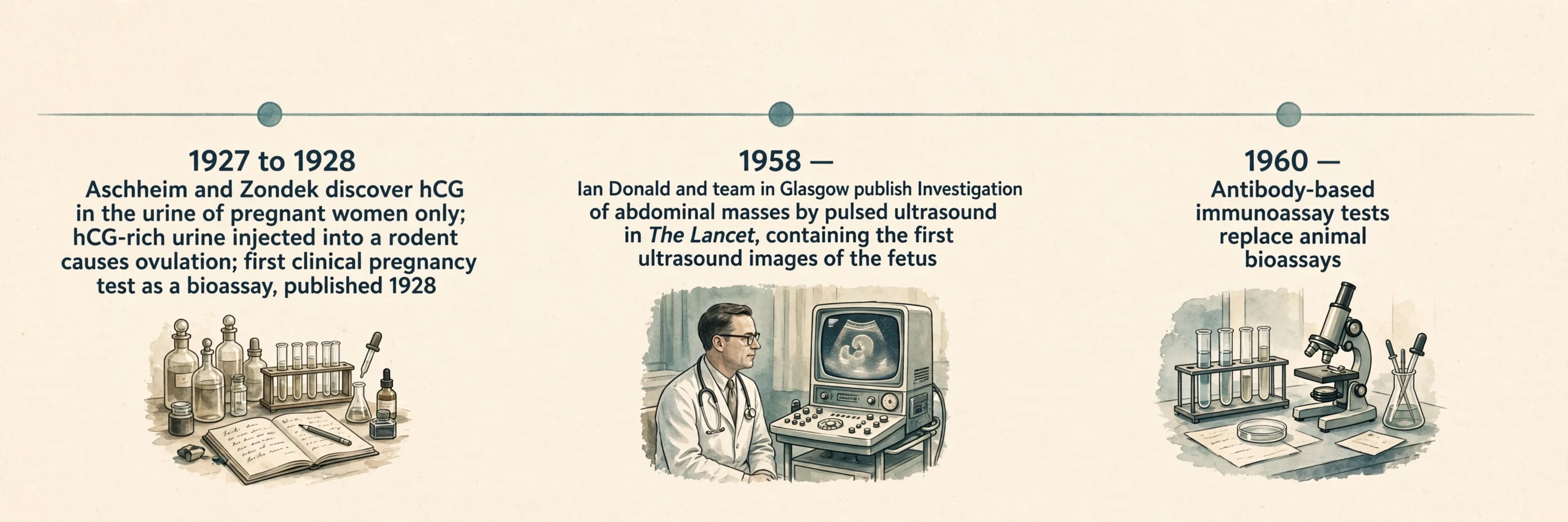
12.4 What history explains about today's tests
The home test descends from hCG, and the scan descends from 1958, and both exist because someone asked what pregnancy leaves behind in urine and what sound can show through tissue. The test and the scan are not separate inventions. They are two answers to the same question — how can we know what is happening inside the uterus without opening it? — given thirty years apart, one in chemistry and one in physics. The hormone found in 1927 became the test of 1960 and the home test that followed. The sound picture of 1958 became the dating scan and the anatomy scan. Each modern check in this article has a history, and knowing the history makes the check easier to understand: a test detects a signal the embryo sends, and a scan reads the echoes of a sound the body returns. Neither is magic. Both are the result of someone, decades ago, asking a precise question and building a tool to answer it.
13. Common Mistakes and Misconceptions About Pregnancy
Six errors recur in conversations about pregnancy, and each one has a verified fact that replaces it. They are worth listing together, because a reader who avoids these six will read every other page about pregnancy more accurately.
13.1 Mistake 1: counting pregnancy from conception
Pregnancy is counted from the first day of the last menstrual period, not from conception, so the gestational count runs about two weeks ahead of fertilisation age. A person who conceived four weeks ago is about six weeks pregnant by the count doctors use. The two-week gap is a convention, not a mistake, and it exists because the last period is observable while conception is not. Count from the last period, and the rest of the timeline lines up.
13.2 Mistake 2: a heartbeat at week 5 means a formed heart
The forming heart's first contractions come around day 21 to 22 after fertilisation — roughly 5 weeks and 1 to 2 days by the gestational count — but the early heart has no chambers or valves yet, which is why the American College of Obstetricians and Gynecologists says "embryonic cardiac activity" before around 10 weeks rather than "heartbeat". The first movements are the start of the heart, not a finished heart. The chambers form as two tubes fuse and fold, and the heart that beats at week 5 is a tube learning to pump, not a four-chambered organ.
13.3 Mistake 3: the placenta is a perfect barrier
The placenta is a partial barrier, not a wall. Maternal and fetal blood do not mix, and exchange happens by diffusion and transport, but alcohol can cross the placental barrier, and other small molecules cross with it. The placenta filters and selects — it carries antibodies across deliberately — but it does not seal the fetus off from the mother's blood. A barrier that lets some substances through is a filter, and filters have limits.
13.4 Mistake 4: a screening result is a diagnosis
NIPT screens; it does not diagnose. A positive screening result from week 10, based on cell-free fetal DNA in maternal blood, is confirmed by chorionic villus sampling or amniocentesis. A screening test places a population into higher and lower likelihood groups. Diagnosis samples fetal tissue directly. Confusing the two turns a reason to test further into a conclusion, and that confusion causes real distress. The correct reading of a positive screen is always the same: ask what the diagnostic test shows.
13.5 Mistake 5: viability is a single week
No one week marks viability: in modern high-resource settings it is generally placed between 23 and 24 weeks of gestational age, with no single threshold holding in all clinical settings. United States academic medical centres recorded, between 2013 and 2018, survival to hospital discharge climbing from 30 percent at 22 weeks to 80 percent at 25 weeks, passing 55 percent at 23 weeks and 70 percent at 24 — a gradient, not a cliff edge, and a cohort figure from a specific place and time. The outcome depends on the hospital, on whether full intensive care is given, and on birth weight and sex. Any page that names one week as the week of viability is simplifying a gradient into a line.
13.6 Mistake 6: pregnancy lasts exactly 40 weeks for everyone
Forty weeks from the last period is the convention, and 38 weeks from conception is the biology, and a due date is an estimate. In days, full term is approximately 270 days from conception, and the due date falls 284 days after the last period, on the usual assumption that conception happened on day 14 of the cycle. Cycles vary in real life, conceptions happen on different days, and babies arrive across a window around the due date. The date on the chart is the centre of that window, not a deadline the body has agreed to meet.
The story told in this article, from a single fertilised cell to a newborn taking a first breath, is a story of two clocks, three layers, one temporary organ, a handful of hormones, and a feedback loop that ends the process it started. Fertilisation in a fallopian tube, cleavage without growth, a blastocyst embedding in the uterine lining, three germ layers becoming every organ, a placenta that exchanges, filters and signals, hormones that hold the pregnancy and then let it go, a body that makes more blood and breathes more deeply and tolerates a half-foreign fetus, and labour driven by oxytocin around a loop that strengthens with every contraction — that is the mechanism, and it is the same mechanism in every pregnancy, however different the experience of it feels from the inside. That is how pregnancy works, told as one mechanism.
I have spent years reading about this process, and the part that still catches me is its ordinariness. It happens millions of times, in bodies that are not doing anything extraordinary, using the same tissues and signals the body uses for everything else — the same genome read three ways, the same immune cells retuned, the same muscle contracting under a hormone it already knows. Pregnancy is not a separate biology. It is ordinary biology, run for nine months toward a single purpose, and that is why it repays close attention: to understand how pregnancy works is to understand the body itself, working at its most coordinated.
14. Frequently Asked Questions
The questions below are the ones readers ask most often, answered from the facts already given in this article — each one another angle on how pregnancy works.
14.1 How does pregnancy work?
How does pregnancy work, in one paragraph? A sperm fertilises an egg, usually in a fallopian tube. The fertilised cell divides as it travels to the uterus, becomes a blastocyst, and implants in the uterine lining about a week after fertilisation. The outer layer of the blastocyst builds the placenta, which joins the fetus to the uterus by the umbilical cord, passing oxygen and nutrients one way and waste and carbon dioxide the other. Hormones — oestrogen, progesterone and hCG above all — hold the pregnancy, stop ovulation and quiet the uterus. Across roughly 38 weeks from conception — 40 weeks on the last-period count — a single cell grows into an embryo, then a fetus, then a newborn, delivered by labour driven by an oxytocin feedback loop. That, in short, is how pregnancy works.
14.2 What happens in each trimester of pregnancy?
First comes the building: fertilisation, implantation, gastrulation, the neural tube closing, the heart starting, and the change from embryo to fetus at week 9 of gestation, with every organ system laid down in rudimentary form by week 10 of gestation. The second trimester grows and moves: first felt movements in about weeks 16 to 20, with vernix and lanugo arriving in weeks 17 to 20, and the anatomy scan at about 18 to 22 weeks. The third trimester gains weight and prepares the lungs: weight gained rapidly, surfactant made, fat laid down from week 31, and birth at about 2.5 to 4 kg and about 51 cm long. That, trimester by trimester, is how pregnancy works.
14.3 When does the baby's heart start beating?
The first contractions of the forming heart arrive around day 21 to 22 after fertilisation — about 5 weeks and 1 to 2 days of gestational age — with the first pumping motions coming in the fourth week after fertilisation, about week 6 gestational. Cardiac activity is usually visible on transvaginal ultrasound at 6 to 7 weeks, and audible on a handheld Doppler at 10 to 12 weeks. ACOG — the American College of Obstetricians and Gynecologists — calls it "embryonic cardiac activity" before around 10 weeks, rather than "heartbeat", since the early heart has no chambers or valves yet. The rate peaks near 170 beats per minute around weeks 9 to 10, then settles to a typical 110 to 160 beats per minute.
14.4 What is the placenta and what does it do?
The placenta is the organ that connects the developing fetus to the wall of the uterus, joined to the fetus by the umbilical cord. It is built from fetal cells and is the first fetal organ to develop. It does three jobs at once: it exchanges oxygen and nutrients for waste and carbon dioxide, without maternal and fetal blood mixing; it acts as a partial barrier, blocking some harmful substances but not all — alcohol can cross it — while carrying antibodies to the fetus; and it acts as an endocrine gland, producing oestrogen, progesterone, hCG, human placental lactogen, placental growth hormone, relaxin and kisspeptin. Those three jobs together are the core of how pregnancy works.
14.5 How long does pregnancy last, and why is it counted as 40 weeks?
A typical pregnancy runs about 38 weeks from conception, or 40 weeks counted from the first day of the last period. The 40-week figure is the convention: gestational age is counted from the last period, which is observable, while conception happens about two weeks later and is not. Counted in days, a full-term pregnancy is approximately 270 days from conception, with the due date set at 284 days from the last period, assuming conception on day 14 of the cycle. Those 40 weeks fall into three trimesters of about 13 weeks each. A due date is an estimate built on an average, not an appointment.
14.6 When can the pregnant person first feel the baby move?
Quickening — the first felt movements — usually begins in approximately weeks 16 to 20, as the fetus's limb movements become more powerful. It is often felt first as a flutter, and it is easy to miss at the start. It is the first movement felt directly, rather than seen on a scan, and it marks the second trimester's change from a fetus that is built to a fetus that moves.
14.7 What is the difference between a screening test and a diagnostic test in pregnancy?
Screening sorts a population into higher and lower likelihood; diagnosis examines fetal tissue directly and can confirm whether a condition is present. NIPT, available from week 10, looks at cell-free fetal DNA in the mother's blood to screen for chromosomal conditions. It is a screening test, not a diagnostic test, and a positive screening result needs confirmation by chorionic villus sampling or amniocentesis. The same distinction applies to other prenatal checks: a screening result that raises a question leads to a diagnostic test, not to a conclusion on its own.
15. Sources
OpenStax Anatomy and Physiology 2e, 28.2 Embryonic Development — OpenStax 28.2
OpenStax Anatomy and Physiology 2e, 28.3 Fetal Development — OpenStax 28.3
OpenStax Anatomy and Physiology 2e, 28.4 Changes During Pregnancy, Labor, and Birth — OpenStax 28.4
World Health Organization, Maternal mortality fact sheet, published 7 April 2025, data year 2023 — WHO maternal mortality
NICE Guideline NG126, Ectopic pregnancy and miscarriage, Context — NICE NG126
Society for Endocrinology, Placenta — Society for Endocrinology placenta page
PMC, A Short History of Sonography in Obstetrics and Gynaecology — PMC short history of sonography
NewYork-Presbyterian Health Library, How Pregnancy Conception Occurs — NewYork-Presbyterian conception explainer
University of Utah, Human Reproduction lectures, Fertilization — University of Utah fertilisation lectures
Clue, What is Conception? — Clue conception explainer
The Bump, When Does a Fetus Have a Heartbeat — The Bump fetal heartbeat page
Americord Registry, When Does a Baby's Heart Start Beating — Americord heartbeat article
Biology Insights, When Does the First Heartbeat Happen — Biology Insights heartbeat review
Cleveland Clinic, Get Prenatal Testing — Cleveland Clinic prenatal testing
CDC, National Center for Health Statistics, Births: Final Data for 2024, released 9 June 2026 — CDC NCHS births page
Diabetes Therapy (Springer), Overlap of Gestational Diabetes Mellitus and Pre-eclampsia — Diabetes Therapy review
Frontiers in Immunology, Regulatory T cells in pregnancy — Frontiers Treg article
Frontiers in Immunology, Trophoblast immune evasion and HLA — Frontiers trophoblast HLA article
UNSW Embryology, Timeline human development — UNSW Embryology timeline
Endowment for Human Development, Prenatal Development Timeline — Endowment for Human Development timeline
LibreTexts, 17.4 Transit of Sperm — LibreTexts transit of sperm
Wikipedia, Fetal viability, for the cited 2013–2018 US academic-centre cohort only — Wikipedia fetal viability
Healthykids.sg, Antenatal checks by week — Healthykids antenatal checks guide
PANCE Review, summary of ACOG gestational diabetes screening — PANCE Review ACOG screening summary
OH Baby, History of Pregnancy Testing — OH Baby history of pregnancy testing
The Berliner, The Berliners who invented the world's first pregnancy test — The Berliner first pregnancy test article