1. A paper cut, a sunburn, and a blister — the organ you live in
A sheet of paper slides across the tip of your finger, and the sting is absurdly out of proportion to the injury — a small, sharp lesson in how the skin works. There is barely a mark — a hairline split, a bead of blood the size of a pinhead — and yet it throbs for hours. A cut that shallow should not hurt that much. It hurts because the skin is not a wrapper. It is wired, supplied with blood, patrolled by immune cells, and built to notice the smallest breach. That sting is the sound of an alarm system doing its job, and the quiet repair that follows over the next few days is the same organ doing a second job, just as competently and with far less fuss.
That is the question this article answers: how the skin works, from the dead cells on its surface to the fat layer that anchors it to muscle, from the pigment that shades it to the sweat that cools it. Three small, ordinary injuries will carry us through the whole story, because each one shows a different system at work before any anatomy has been named.
Take the sunburn first. You spend an afternoon outside without thinking about it, and the bill arrives the next morning: skin hot to the touch, tight when you move, tender under a shirt. A few days later it peels in thin flakes. What you are feeling is a barrier that did its job and failed at it at the same time. Ultraviolet light got past the pigment meant to absorb it, damaged the DNA in living skin cells, and the body answered with inflammation — extra blood flow, which is why the skin turns red and feels hot — and then with a controlled demolition, shedding the damaged surface cells in sheets. The peeling is not the damage. The peeling is the repair.
Then there is the blister after a long walk. Mile after mile, the same patch of skin rubs against the same seam of a shoe, and the layers shear apart. Fluid gathers in the gap, pressure builds, and every step hurts. That pocket of fluid looks like a problem, but it is a cushion: the body has inflated a small, sterile pillow between the damaged surface and the living tissue below, protecting the raw layer underneath while new cells grow across it. Pop it, and you remove the protection. Leave it, and it reabsorbs once the job is done. Three everyday events — a cut, a burn, a blister — and already three different skin systems have shown themselves: barrier, pigment, and repair, all running at once, all in tissue only a few millimetres thick.
Here is the short version of what follows. Your skin is a living, renewing organ built in three layers. The epidermis, the thin outer layer, waterproofs you and keeps microbes out. The dermis, the thick middle layer, carries the blood vessels, nerves, glands, and the collagen and elastin that give skin its strength and stretch. The hypodermis, the fatty layer underneath, insulates you, cushions you, and stores energy. Across those layers, the skin senses the world through four kinds of touch receptor, cools you with sweat and blood flow, makes a hormone from sunlight, fights infection with its own immune sentinels, and heals its own wounds in four overlapping phases. It covers about 1.5 to 2 square metres in an adult — roughly 15 percent of body weight — and it carries about 2 to 4 million sweat glands. It does all of this while replacing its own surface, cell by cell, roughly every month.
I first understood that this was an organ, not a covering, under a microscope in a teaching lab, looking at a thin slice of skin stained pink and purple. I had expected something simple, a sheet of cells. What I saw instead was a small city in cross-section: a wall of flattened cells at the top, a busy middle layer threaded with vessels and coiled glands, and fat cells below like a row of pale balloons. A hair follicle cut straight down its middle looked like a tunnel with a root at the bottom. I remember thinking that none of it looked like packaging. Packaging does not have a blood supply. That slide is still the image I carry when I think about skin — not a surface, but a place where things live and work.
The rest of this article walks through that place layer by layer, then function by function: the barrier, the pigment, the renewal cycle, temperature control, touch, healing, vitamin D, immunity, ageing, and the mistakes people make about all of it. It ends with the people who first mapped the skin under their own microscopes, one of them only twenty-one years old. By the end, the next time your skin itches, sweats, raises goosebumps, or closes a cut, you will be able to name the layer and the mechanism behind it.
2. What is skin? How the skin works as an organ
Call it a covering and you miss almost everything about it. Skin is an organ — the largest organ in the human body — and it earns that title the same way the heart or the liver does: it is made of several tissues working together as one system, and it performs jobs no other organ can do. The integumentary system, the name anatomists give to the whole arrangement, is the skin plus its appendages: hair, nails, and glands. Together they form the boundary between you and the world, and unlike a wall, that boundary is alive, sensing, secreting, and repairing itself around the clock.
The largest organ you own
OpenStax, the open anatomy textbook used in classrooms around the world, describes the skin as the body's largest organ, covering the whole body. That is not a piece of trivia. It is a clue to how much work the organ does. An organ that covers everything has to handle everything — heat, cold, sunlight, microbes, knocks, friction, dryness, and water — and it has to do so while staying flexible enough for you to move, sensitive enough for you to feel, and tough enough to keep your insides in. No other organ faces the outside world so directly, and no other organ is asked to be a barrier, a sensor, a radiator, a hormone factory, and a first line of immune defence all at once.
Part of what makes skin strange as an organ is that most of it is not one tissue. The surface is epithelium, the middle is connective tissue, the bottom is fat, and running through all of it are blood vessels, lymph vessels, and nerves. A heart is mostly muscle. A liver is mostly one kind of cell. Skin is a committee, and its members do very different jobs in very close quarters. That is why the layer-by-layer picture matters so much: you cannot understand what skin does until you can see where each job happens.
How big is it, really?
In an adult, the skin measures about 1.5 to 2 square metres — roughly the area of a single bed sheet, or a door. It accounts for around 15 percent of total body weight, with the literature giving a range of about 12 to 16 percent. Area and share of body weight are the size figures this article gives, and there is an honest reason for that restraint. Sources disagree widely on a single weight in kilograms, because authors differ on whether the fatty hypodermis counts as part of the skin at all. Some count it, some do not, and the number swings several-fold depending on the answer. Rather than pick one figure and pretend the disagreement does not exist, it is better to say what is solid: about 1.5 to 2 square metres, about 15 percent of you. That is a large organ by any measure, and the disagreement about the kilograms is itself a useful warning — even the experts argue about where this organ ends.
A thickness map, not a uniform sheet
Skin is not one thickness. It is a map of thicknesses, and the map follows function. The skin under your eyes and around your eyelids is the thinnest on the body, about 0.5 millimetres thick — thin enough to show the blood beneath it, which is why tired eyes look dark. The skin on your palms and the soles of your feet is the thickest, about 4 millimetres. Between those extremes, thickness changes with the work each patch of skin has to do: delicate where it needs to flex and sense, thick where it takes punishment.
Look closer and the layers tell the same story. The epidermis, the outer layer, is only about 0.05 millimetres thick over the eyelids, yet it thickens to about 1.5 millimetres on the palms and soles. The dermis, the middle layer, measures 1.5 to 4 millimetres and accounts for close to 90 percent of the skin's total thickness. That proportion matters. When you press your fingertip into your forearm, most of what you are pressing is dermis — the living core — with only a thin epidermal sheet on top of it. The thin places are built for sensitivity and movement. The thick places are built to take a beating. Both are the same organ, tuned differently.
What one square inch contains
The Cleveland Clinic offers a way to feel the density of this organ rather than just its size: one square inch of your skin contains approximately 19 million skin cells and 60,000 melanocytes, the pigment-making cells. It also contains about 1,000 nerve endings and 20 blood vessels. Read that again slowly. In a patch of skin the size of a postage stamp, a million-scale crowd of cells is being fed by twenty blood vessels and watched over by a thousand nerve endings. That is why a paper cut stings out of proportion to its size: the cut does not have to go deep to reach nerves, because the nerves are packed in just below the surface, waiting. It is also why skin heals as well as it does: the blood supply is dense, the cells are numerous, and the repair crews are already on site. Density is the point here, not trivia. Skin is not a thin sheet with a few things in it. It is a crowded, well-supplied tissue, and its crowdedness is what makes it so capable.
Those blood vessels belong to the same circulatory system that serves the rest of the body, and the skin's vessels are part of that larger story — how blood reaches every tissue, and how the heart drives it there — which is told in full in How the Human Heart Works. For now, it is enough to know that the vessels in the dermis do two jobs at once: they feed the skin itself, including the avascular epidermis above them, and they serve the whole body as a radiator, carrying heat to the surface or holding it in the core. That double duty will return in the section on temperature, where the same vessels become the body's thermostat.
3. How the skin works, layer by layer: epidermis, dermis, hypodermis
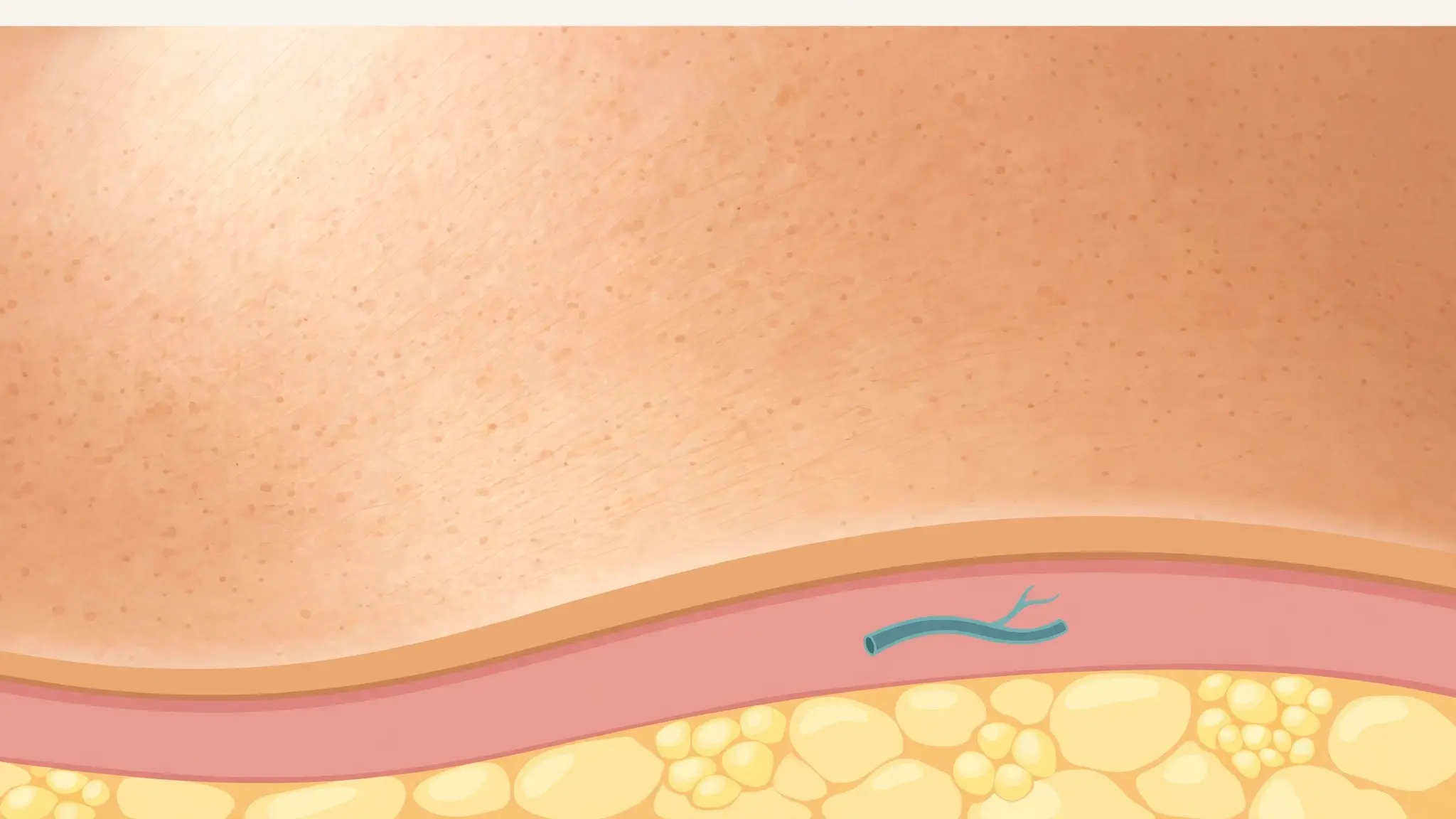
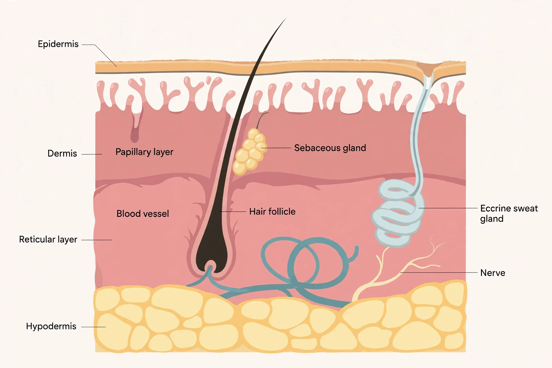
Everything that follows hangs on one picture, so it is worth getting that picture right before any detail arrives. Skin is built in three layers. The epidermis is the thin top layer, the one you can see and touch. The dermis is the thick middle layer, where the blood, nerves, glands, and strength live. The hypodermis is the bottom layer, mostly fat, which anchors the skin to the muscle and bone beneath it and insulates the body. That is the standard three-layer framing used throughout this article, and it is the framing most clinics and textbooks use.
There is a nuance worth one sentence, because it explains why you will sometimes read about two layers instead of three. Some anatomy texts count two main layers — epidermis and dermis — and treat the hypodermis as an associated layer beneath them, noting that it is not strictly part of the skin itself. The distinction is a matter of definition, not anatomy: the tissue is the same either way, and the fat layer does the same jobs. This article uses the three-layer picture because it is the clearest map of where things happen, and it says so once, here, so the reader is never confused by a textbook that counts differently. Every function that follows — barrier, pigment, sweat, sensation, healing — happens in one of those three layers, and that map is the foundation for understanding how the skin works.
Layer | Depth / position | Main tissue | Key cells / structures | Main jobs |
|---|---|---|---|---|
Epidermis | Top layer, the surface you see | Keratinised stratified squamous epithelium | Keratinocytes, melanocytes, Merkel cells, Langerhans cells | Waterproof barrier, microbe barrier, pigment, touch sensing |
Dermis | Middle layer, about 90% of skin thickness | Connective tissue: collagen and elastin mesh | Fibroblasts, blood and lymph vessels, nerves, hair follicles, sweat and oil glands | Strength and stretch, blood supply, sensation, sweating, hair growth |
Hypodermis | Bottom layer, beneath the dermis | Loose connective tissue and adipose (fat) tissue | Fat cells, larger blood vessels and nerves | Insulation, cushioning, anchoring skin to muscle and bone, energy storage |
3.1 The epidermis: the wall you can see
The epidermis is the part of the skin you actually meet. It is made of keratinised stratified squamous epithelium — a mouthful that breaks down into something simple. "Stratified" means layered. "Squamous" means the surface cells are flat, like scales. "Keratinised" means those cells are filled with keratin, the tough protein that also makes hair and nails hard. And the whole layer is avascular: it contains no blood vessels at all. Everything the epidermis needs arrives by diffusion from the blood vessels in the dermis below, which is one reason a shallow scrape that stays in the epidermis does not bleed, while a deeper cut that reaches the dermis does. The boundary between bleeding and not bleeding is the boundary between these two layers.
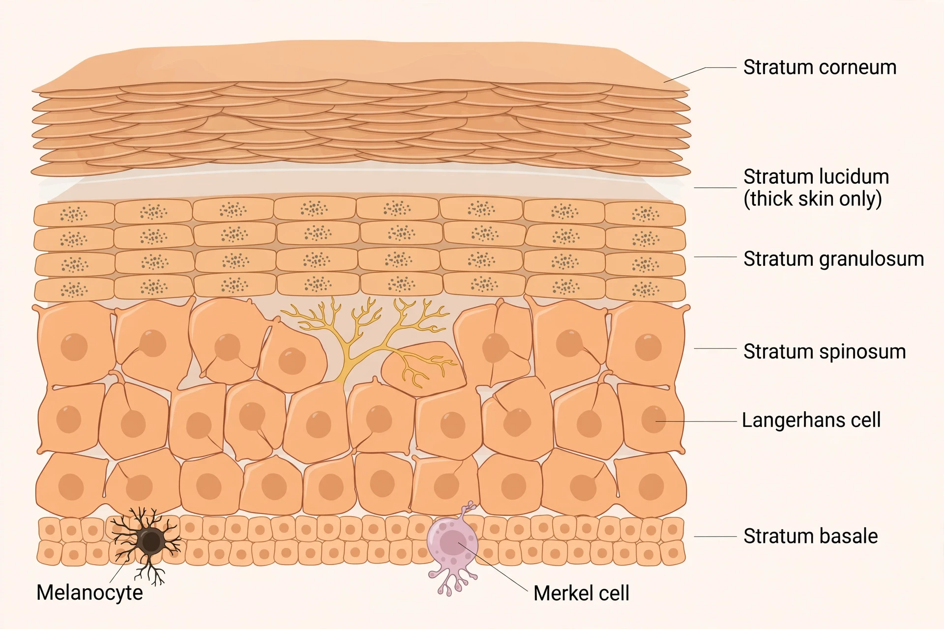
Thin skin — most of the body — has four strata, or sub-layers, in the epidermis. Thick skin — the palms, the soles, and the digits — has five, adding an extra layer called the stratum lucidum. The difference exists because thick skin takes more punishment, and it needs the extra reinforcement. From deep to superficial, the layers are the stratum basale, stratum spinosum, stratum granulosum, stratum lucidum (thick skin only), and stratum corneum.
The strata, bottom to top
The journey starts at the bottom, in the stratum basale, and it is worth pausing there because this single layer is where the whole epidermis is born. The stratum basale is a single row of cuboidal stem cells — small, block-shaped cells that divide constantly by mitosis, producing the new keratinocytes that will become the rest of the epidermis. Two other residents live here as well. Merkel cells sit in the basale and act as touch receptors, stimulating the sensory nerves that the brain reads as touch; they are especially abundant on the hands and feet, where fine touch matters most. Melanocytes also sit here, making the pigment melanin that gives skin and hair their colour and shields the living cells above from ultraviolet damage. One layer, three jobs: birth, touch, and pigment.
Above the basale sits the stratum spinosum, eight to ten layers of keratinocytes deep. Under a microscope, these cells look spiny, as if they bristle with tiny thorns — but that spiny look is a staining artefact, not the shape of the living cell. Unstained samples do not show it. The spinosum is also where the skin's immune sentinels live: Langerhans cells, a type of dendritic cell, are interspersed among the keratinocytes here, engulfing bacteria, foreign particles, and damaged cells. The skin's immune sentinels are introduced properly in the section on immunity, and their full story — including the immune-system article that explains what happens once a threat gets past the skin — is linked there: How the Human Immune System Works. Waterproofing begins in this layer too: the keratinocytes start making keratin and release a water-repelling glycolipid that helps prevent water loss from the body, making the skin relatively waterproof.
Next comes the stratum granulosum, three to five layers deep. Here the cells begin to die in an orderly way. Their nuclei and other organelles disintegrate, leaving behind keratin, keratohyalin, and cell membranes that will form the layers above. It is a strange kind of dying — programmed, useful, and essential. The granulosum is the last layer in which the cells are truly alive in the ordinary sense. Everything above it is a product of that controlled death.
In thick skin only, the next layer is the stratum lucidum, a thin band of dead, flattened keratinocytes densely packed with eleidin, a clear protein derived from keratohyalin. You will not find it on your forearm. You will find it on your palms and soles, where the extra layer adds to the toughness of skin that is walked on and gripped with all day.
The top of the stack is the stratum corneum, usually 15 to 30 layers of dead, keratinised cells. This dry, dead layer is the skin's front line. It helps prevent microbes from getting in, stops the tissues beneath from dehydrating, and provides mechanical protection against abrasion. It is easy to dismiss dead cells as unimportant, but the corneum is arguably the most important layer in the whole epidermis. It is the wall. Everything below it is the city the wall protects.
Keratin, the protein that makes it tough
Keratin is an intracellular fibrous protein, and it is the reason the epidermis works at all. It gives hair, nails, and skin their hardness and water-resistant properties. Inside a keratinocyte, keratin filaments form a tough internal scaffold. As the cell rises through the strata and dies, that scaffold remains, packed into the flattened corneocyte like steel bars in concrete. The result is a surface that is hard to tear, hard to soak through, and hard for microbes to cross — and it is made entirely from a protein the body produces itself, cell by cell, in the stratum basale. The full story of the keratinocyte's journey from birth to shedding, including how long the trip takes, belongs to the section on renewal, which follows the pigment section. The two are linked: keratin builds the wall, and the renewal cycle rebuilds the wall, month after month, for a lifetime.
3.2 The dermis: the living core
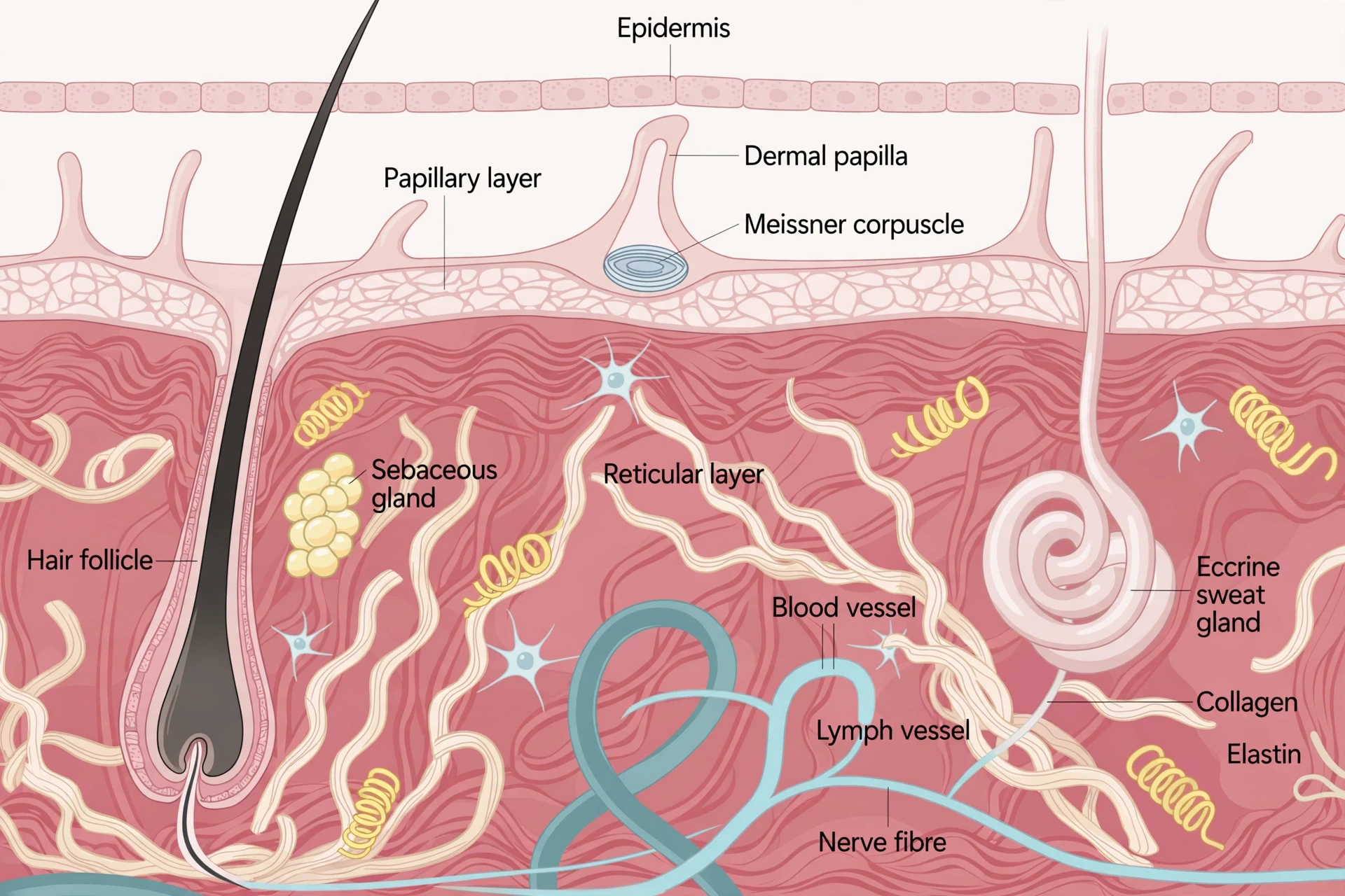
If the epidermis is the wall, the dermis is the city inside it. It is made of two layers of connective tissue that form an interconnected mesh of elastin and collagen fibres, produced by cells called fibroblasts, and it contains the blood and lymph vessels, nerves, hair follicles, and sweat glands that do the skin's living work. The epidermis has no blood supply and no nerves of its own. The dermis has both, in abundance, and it lends them upward: the vessels feed the epidermis from below, and the nerve endings reach up toward the surface to sense the world. Almost everything the skin does that feels alive — bleeding, sweating, feeling, growing hair — happens here, or is supplied from here.
Papillary layer — the fingerprint layer
The upper dermis is the papillary layer, a thin band of loose areolar connective tissue that forms finger-like projections called dermal papillae, reaching upward into the epidermis. Inside those papillae and the tissue around them sit fibroblasts, small fat cells, small blood vessels, phagocytes, lymphatic capillaries, nerve fibres, and Meissner corpuscles, the touch receptors that detect light touch and skin motion. The papillae do two things at once. They increase the strength of the connection between the epidermis and the dermis, interlocking the two layers like the fingers of two clasped hands, so the surface does not shear off easily. And in a growing fetus, fingerprints form where the cells of the stratum basale meet the papillae of the underlying dermal layer — which is why your fingerprints are not just a pattern on the surface, but a record of how your two upper layers grew together before you were born.
Reticular layer — strength and stretch
Below the papillary layer lies the reticular layer, the lower and much thicker part of the dermis. It is dense irregular connective tissue, well vascularised, with a rich sensory and sympathetic nerve supply. Two fibre types give it its character. Elastin fibres provide elasticity, letting the skin stretch and snap back. Collagen fibres provide structure and tensile strength, resisting tearing, and they also bind water, which helps keep the skin hydrated. The difference between the two layers is easy to feel in the language: the papillary layer is loose and delicate, built for sensing and connection; the reticular layer is dense and tough, built for strength. Together they make a dermis that is both sensitive and hard to damage, which is a difficult combination, and the reason the skin can be both your most sensitive sense organ and your toughest barrier.
Everything the dermis houses
The list of what the dermis contains reads like the inventory of a small town. Blood and lymph vessels run through it, feeding the skin and draining it. Nerves run through it, carrying signals for touch, pain, temperature, and itch. Hair follicles are rooted here, with their bulbs deep in the reticular layer. Sweat glands sit here, coiled, with ducts rising to the surface. Sebaceous glands — oil glands — sit beside the hair follicles, releasing oil that keeps the skin soft and supple and limits how much water the skin absorbs when you swim. The Cleveland Clinic's summary of the dermis's functions covers the same ground in plain terms: collagen and elastin give the skin strength and flexibility; the follicles grow hair; the nerves sense touch, pain, temperature, and itch; the oil glands keep the skin soft; the sweat glands release sweat through pores; and the blood vessels nourish the epidermis above. Nothing in that list happens in the epidermis. All of it is supplied from the core.
3.3 The hypodermis: fat with jobs
Beneath the dermis lies the hypodermis, also called the subcutis, made of well-vascularised loose areolar connective tissue and adipose tissue — fat. It serves to connect the skin to the underlying fascia, the fibrous tissue of the bones and muscles, so the skin moves with the body rather than sliding loosely over it. Its fat cushions the muscles and bones beneath, absorbing knocks that would otherwise reach them directly. Nerves and blood vessels enlarge here and branch out into the dermis above. And the fat itself is insulation: it keeps you from getting too cold or too hot, slowing the loss of body heat in winter and buffering temperature swings all year round.
Fat distribution in the hypodermis is hormone- and gene-dependent, and it differs by sex. In men, fat tends to accumulate around the neck, arms, lower back, and abdomen. In women, it tends to accumulate around the breasts, hips, thighs, and buttocks. That is a factual description of a pattern shaped by hormones and genetics, and it is worth stating plainly because it explains why two people can have the same skin, the same layers, and very different silhouettes. The hypodermis is not inert padding. It is a living tissue with its own blood supply, its own nerves, and its own role in how the body stores and uses energy.
4. The barrier: how skin keeps water in and germs out
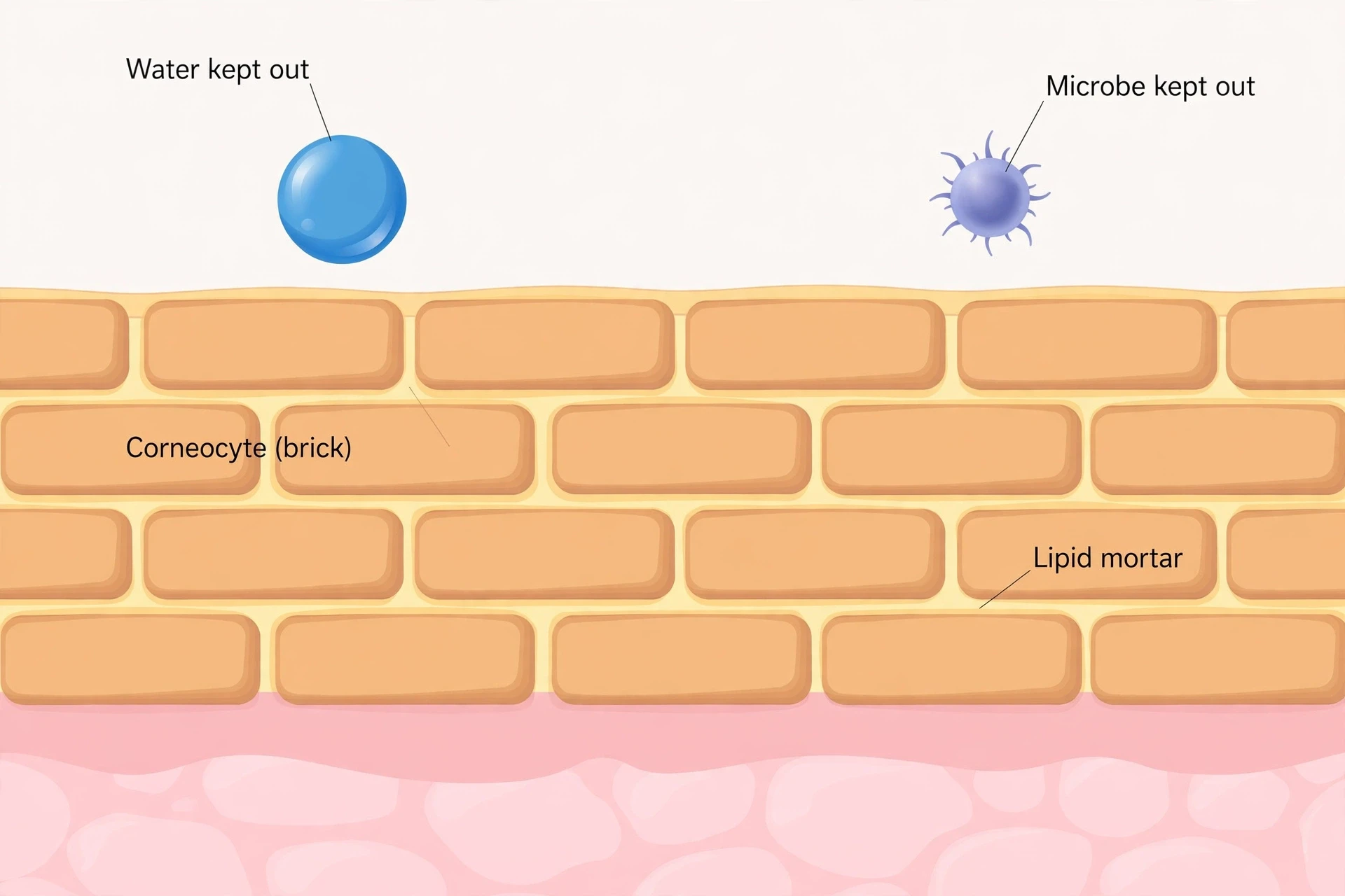
A wall is only as good as its mortar, and the skin's outer wall is no exception. The stratum corneum is often described as bricks and mortar, and the image is worth taking seriously because it explains how the barrier actually works — and how it fails. The bricks are corneocytes, the dead, flattened, keratin-filled cells of the corneum. The mortar is a lipid matrix: a mixture of ceramides, non-esterified fatty acids, and cholesterol, organised into cohesive lamellar layers that fill every gap between the bricks. Water cannot pass easily through that mortar. Microbes cannot pass easily through it either. And when the mortar is damaged — by harsh soaps, by dry air, by eczema — the wall leaks, even though the bricks themselves are still in place.
Bricks and mortar
The 2025 review of skin surface pH published in the MDPI journal Cosmetics describes the arrangement in the careful language of a peer-reviewed paper: corneocytes embedded in an intercellular lipid matrix enriched in ceramides, non-esterified fatty acids, and cholesterol, organised as cohesive lamellar lipid layers. The lipid mortar is roughly half ceramides by weight, with cholesterol and free fatty acids making up most of the rest — the published bands run around 40 to 50 percent ceramides, 20 to 25 percent cholesterol, and 10 to 15 percent free fatty acids, and those figures should be read as approximate, not as a recipe. What matters is the structure, not the exact percentages. The mortar is laid down in smooth, continuous sheets between and around every brick, and it is the mortar, not the cells, that is the main brake on water loss. Water constantly diffuses outward through the skin — a process called transepidermal water loss, or TEWL — and the lipid sheets slow that diffusion to a trickle. Damage the sheets and the trickle becomes a flow. That is why a damaged barrier feels dry no matter how much water you put on it: the water leaves as fast as it arrives, because the mortar is broken.
"This specific arrangement of corneocytes and the lipid matrix is known as the "bricks and mortar" construction model, which is responsible for the skin barrier's structural cohesion, permeability, stability, and strength." — Review of skin surface pH and barrier integrity, Cosmetics (MDPI), 2025
The acid mantle
There is a second, chemical part to the barrier, and it is measured in pH. Healthy skin has a slightly acidic surface, with the stratum corneum typically ranging from about pH 4.0 to 6.0, and most often quoted at around 4.5 to 5.5. The exact value depends on the body site, hydration, sebum production, genetics, age, and sex — the skin in the armpits and groin, for instance, runs a little higher. Deeper in the stratum corneum the pH rises, reaching neutral values around 7.4 in the viable epidermis below. That is a 100- to 1,000-fold difference in hydrogen-ion concentration across a distance of only 15 to 20 micrometres, a steep gradient packed into a layer thinner than a sheet of paper.
That acidity — the so-called acid mantle — does three jobs. It inhibits pathogenic bacteria, which mostly prefer a neutral pH. It favours the resident flora, the microbes that belong on the skin and help defend it. And it enables the enzymes that build the lipid lamellae and shed old corneocytes in an orderly way. Take the acidity away and all three jobs suffer. Alkaline soaps, which often have a pH of 9 to 10, temporarily neutralise this protective acidity and shut down the repair enzymes for hours. That is general guidance about how the barrier works, not a prescription for any individual — but it explains why skin that is washed with harsh alkaline soap often feels tight and dry afterwards, and why it takes time to recover. The chemistry of what soap does to skin, and why some soaps are gentler than others, is explained in full in How Soap Works: The Chemistry of Soap and Detergents.
The second wall beneath the surface
The stratum corneum is not the only barrier. Beneath it, in the stratum granulosum, tight junctions — seals formed by transmembrane proteins including claudins and occludin — provide an additional barrier to anything trying to slip between cells. Think of it as a second wall behind the first: if something gets past the bricks and mortar, it still has to cross a sealed layer where the cells are zipped together. Modern dermatology describes the barrier as four interlocking parts rather than a single wall: a physical component (the stratum corneum), a chemical component (the acid mantle and the skin's own antimicrobial substances), a microbiological component (the resident microbes), and an immunological component (the immune cells in the skin). They work together, and a failure in one puts pressure on the others. Dry, cracked skin is not just a cosmetic problem. It is a physical breach, a chemical shift, and an invitation to microbes, all at once.
Water loss, and the microbes who live on the wall
Water is always leaving through the skin. That is normal. Transepidermal water loss is a constant, quiet diffusion, and the lipid mortar keeps it slow. No single normal value for that loss is given here, because normal values vary by body site and by measurement method, and a single number would be misleading. What can be said plainly is that the mortar is the brake, the acid mantle keeps the brake working, and the tight junctions are the backup. When all three are intact, the skin holds its water. When they are not, it does not — and no amount of moisturiser can substitute for a repaired mortar, because the moisturiser sits on top of the wall while the leak runs through it.
Living on that wall is a whole community. Each square centimetre of skin carries roughly one million to one billion microorganisms, a peer-reviewed range that replaces a far higher figure sometimes seen in beauty blogs, which appears to confuse total-body counts with per-centimetre density. About a thousand bacterial species dominate, led by Staphylococcus, Cutibacterium — formerly called Propionibacterium — and Corynebacterium, along with roughly a hundred fungal species, mostly Malassezia, plus viruses and Demodex mites. They are not evenly spread. Dry sites, moist sites, and sebaceous sites each have their own mix, because the microbes that thrive in the oily skin of the forehead are not the same ones that thrive in the dry skin of the forearm. Their jobs are real: they regulate inflammation, fortify the barrier, and modulate the immune response. They compete with invaders for space and food, and in doing so they defend the wall they live on. The skin microbiome is the body's other great microbial community, and its companion story — the trillions of microbes inside the gut — is told in The Gut Microbiome Explained.
5. What is melanin?
Melanin is the pigment that colours skin, hair, and eyes. It is also one of the most misunderstood substances in the human body — credited with powers it does not have, blamed for problems it does not cause, and treated as if it were a simple dye. It is not a dye. It is a protective polymer, made in one cell type, delivered to another, and arranged with a precision that looks almost deliberate. Understanding what melanin is, and what it is not, clears up half the myths about skin colour, tanning, and sun protection in a single stroke.
Made in the basement, delivered upstairs
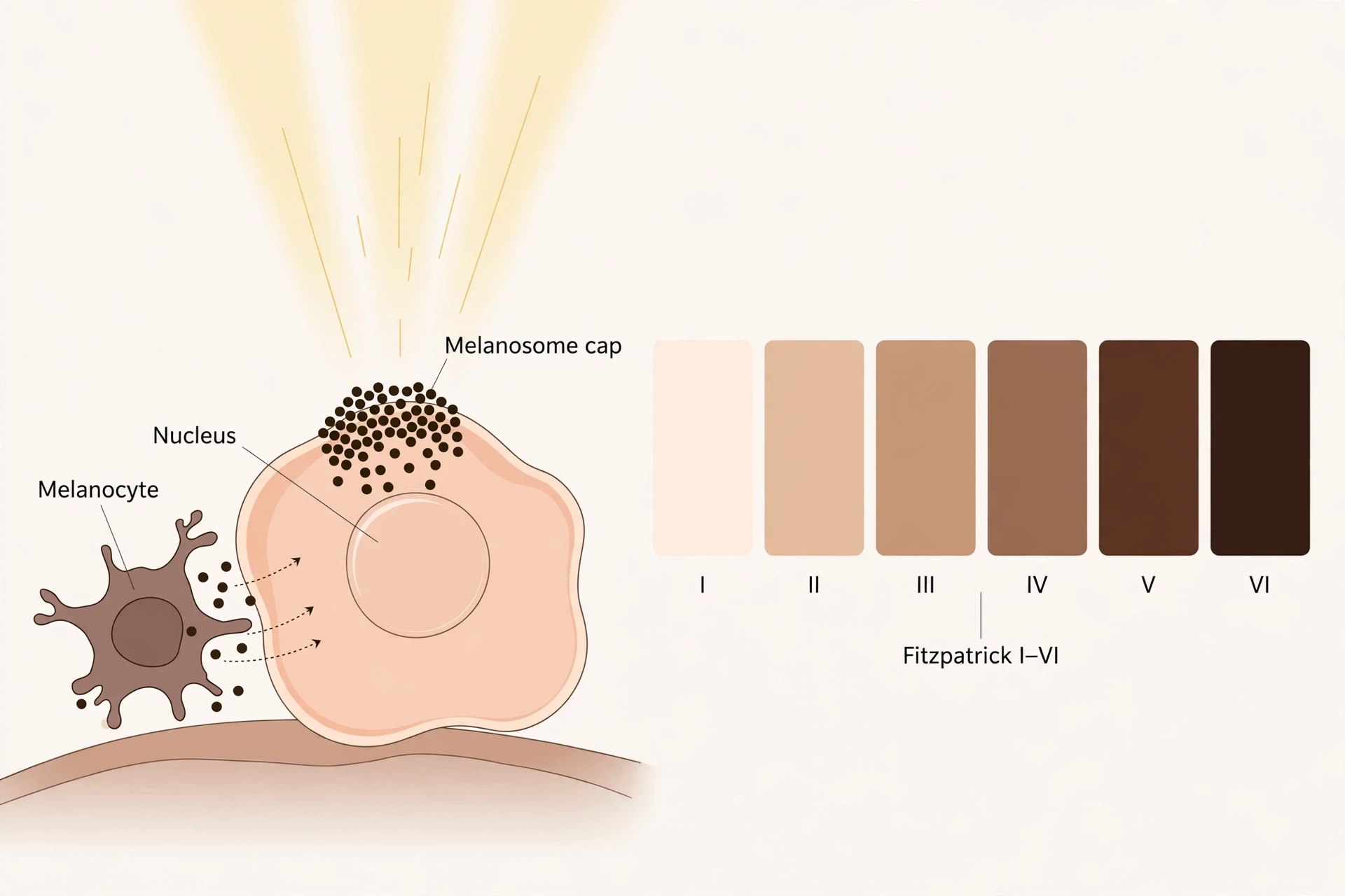
Melanin is made by melanocytes, the pigment cells that live in the stratum basale, the bottom layer of the epidermis. A melanocyte makes melanin from the amino acid tyrosine, using an enzyme called tyrosinase, inside small packages called melanosomes. It then passes those melanosomes to its neighbouring keratinocytes — one melanocyte supplies roughly 35 to 40 neighbouring keratinocytes, a figure that should be read as approximate. Inside the keratinocyte, the melanin is arranged like a cap over the nucleus, sitting on the sun-facing side of the cell, shielding the DNA inside from ultraviolet radiation. Picture a tiny umbrella held over the cell's most precious contents. That is not a metaphor invented for this article. It is the standard textbook description of how melanin is positioned, and it is the reason the pigment protects what it protects: the nucleus holds the DNA, and the cap sits between the DNA and the sun.
Two melanins, opposite personalities
There are two kinds of melanin, and they behave almost like opposites. Eumelanin is brown-black, strongly photoprotective, and absorbs broadly across both kinds of ultraviolet light — UVA, at 315 to 400 nanometres, and UVB, at 280 to 315 nanometres. Pheomelanin is red-yellow, weakly protective, and photo-unstable: it does not have eumelanin's free-radical-scavenging property, and under ultraviolet light it can itself become a source of free radicals, potentially promoting the very damage it fails to prevent. A review in Photochemistry and Photobiology puts the contrast bluntly: eumelanin is UV-absorbent, whereas pheomelanin is photo-unstable and may even promote carcinogenesis. People with red hair and fair skin carry more pheomelanin, which is part of why they burn so readily — their pigment is not just weaker protection, it is a different substance with different chemistry.
Why skin colour differs
Skin colour differs mostly because of the amount and type of eumelanin, and the way melanosomes are packaged — not because of the number of melanocytes. People of all skin colours have roughly similar numbers of melanocytes. What differs is how much melanin those cells make, what kind they make, and how the melanosomes are arranged inside the keratinocytes. The ratio of eumelanin to total melanin is one of the most significant factors determining skin colour, and the packaging differs too: in darker skin, most melanosomes sit singly inside the keratinocytes, while in lighter skin they tend to sit in clumps. The single-versus-clumped figures — about 89 percent singly in dark skin versus about 85 percent clumped in light skin — come from secondary sources and should be read as approximate. The core point is solid and worth stating plainly: darker skin is not skin with more pigment cells. It is skin whose pigment cells make more eumelanin and package it differently.
How much protection does melanin actually give?
The honest answer is: some, but far less than most people assume. Often-quoted laboratory estimates put melanin's natural protection at about SPF 13 for darker skin — Fitzpatrick types IV to VI — versus about SPF 3 for lighter skin, types I to III. The figures come from Kaidbey and colleagues (1979), who measured them in the laboratory; they are estimates, not a guarantee of protection in real sun. (Brenner and Hearing's 2008 review, often wrongly credited with these numbers, actually puts melanin's efficacy much lower, at about SPF 1.5 to 2, possibly as high as 4.) But even if the numbers are taken at face value, the conclusion is the same: even the higher figure, about SPF 13, is far below the SPF 30 or higher that dermatologists recommend for everyone. Melanin is a real shield, and it is not enough. That single fact dismantles the idea that dark skin does not need sunscreen, which is taken up properly in the myths section. For now, the point is arithmetic: a natural SPF of around 13 is better than 3, and both are a long way from 30.
The Fitzpatrick scale
Dermatologists needed a way to talk about sun response without talking about race, and in 1975 Thomas Fitzpatrick, a dermatologist and professor at Harvard University, gave them one. The Fitzpatrick scale classifies skin into six phototypes, I to VI, by how it responds to ultraviolet radiation — whether it burns, whether it tans, and how readily. Type I always burns and never tans. Type VI is deeply pigmented and rarely burns. The scale was originally developed for lighter skin, and types V and VI were added later. It measures sun response, not ethnicity, and it is a crude scale — a useful clinical shorthand, not a precise measurement of anything. Those three cautions matter, because the scale is often misused as a way of sorting people rather than a way of predicting sunburn. Used properly, it is a quick guide to how much protection a given person's skin offers on its own, and how much help it needs from sunscreen and shade.
Type | Description | Burns / tans | Natural melanin protection |
|---|---|---|---|
I | Very fair, often with red or blond hair and freckles | Always burns, never tans | Lowest |
II | Fair skin, light eyes | Usually burns, tans with difficulty | Low |
III | Medium skin, sometimes called white-olive | Sometimes burns, tans gradually | Moderate |
IV | Olive-brown skin | Rarely burns, tans easily | Moderate to high |
V | Brown skin | Very rarely burns, tans deeply | High |
VI | Deeply pigmented dark brown to black skin | Almost never burns, tans deeply | Highest |
Tanning is a damage response, not a shield
A tan feels like protection, and it looks like it too: skin darkens after sun exposure, as if it were building a shield. What is actually happening is a damage response. Ultraviolet light — and the skin damage it causes — stimulates keratinocytes to release a hormone called alpha-melanocyte-stimulating hormone, or alpha-MSH. That hormone acts on a receptor called MC1R on the melanocytes, switching on the enzymes that make eumelanin. The darkening that follows is the visible evidence that the skin's DNA has been stressed and the body is responding. It offers some extra protection after the fact, but a tan is not a safe shield and it is not a substitute for sunscreen. The full correction of the "healthy tan" idea, including the "base tan" myth, is in the myths section. Here, the mechanism is enough: tanning is the body reacting to damage, not preventing it.
The vitamin D trade-off
Melanin's protection comes with a cost, and it is a cost worth naming honestly. Melanin absorbs UVB, and UVB is the same wavelength the skin needs to make vitamin D. More melanin means less UVB reaching the machinery that starts vitamin D synthesis, so darker skin needs more sun exposure than lighter skin to make the same amount of vitamin D. One secondary estimate puts the difference at roughly 1.3 to 1.4 times as much sun — an approximate figure, not a precise one. The underlying mechanism is well established: increases in either latitude or melanin concentration diminish the skin's synthesis of previtamin D3, as Holick showed in 1981. The full pathway — how sunlight becomes a hormone, step by step — is explained in the vitamin D section later in this article, and what happens when that factory under-produces is covered in Vitamin D Deficiency: Symptoms & Treatment. The trade-off is real, and it is one of the reasons the same pigment that protects against sunburn can leave darker-skinned people more vulnerable to low vitamin D, especially in northern winters.
6. Keratin and the skin's renewal cycle
The epidermis is a wall that rebuilds itself while you use it. Every keratinocyte in it is on a one-way trip: born at the bottom, pushed upward through the strata, dying and flattening as it goes, and finally shed from the surface as a dead flake. The wall never stops being replaced, and it never closes for repairs. That is the renewal cycle — a quiet, constant part of how the skin works — and it is one of the two numbers in skin writing most often mangled — the other being how fast we shed, which is handled below with the care it needs.
A cell's month-long journey upward
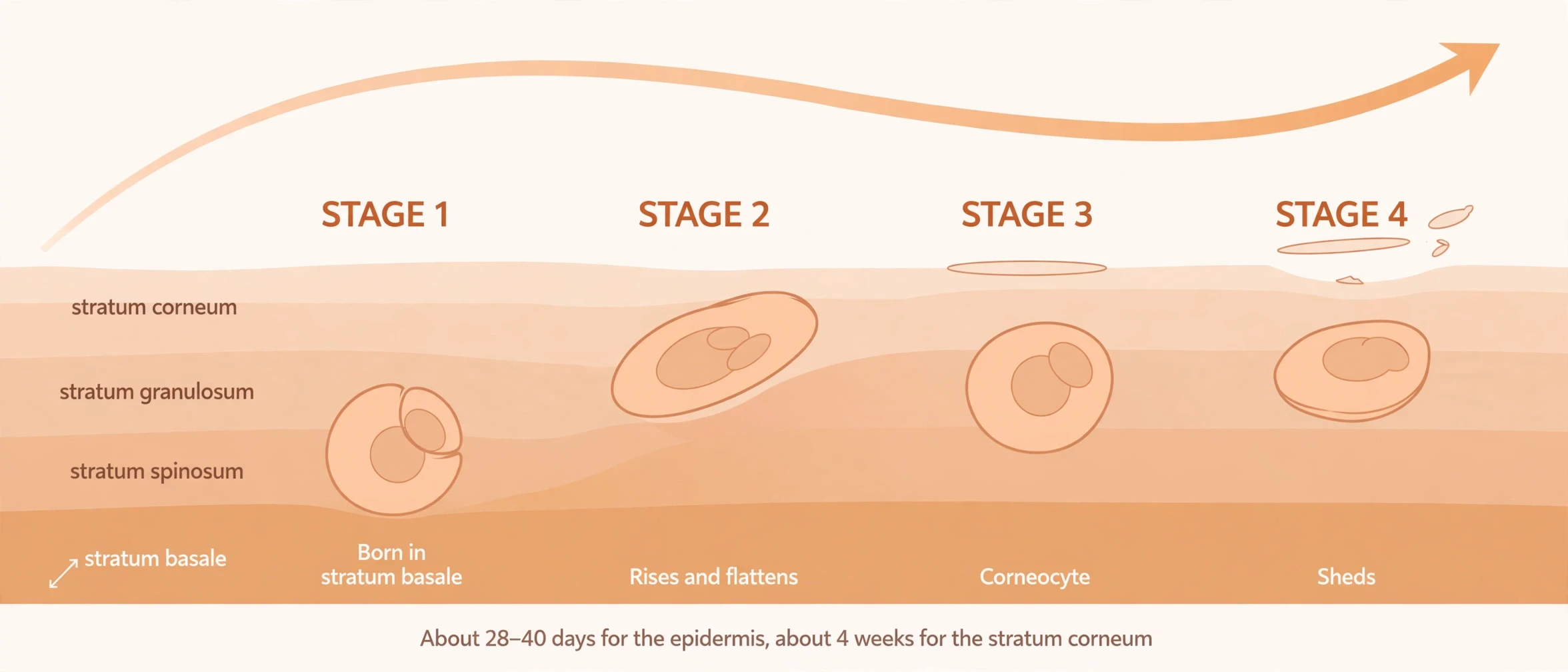
A keratinocyte is born in the stratum basale, the single row of dividing stem cells at the bottom of the epidermis. From there it is pushed upward as new cells are born beneath it, rising through the stratum spinosum and the stratum granulosum, growing flatter and filling with keratin as it goes. In the granulosum its nucleus and organelles disintegrate. By the time it reaches the stratum corneum it is a dead, flattened corneocyte, packed with keratin and wrapped in a tough envelope. It stays in the corneum for a while, doing its job as a brick in the wall, and then it sheds — desquamation, the controlled loss of corneocytes from the surface.
How long does the whole trip take? The full epidermal renewal takes roughly 28 to 40 days in adults. The outermost layer alone, the stratum corneum, is replaced in about four weeks. Those two numbers describe two different things, and keeping them apart matters. The "about four weeks" figure is for the corneum only. The "28 to 40 days" figure is for the whole epidermis, from a cell's birth in the basale to its shedding from the surface. What must never be written is the sloppy version — that the whole skin is replaced on a monthly cycle. The dermis is not replaced on that cycle. The hypodermis is not replaced on that cycle. Only the epidermis renews on a monthly rhythm, and even that rhythm slows with age — which is why older skin looks thinner and heals more slowly, a thread picked up again in the ageing section.
How fast do we actually shed?
Here is where skin writing usually goes wrong, so the numbers in this section come with their provenance attached. The figure you will see everywhere is 30,000 to 40,000 skin cells shed per minute. It appears in dermatology blogs, textbooks, and popular science pages. It is also a figure whose primary source could not be located: no peer-reviewed measurement was found tracing it to a single study. It is consistent with the arithmetic of skin area and turnover, and it is safe to repeat as what it is — an often-quoted estimate of around 30,000 to 40,000 cells a minute — but it must never be presented as a measured laboratory fact, and no fake citation should be attached to it.
One wording must never be used at all. A Cleveland Clinic page on skin gives the same figure with a different time unit from the per-minute figure used everywhere else, and it reads like a typo — a missing "per minute". The per-minute figure is the one used here.
The annual figures are worse, and they are omitted entirely. Published annual figures disagree with each other by a wide margin, and the discrepancy comes from different counting methods — counting cells versus weighing dust. No annual shed-weight figure is given in this article, by design. The mechanism is what matters: cells are born, rise, die, flatten, and shed, continuously, at a pace of tens of thousands a minute by the most commonly quoted estimate, and the pace slows with age.
Why renewal slows with age
The direction is well established even where the exact numbers are not: renewal slows as we get older. The epidermis takes longer to replace itself, the stratum corneum holds onto its cells longer, and the skin looks duller and feels rougher as a result. The exact number for older skin could not be verified against a primary source, so no figure is given here. The direction is the point, and it is enough: slower renewal means a thinner, less resilient surface, slower healing, and skin that shows its age in texture before it shows it in wrinkles. The ageing section takes up the full picture — collagen loss, sun damage, and the numbers that do hold up.
I learned what renewal looks like the unglamorous way, watching a sunburn peel. Years ago, after a long day outdoors without enough sunscreen, the skin on my shoulders came off in thin, translucent sheets over the course of a week. It was not painful by then, just strange — proof, in my own hands, that the surface I lived in was being replaced on a schedule I could not feel and could not stop. A callus tells the same story from the other side: press the same patch of skin again and again, and the epidermis thickens there, laying down extra corneocytes faster than they shed. The wall adapts to the work asked of it. Peel after a burn, thicken under pressure — the renewal cycle is not an abstraction. It is the reason your skin looks different in September than it did in June, and the reason a blister on a new shoe is gone in a week.
7. How does skin regulate temperature?
Your body runs hot. The chemical reactions that keep you alive generate heat constantly, and your core temperature sits near 37 degrees Celsius, held within a narrow band whether you are running in summer or standing in snow. The skin is the main instrument that holds it there. It does the job with two tools — sweat and blood flow — both controlled by a thermostat in the brain, and with a small, vestigial third act that most people know by its oddest symptom: goosebumps.
The thermostat in your brain
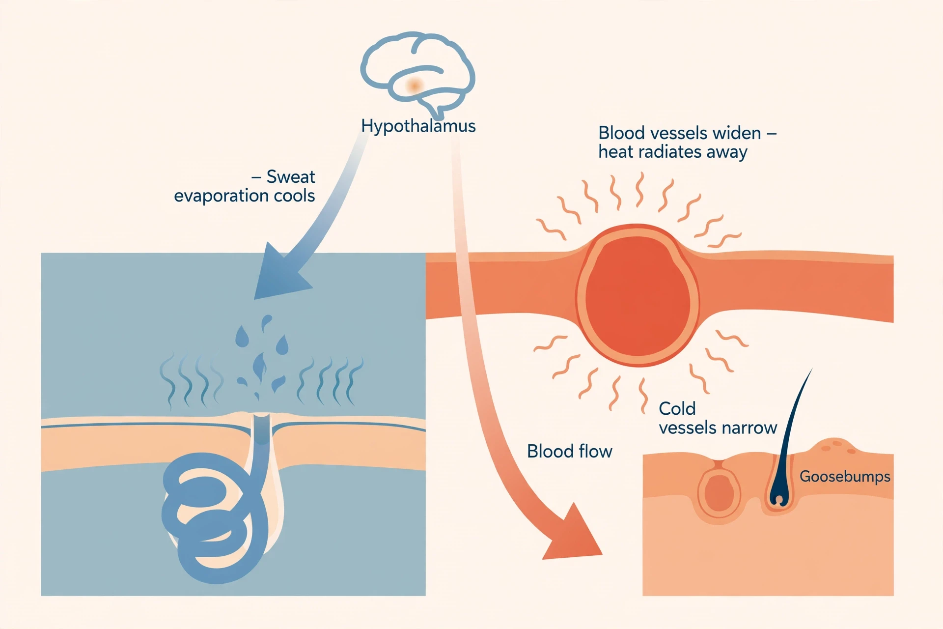
The thermostat is the hypothalamus, a small region at the base of the brain that measures blood temperature and holds a set point of about 37 degrees. When the blood running past it is warmer than the set point, the hypothalamus sends nerve impulses to the skin, ordering cooling. When the blood is cooler, it orders heat conservation. Sweating is a reflex sympathetic autonomic function, which sounds technical until you unpack it: the sympathetic nervous system, the same system that drives the fight-or-flight response, controls sweating — but through cholinergic neurons, which use acetylcholine as their signal, an unusual arrangement for the sympathetic system. The nerve impulses travel from the anterior hypothalamus down the reticulospinal tracts to the secretory cells of the eccrine sweat glands. You do not decide to sweat. The hypothalamus decides, and the glands obey, which is why sweat arrives unbidden on a hot day and why it can also arrive with nerves, when the same sympathetic system fires for a different reason.
Sweat glands — two kinds, two jobs
There are two kinds of sweat gland, and they do different jobs. Eccrine glands are the cooling system. There are about 2 to 4 million of them spread across nearly the entire body, with some counts running to 5 million, and they make up about 90 percent of the roughly 4 million sweat glands in total. They open directly onto the skin surface, produce a watery sweat, and have been active since birth. They are densest on the palms and soles, at 250 to 550 glands per square centimetre — which is why your hands get clammy before a presentation, even when the rest of you is cool. Apocrine glands are the other kind. They sit in the armpits, groin, and around the nipples, open into hair follicles rather than directly onto the skin, and only activate at puberty. They produce a thicker fluid, and body odour comes not from that fluid itself but from bacteria breaking it down on the skin surface. The sweat that cools you is eccrine. The sweat that smells is apocrine, and only after bacteria have had their say.
How sweat actually cools you
The most common misunderstanding about sweat is that the sweat itself cools you. It does not. Sweat cools you only when it evaporates. Sitting on the skin, a bead of sweat does nothing for your temperature. Evaporating, it carries heat away with it: evaporating one gram of sweat removes approximately 2.4 kilojoules of heat. That is why humid days feel so much worse than dry ones at the same temperature — in humid air, sweat evaporates slowly, the cooling fails, and the body keeps pumping out sweat that does no good. It is also why a fan helps even when the air is warm: moving air speeds evaporation, and evaporation is the cooling step.
The sweat itself starts as a plasma-like fluid, with a sodium concentration around 135 to 145 millimoles per litre, and as it travels up the duct the gland reabsorbs salt, so the sweat that reaches the surface carries only about 10 to 70 millimoles per litre of sodium. That is why sweat tastes salty but not as salty as blood. The average healthy adult can produce over half a litre of sweat per hour, and sweat is about 99 percent water. The other one percent is salts and trace substances — and it is not, despite what detox marketing claims, a meaningful route for removing toxins. That myth gets its own correction later.
Cooling, then, has two mechanisms working together. The first is radiative cooling through cutaneous vasodilation — blood vessels widening near the surface so heat radiates away. The second is evaporative cooling through sweat. The hypothalamus runs both at once, and both are needed: blood flow brings the heat to the surface, and evaporation carries it away. Exercise is the everyday context in which most people meet heavy sweating, and the body's wider response to exercise — muscles, heart, brain, and mood — is told in How Exercise Changes the Body.
Blood flow — the radiator
The radiator is the skin's blood supply. When you are hot, the capillaries in the skin dilate — vasodilation — and hotter blood flows near the skin surface, where its heat can radiate away into the air. That is why your face flushes when you exercise: the vessels are wide open, carrying heat to the surface on purpose. When you are cold, the opposite happens. The skin capillaries narrow — vasoconstriction — and warm blood is kept in the core, away from the surface where it would lose heat. That is why your hands go pale and cold first: the body is sacrificing the extremities to protect the core. The skin's vessels are not just feeding the skin. They are a valve system for the whole body's heat, opening and closing on orders from the hypothalamus, and they can move a large share of your blood to the surface or away from it within minutes.
Goosebumps — a leftover with a second job
Goosebumps are the visible sign of a reflex that once mattered a great deal and now mostly does not. Tiny smooth muscles called arrector pili are attached to each hair follicle. Under sympathetic control, they contract and pull the follicle upright, puckering the skin around it into a small bump. In a furry mammal, that raised hair traps a layer of insulating air against the skin, and the animal gets warmer. In humans, the hair is too short and too sparse for the trapped air to matter, so the insulation effect is vestigial — a leftover from furrier ancestors. Adults carry roughly five million hairs, each sitting in a follicle with its own miniature arrector pili muscle attached, and every one of them can still stand up on command. The command still fires when you are cold, and it also fires with strong emotion — fear, awe, a piece of music — because the same sympathetic system drives both.
There is a second job, though, and it is newer than the insulation story. Research in mice has found that the sympathetic nerve, the arrector pili muscle, and the hair-follicle stem cells form a single unit: the muscle holds the nerve-to-stem-cell connection in place, and prolonged cold stimulates the stem cells to grow new hair. The specific 2020 Cell paper behind that finding is Shwartz and colleagues' study of the cell types that promote goosebumps forming a niche to regulate hair-follicle stem cells — a mouse study, so the human relevance is still being worked out — but the finding itself, reported in secondary sources, is a useful reminder that a structure can lose one job and keep another. Goosebumps may no longer keep you warm, but the muscle that makes them is still part of how your hair grows. That is also the answer to a question many people ask: why do we get goosebumps? Because a muscle attached to each hair follicle contracts under sympathetic control, pulling the hair upright and puckering the skin — an old reflex, kept on for a new reason.
8. How skin feels: touch, pain, temperature
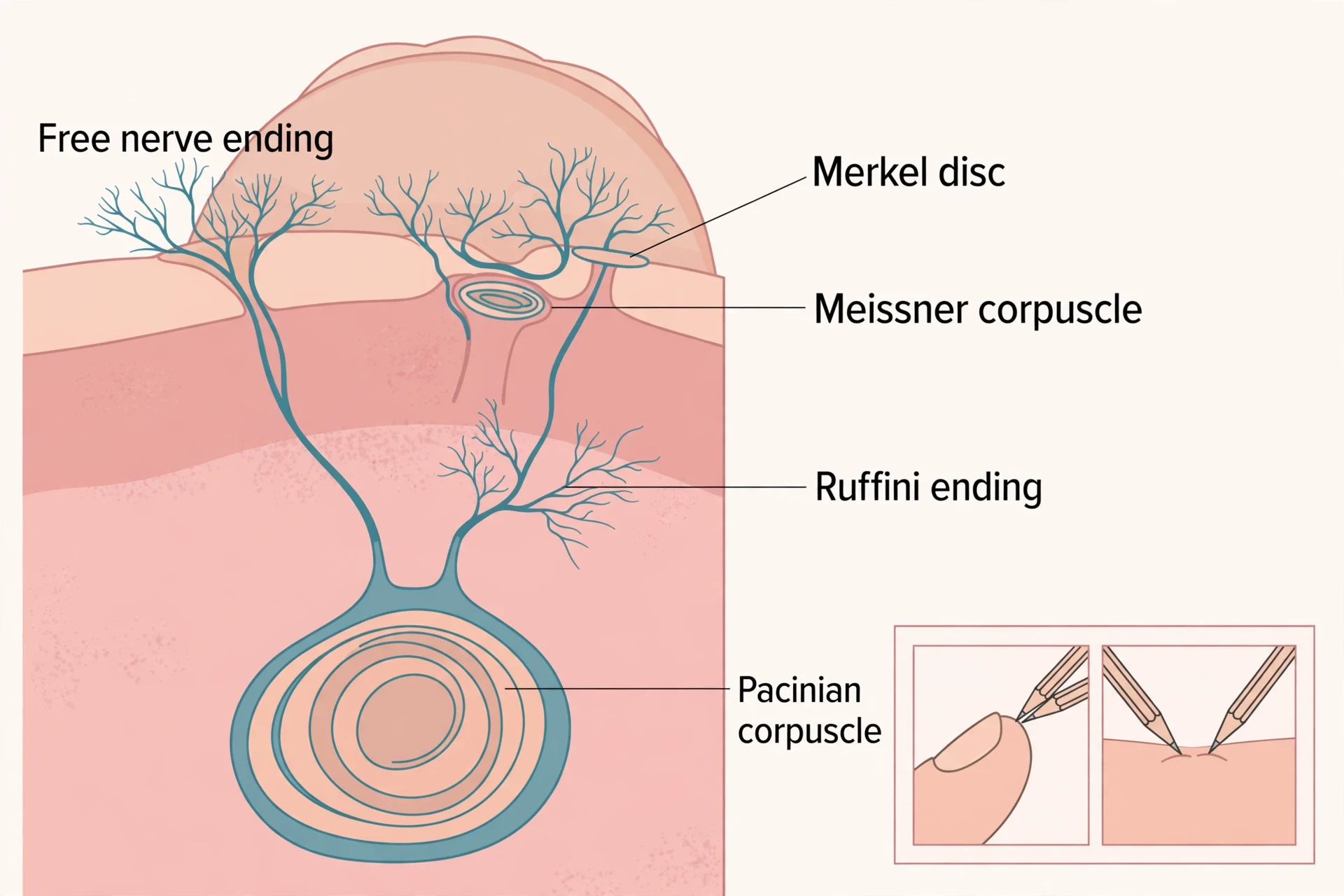
Touch is the skin's most intimate job, and it is also the most precisely engineered. It is one of the clearest demonstrations of how the skin works as a sense organ, turning pressure on the surface into information the brain can use. The skin does not feel with one kind of sensor. It feels with four kinds of mechanoreceptor, each tuned to a different aspect of contact, plus a separate set of free nerve endings for pain, itch, and temperature. The four receptors are organised by two questions: how fast does the receptor adapt, and how large is the patch of skin it watches? Those two questions generate four answers, and together the four cover almost everything your hands need to know about the world.
Four receptors, two questions each answers
Merkel discs are slow to adapt and watch a small patch of skin. They detect edges and points, fine detail, and sustained pressure — the shape of a coin, the dot of a Braille cell, the difference between a smooth surface and a rough one. They are the most precise of the four, with a spatial resolution of about half a millimetre, which is why you can read Braille with your fingertips and tell two close points apart. Meissner corpuscles are fast to adapt and also watch a small patch. They detect light touch, low-frequency flutter, and skin motion — the brush of a sleeve, the slip of an object starting to fall from your grip. Ruffini endings are slow to adapt and watch a large patch. They respond to skin stretch, telling the brain how the skin is being pulled and how the fingers are positioned. Pacinian corpuscles are fast to adapt and watch a large patch. They detect vibration and deep pressure — the buzz of a phone, the rumble of a passing truck through the floor, the deep thud of a knock on a door.
The organising idea is worth holding onto, because it makes the whole system memorable: adaptation speed crossed with receptive-field size gives four receptors, and each one answers a different question about contact. Slow and small means fine detail held still. Fast and small means light touch and movement. Slow and large means stretch. Fast and large means vibration. Your fingertips are rich in the two small-field receptors, Merkel and Meissner, which is why they are so good at detail. Your palms and soles carry the larger-field receptors, which is why they are better at pressure and vibration than at reading fine print by touch.
Receptor | Adapts | Receptive field | Where it sits | What it detects best |
|---|---|---|---|---|
Merkel discs | Slow | Small | Dermis–epidermis border, densest under fingertips | Edges, points, fine detail, sustained pressure |
Meissner corpuscles | Fast | Small | Dermal papillae, concentrated at fingertips | Light touch, low-frequency flutter, skin motion |
Ruffini endings | Slow | Large | Dermis | Skin stretch, finger position |
Pacinian corpuscles | Fast | Large | Deep in the dermis | Vibration, deep pressure |
Where they sit
Depth is part of the design. Merkel discs sit right at the border between dermis and epidermis, densest under the fingertips, where they can catch the finest detail of whatever the finger touches. Meissner corpuscles sit in the dermal papillae, just below the surface, concentrated at the fingertips, where they catch light touch and the first hint of slip. Ruffini endings sit in the dermis itself, where they can sense the stretch of the skin as the hand moves. Pacinian corpuscles sit deep in the dermis, where they pick up vibration and deep pressure that the surface receptors miss. Small, finely calibrated receptors sit in the upper layers. Large ones sit deeper. The map is not random: the closer a receptor is to the surface, the finer the detail it reports, and the deeper it sits, the broader and coarser the signal it carries.
Pain, itch and temperature travel separately
Pain, itch, and temperature do not use the four mechanoreceptors. They use separate free nerve endings — nociceptors — that sit near the surface of the skin, distinct from the touch receptors. That separation is why you can feel a pinprick and a feather as different kinds of event, even on the same patch of skin: different endings, different signals, different pathways to the brain. The free endings are the skin's alarm system, and they are tuned to damage and danger rather than to texture. Touch tells you what something is. Pain tells you it is harming you. The skin runs both systems at once, on the same surface, without confusing them — and those endings are the start of the nervous system's story, which is told in full in How the Nervous System Works.
The two-point test you can do right now
Here is a test you can do in thirty seconds with two pencil tips. Touch the pad of your fingertip with both tips at once, about two millimetres apart. You will feel two distinct points. Now do the same on the skin of your back, with the tips about two millimetres apart. You will feel one point. On the back, the two tips have to be about 40 millimetres apart — a twenty-fold difference — before they register as two. Those values are textbook approximations, not precise measurements, and they should be read as "about": about 2 millimetres on the fingertip, about 40 millimetres on the back. The concept is the point, and it is the concept the receptor map explains: small receptive fields mean fine discrimination, and the fingertips are packed with small-field receptors, while the back is not. The test works because your fingertips are built for reading the world and your back is built for covering it.
One sentence, for the curious, on how the signal starts: the mechanically gated ion channel Piezo2 converts skin deformation into electrical signals in these receptors, turning a press on the skin into a message the brain can read. That is the molecular beginning of touch — a channel that opens when the skin bends, and closes when it straightens. Everything else, from Braille to a handshake, is built on that opening and closing.
9. How does skin heal?
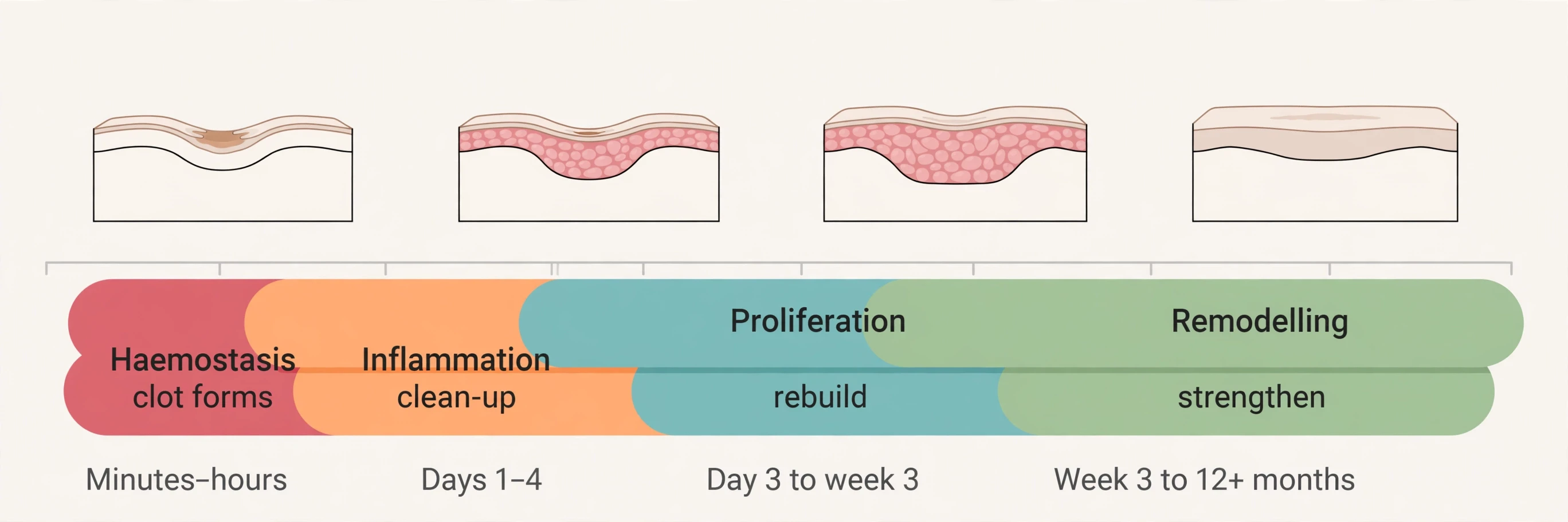
Healing is the skin's most reassuring trick, and it is also its most complex. A cut is not repaired by one event but by four overlapping phases, each with its own cells, its own timing, and its own visible signs. The phases are haemostasis, inflammation, proliferation, and remodelling — and the word "overlapping" matters more than the list. They are not a relay race, with one phase finishing before the next begins. They bleed into each other, start early, and run together, which is why a wound can be inflamed and rebuilding at the same time. Typical timings, from wound-care guidance, run like this: haemostasis over minutes to hours, inflammation over days one to five, proliferation from about day three or four to week three, and remodelling from week three to twelve months or more. The long tail is the surprise. The cut looks closed in a week. The repair underneath goes on for a year.
Four phases, overlapping — not a relay race
The four phases are easy to name and worth learning in order, because everything else in this section hangs on them. First, haemostasis stops the bleeding. Second, inflammation cleans the site. Third, proliferation rebuilds the tissue. Fourth, remodelling strengthens and tidies what was rebuilt. Each phase has its own cast of cells and its own visible signs, and each one sets up the next. A failure in any phase — an infection during inflammation, poor blood supply during proliferation — slows the whole sequence, which is why the factors that slow healing are best understood as factors that break one of these four phases.
Phase | When | Main cells / actors | What happens | What you see |
|---|---|---|---|---|
Haemostasis | Minutes to hours | Platelets, clotting factors | Vessels constrict, platelets aggregate, a clot forms | Bleeding stops, a scab forms |
Inflammation | Days 1–5 | Neutrophils, then macrophages | Bacteria and debris cleared, growth factors released | Redness, warmth, swelling, soreness |
Proliferation | About day 3/4 to week 3 | Fibroblasts, new blood vessels, keratinocytes | Collagen and granulation tissue laid down, wound contracts, surface re-covered | Pink new tissue, wound closes and shrinks |
Remodelling | Week 3 to 12+ months | Fibroblasts, collagen fibres | Weak collagen replaced by stronger collagen, fibres realign, excess vessels regress | Scar pales, flattens and softens |
Phase 1 — haemostasis: stop the bleeding
Within seconds to minutes of an injury, the blood vessels at the cut constrict to reduce bleeding. Platelets flood the area and aggregate into clumps. Clotting factors in the blood form a clot, a mesh that plugs the gap. The clot then dries into a scab — the dark, hard crust that covers a healing cut. A scab is not dirt and it is not dead skin to be picked off. It is a dried clot, and it is doing a job: sealing the wound, keeping microbes out, and protecting the fragile repair underneath while the next phases get to work. Pick it off and you reopen the wound to the stage before haemostasis finished, and the whole sequence has to start again.
Phase 2 — inflammation: clean the site
Once the bleeding has stopped, white blood cells arrive to clean up. Neutrophils come first, then macrophages, engulfing bacteria and debris and clearing the damaged tissue. The macrophages also release growth factors, the chemical signals that call the rebuilding cells to the site. The visible signs — redness, warmth, swelling, and soreness — are the outward marks of this clean-up in progress. Redness is extra blood flow. Warmth is the same blood, close to the surface. Swelling is fluid and cells arriving at the site. Soreness is the nerves reporting the activity. Inflammation here is repair, not failure. A wound that is not inflamed at all is a wound that is not being cleaned, and a wound that stays inflamed too long is a wound whose clean-up has stalled. The redness around a fresh cut is the body doing its job in plain sight.
Phase 3 — proliferation: rebuild
From about day three or four, the rebuilding begins in earnest. Fibroblasts lay down collagen and extracellular matrix, forming granulation tissue — the soft, pink, grainy tissue that fills the wound bed. New blood vessels form, a process called angiogenesis, bringing oxygen and nutrients to the hard-working cells. The wound contracts, pulled smaller by specialised cells, and keratinocytes migrate across the surface to re-cover it, a step called re-epithelialisation. This phase can last from about four days to three weeks or more, depending on the size and depth of the wound. It is the phase you can watch: the pink tissue filling in, the edges drawing together, the surface closing over. A shallow scrape that stays in the epidermis closes quickly, because there is less to rebuild. A deep cut that reaches the dermis takes longer, because the dermis has to be rebuilt as well as the surface.
Phase 4 — remodelling: the long tidy-up
Remodelling is the phase nobody watches, because by the time it starts, the wound looks healed. It begins around week three and runs for twelve months or more. The early collagen laid down during proliferation is type III collagen — quick to make, weak in strength. During remodelling it is gradually replaced by type I collagen, which is stronger, and the fibres reorganise themselves along the lines of tension in the skin. Excess capillaries that were needed during rebuilding regress, which is why a scar fades from red to pale. The scar softens, flattens, and pales over months. Nothing about this phase is fast, and nothing about it is visible day to day — but it is the phase that decides how strong the healed skin will be, and how much the scar will show a year later.
Scars — and why they never fully disappear
Here is the fact that surprises most people, and the answer to a question many ask: why do scars never fully disappear? Healed skin reaches a ceiling of roughly 80 percent of its original tensile strength, and never quite 100 percent. Scar tissue is less elastic than the skin it replaces, it has no hair and no sweat glands, and it may be a slightly different colour from the surrounding skin. That is the price of a repair that is fast rather than perfect. The body rebuilds with collagen laid down in a hurry, and the result is strong enough to hold, but never as strong, never as flexible, and never as complete as the original tissue. A scar is repaired skin, not restored skin, and the distinction matters: the wound is closed, the barrier is back, but the tissue is different, and it stays different.
Wounds also heal from the inside out and from the edges inward, and depth dictates how they heal. A scrape that reaches only the epidermis will not draw blood, because the epidermis has no vessels, and it heals easily and quickly. A cut that reaches the dermis draws blood and takes longer, because the dermis has to be rebuilt. That is why two cuts of the same length can heal on very different schedules: it is not the length that matters, it is the depth.
I have a scar on the back of my left hand, thin and pale now, from a cut I got years ago opening a box with a knife that slipped. It has been there long enough that I forget about it for months at a time, and then I notice it again in some slant of light and remember the week it took to close and the months it took to fade. It does not hurt. It does not itch. It has no hair on it, and it sweats no more than the skin around it does — which is to say, not at all, because the glands never grew back. It is a small, permanent record of a repair that stopped at about four-fifths of the original strength and called that good enough. Most of us carry a few such records, and they are worth reading as evidence: the skin heals, and it heals honestly, leaving the receipt on the surface.
What slows healing down
Healing can be slowed, and the factors that slow it fall into two groups — local factors at the wound itself, and systemic factors in the whole body — a framework set out by Guo and DiPietro in a 2010 review in the Journal of Dental Research. Infection is the most direct local problem: bacteria in the wound keep the inflammation phase running and prevent rebuilding from starting properly. Poor blood supply starves the repair of oxygen and nutrients. Diabetes slows healing because high blood sugar impairs circulation, nerves, and immune response — the mechanism behind that slower healing is explained in Type 2 Diabetes Explained. Smoking narrows the blood vessels and cuts the oxygen reaching the wound. Poor nutrition — low protein, low vitamin C, low zinc — deprives the fibroblasts of the materials they need to make collagen. Age slows healing because the skin is thinner, the cell response is slower, and there is less collagen to work with. Stress raises cortisol, and studies have found that stress slows healing — the direction is clear, even though the exact figures in secondary summaries were not checked against the primary papers while this article was being researched, so no percentage is given here. Obesity and medicines such as corticosteroids slow healing too. The list is long, but the pattern is simple: anything that weakens blood flow, weakens the immune response, or deprives the rebuilding cells of materials will slow the four phases down.
One practical note, offered as general educational information rather than personal advice: a cut that is not improving after about two weeks warrants a check. That is a common general prompt in wound-care guidance — a rule of thumb, not a diagnostic threshold — and it applies here with the Medical Advice Disclaimer in force for the whole article. Most small cuts close in days. One that does not is telling you something, and the telling is worth listening to.
10. Vitamin D: the skin as a hormone factory
Of all the skin's jobs, the strangest is this one: the skin makes a hormone out of sunlight. Not a vitamin in the dietary sense, though we call it one. A hormone — a chemical messenger that travels through the blood and tells distant organs what to do. The skin is where that hormone's story starts, and it is the only tissue in the body where the full pathway from sunlight to active hormone happens under normal physiological conditions. That is a remarkable claim for an organ most people think of as a wrapper, and it is the most surprising chapter in how the skin works — worth following step by step.
Sunlight in, hormone out
The pathway runs in four steps. First, ultraviolet B light — UVB, at 290 to 315 nanometres — strikes 7-dehydrocholesterol, a cholesterol-like molecule stored in the skin. The UVB photons break it apart into previtamin D3. Second, heat from the skin itself isomerises that previtamin D3 into vitamin D3, also called cholecalciferol, over a period of about three days. Third, the vitamin D3 travels in the blood to the liver, where it is hydroxylated into 25-hydroxyvitamin D. Fourth, it travels to the kidney, where it is hydroxylated again into 1,25-dihydroxyvitamin D — calcitriol — the active hormone. Two details make the system elegant. The accumulation of previtamin D3 is capped at about 10 to 15 percent of the original 7-dehydrocholesterol, because the previtamin photoisomerises into two biologically inert products, lumisterol and tachysterol, when there is already enough of it. And the skin is the only tissue yet known where the complete UVB-induced pathway, from 7-dehydrocholesterol to hormonally active calcitriol, occurs under physiological conditions — a finding reported by Lehmann in 2005. The liver's hydroxylation step is the third stop on that journey, and the kidney's is the fourth.
Calcitriol is a hormone in the full sense: it regulates calcium, supports bone, and influences cells far from the skin. The endocrine system is the wider story of hormones like it, and it is told in How the Endocrine System Works.
You cannot overdose from sun alone
One of the most reassuring facts in this whole article is also one of the least known: excessive exposure to sunlight will not cause vitamin D intoxication. The system is self-limiting. When previtamin D3 and vitamin D3 build up, sunlight photolyses them — breaks them apart with light — into several noncalcemic photoproducts that do not raise calcium. The cap on previtamin D3 accumulation, the inert byproducts, and the photolysis of the excess together form a built-in safety valve. Sunlight cannot make you overdose on vitamin D, because sunlight is also what dismantles the excess. Vitamin D supplements are a different matter, and they can be overdosed — but that is a question of pills, not of sun, and it belongs to the vitamin D article rather than this one.
The latitude rule
There is a catch, and it is written in geography. During the winter at latitudes above approximately 35 degrees, there is minimal, if any, previtamin D3 production in the skin. The sun sits too low in the sky, its UVB is filtered out by the longer path through the atmosphere, and the skin's factory effectively closes for the season. New York sits at about 40.7 degrees north — well above that line — so winter sun there makes little vitamin D, no matter how long you stand in it. The same applies across much of Europe and North America. That is not a failure of the skin. It is physics: the angle of the sun decides how much UVB reaches the ground, and the skin can only work with the light it receives. What happens when this factory under-produces — the symptoms, the causes, and what deficiency looks like — is covered in Vitamin D Deficiency: Symptoms & Treatment.
11. Skin and the immune system
The skin is not just a wall that immune cells hide behind. It is part of the immune system itself — its most peripheral outpost, in the phrase this article will return to in the history section. The defence is layered, like the barrier, and each layer does a different job. Remove any one of them and the others have to work harder. Together they make the skin the first place an invader meets resistance, and the place where the immune response often begins. That layered defence is a central part of how the skin works as an immune organ.
The sentinels in the epidermis
The sentinels are the Langerhans cells, the dendritic cells that live mainly in the stratum spinosum of the epidermis. They are antigen-presenting cells: they engulf bacteria, foreign particles, and damaged cells, process what they have captured, and present fragments of it to the immune system, sounding the alarm and shaping the response. The Cleveland Clinic describes them simply: Langerhans cells in the epidermis are part of the body's immune system, and they help fight off germs and infections. What is remarkable is how long it took anyone to realise that. Langerhans described these cells in 1868, when he was twenty-one, and believed they were nerve cells. Their immune function was not recognised for over a century — until 1973, when Inga Silberberg showed what they really were. The full story of that century-long misunderstanding is told in the history section. For now, the point is the job: the epidermis is not just a wall with guards behind it. The guards live in the wall.
Four layers of defence
The skin's immunity can be described as four layers of defence, matching the four-part barrier described earlier. The first is the physical barrier: the stratum corneum, the bricks and mortar that keep most invaders out by simply being in the way. The second is the chemical barrier: the acid mantle, which discourages pathogens, and the antimicrobial peptides the skin produces, such as defensins and cathelicidin, which attack microbes directly. The third is the cellular sentinels: the Langerhans cells in the epidermis, and the dermal macrophages and mast cells deeper down, which capture, kill, and signal. The fourth is microbiome competition: the resident microbes that occupy the skin's surface and deny invaders space and food. Four layers, four different mechanisms, one organ. The skin is the immune system's outermost post, and what happens once a threat gets past it — the antibodies, the T cells, the memory that makes vaccines work — is explained in How the Human Immune System Works. The same class of cell that Langerhans cells belong to, the antigen-presenting cells, are the cells vaccines are designed to train, and that story is told in How Vaccines Work.
12. Ageing, sun damage and skin cancer basics
Skin shows age more visibly than any other organ, and it shows sun damage more visibly than any other organ too — because it is the only organ that faces the sun directly, every day, for a lifetime. This section is educational only: mechanisms and numbers, no personal medical advice, no diagnosis language. It covers what changes as skin ages, how sunlight accelerates those changes, what one careful experiment showed about sunscreen, and the latest numbers on skin cancer in the United States.
What changes as skin ages
The most often quoted figure for collagen loss is roughly 1 percent per year from the mid-twenties onward. It should be read as what it is: a rough population average, repeated across dermatology literature, whose exact primary source was not pinned down while this article was being researched — not a precise personal prediction. Nobody loses exactly 1 percent a year, and the rate is shaped by genetics, sun exposure history, hormonal status, and lifestyle. What matters is the direction, not the decimal: collagen declines with age, the skin gets thinner, cell responses slow down, and renewal and healing both take longer, as described in the sections on the renewal cycle and wound healing.
After the menopause, that decline accelerates. The specific figures sometimes quoted for the first years after the menopause trace to a 1987 study by Brincat and colleagues, but the numbers could not be verified against a primary source in the research for this article, so they are not stated here. The direction is well established and is stated plainly: collagen loss speeds up after the menopause, and the skin thins faster as a result. The evolutionary and hallmark-level view of why ageing happens at all — the deeper story behind these skin-level changes — is told in Why Do We Age?.
Photoageing — sun as a collagen-eating signal
Sunlight does not just burn skin. It ages it, through a mechanism that is now well understood. Ultraviolet light activates enzymes called matrix metalloproteinases, or MMPs, which degrade collagen and elastin — the two fibres that give skin its strength and stretch. In chronically sun-exposed skin, that degradation can outrun new synthesis, even in younger people. The result is the familiar picture of photoageing: skin that is thinner, less elastic, more wrinkled, and slower to heal than skin of the same age that has been protected from the sun. It is not the sun making the skin old. It is the sun switching on the enzymes that eat the structures that keep skin young. The difference between a sun-exposed forearm and a sun-protected inner arm, in the same person at the same age, is the visible proof: one has spent decades with its MMPs activated, and the other has not.
The sunscreen experiment
There is a rare thing in dermatology: a randomised controlled trial of sunscreen and ageing. In a study published in the Annals of Internal Medicine in 2013, Hughes and colleagues followed 903 adults over four and a half years. Those assigned to use sunscreen daily showed 24 percent less skin ageing than those who used it only at their own discretion. That is one study's result, not a guarantee — but it is a measured result, from a trial rather than a survey, and it is the closest thing the field has to a direct answer to the question of whether daily sunscreen measurably slows visible ageing. The answer it gave was yes, by about a quarter, over four and a half years. The DOI for that study is 10.7326/0003-4819-158-11-201306040-00002, and it is listed in the Sources.
Skin cancer in numbers — the latest figures
Skin cancer is by far the most common cancer in the United States. Melanoma accounts for only about 1 percent of skin cancers, but it causes a large majority of skin-cancer deaths — a mismatch that explains why melanoma gets so much attention despite being a small share of cases. The latest figures come from the American Cancer Society, whose key-statistics page was last revised on 12 August 2026. For 2026, the Society estimates about 112,000 new melanomas in the United States — 65,400 in men and 46,600 in women — and about 8,510 deaths, 5,500 men and 3,010 women. Those are the figures to use. The 2025 projections quoted in many sources from last year — 104,960 new invasive cases and 8,430 deaths — have been superseded by the 2026 estimates, and this article uses the newer numbers.
The lifetime risk of melanoma is about 3 percent — roughly 1 in 33 — for White people, about 0.1 percent — 1 in 1,000 — for Black people, and about 0.5 percent — 1 in 200 — for Hispanic people. The average age at diagnosis is 67, and melanoma is one of the most common cancers in young adults, especially young women. More than 80 percent of cutaneous melanoma cases are attributable to ultraviolet radiation exposure, a figure drawn from secondary synthesis of global cancer data and stated here with that caveat. Non-melanoma skin cancers — basal cell and squamous cell cancers — occur roughly 18 to 20 times more often than melanoma, an order-of-magnitude comparison rather than a precise count. Melanoma is cancer, and the mutation and metastasis biology behind these statistics — what makes a skin cell become a cancer cell — is explained in How Cancer Works.
UVA vs UVB
Ultraviolet light comes in two kinds that matter for skin. UVB, at 280 to 315 nanometres, mostly affects the epidermis: it is the wavelength that causes sunburn, and the wavelength the skin uses to make vitamin D. UVA, at 315 to 400 nanometres, reaches deeper, into the dermis, where it drives photoageing and damages the collagen and elastin there. Both matter, and both are covered by the same protection. The World Health Organization's International Agency for Research on Cancer classifies solar radiation and ultraviolet radiation as Group 1 carcinogens — substances known to cause cancer in humans — a classification reported in secondary reviews and stated here with that caveat.
The standard sun-protection baseline (general information, not advice)
The standard baseline for sun protection, as set out in guidance from the American Academy of Dermatology, is straightforward: broad-spectrum, water-resistant sunscreen of SPF 30 or higher on uncovered skin, plus shade, protective clothing, a wide-brimmed hat, and UV-blocking sunglasses, and avoiding indoor tanning. That is general educational information, not personal medical advice, and it is stated here in plain language with the Medical Advice Disclaimer in force. The number that matters most in that list is 30 — the SPF that dermatologists recommend for everyone, and the figure that melanin's natural protection, at an estimated 13 at best, does not reach.
13. Common mistakes and misconceptions
Skin attracts myths the way a wound attracts attention — because everyone has skin, everyone has opinions about it, and most of those opinions were formed long before anyone checked them. Each one below is a small misunderstanding of how the skin works, and each one is correctable with the evidence. Six myths follow, each stated plainly and then corrected with the evidence. One of them deserves a direct disagreement, in the writer's own voice, because the data on it is unambiguous and the myth is genuinely harmful.
"Skin breathes"
Most people assume the skin breathes, in the sense that it takes in oxygen and gives out carbon dioxide the way the lungs do. What the anatomy actually shows is something else entirely. The skin does exchange tiny amounts of gases and water vapour with the air — that is part of transepidermal water loss, described in the barrier section — but it does not perform respiration. The lungs do that. OpenStax lists the skin's functions as protection, thermoregulation, sensation, and vitamin D synthesis. Respiration is not on the list, and it never was. Your skin is not a lung. It is a barrier that happens to leak a little water vapour, and calling that leak "breathing" confuses a minor side effect with a major function.
"You absorb 60% of what you put on your skin in 26 seconds"
This claim circulates widely in skincare marketing, and there is no evidence for it — neither the 60 percent nor the 26 seconds. The stratum corneum blocks most substances, and absorption through the skin is highly selective, depending on the molecule's size, its lipid solubility, and the condition of the skin. A large molecule sitting on an intact barrier mostly stays where it is. A small, fat-soluble molecule may get through. Some substances do penetrate — that is how nicotine patches and some medicines work, delivering drugs through the skin into the blood. So the correction is not "nothing gets in". The correction is "selective, not everything, and not 60 percent in 26 seconds". The barrier is a filter with rules, not an open door with a timer.
"Sweating detoxes you"
Sweat is about 99 percent water, plus salts. There are no studies showing that perspiration removes toxins from the body. The liver and kidneys have the primary responsibility for detoxifying the body — a point one dermatologist puts plainly, and one worth stating without hedging. Sweat does contain trace amounts of some substances, and a 2011 study in the Archives of Environmental Contamination and Toxicology is sometimes cited on this point — but the research for this article did not verify its DOI against Crossref, so it is not cited here, and the trace amounts do not change the conclusion. The organ that actually does the detoxifying is the liver, and its chemistry is explained in How the Liver Works: The Body's Chemical Processing Plant. A sauna may feel cleansing. What it is actually doing is making you sweat water and salts, while your liver and kidneys do the work the marketing attributes to the sweat.
"Dark skin doesn't need sunscreen"
This is the myth with the clearest numbers against it, and it deserves the direct disagreement. Most people assume that melanin is enough protection on its own, but what the evidence actually shows is that it is not — not by a wide margin. Melanin's natural protection is estimated at about SPF 13 for darker skin, against the SPF 30 or higher that dermatologists recommend for everyone. Ultraviolet damage, photoageing, hyperpigmentation, and skin cancer still occur in darker skin. Melanoma is less common in Black patients, but when it does occur it is more often diagnosed at later, more serious stages, and the five-year survival reflects that: about seven in ten for Black Americans compared with about nineteen in twenty for white Americans, a gap driven primarily by later-stage diagnosis. The figures are from the American Cancer Society's Cancer Facts & Figures 2025, for melanoma diagnosed in 2014–2020 — but the gap itself is consistent across sources. There is also a form of melanoma that has little to do with the sun at all: acral lentiginous melanoma, which appears on the palms, the soles, and under the nails, disproportionately affects darker skin, and is not driven by ultraviolet exposure. Sunscreen does not prevent that form. But the sun-driven forms, and the photoageing and hyperpigmentation that come with them, are preventable in every skin tone, and the idea that dark skin is exempt from them is wrong in a way that costs lives.
"A tan is healthy — a base tan protects you"
A tan is visible evidence of a DNA-damage response, not a sign of health. Ultraviolet light damages the DNA in skin cells, the skin releases alpha-MSH, the melanocytes make more eumelanin, and the skin darkens. That darkening is the body reacting to injury. It offers only modest extra protection afterwards — and no SPF-equivalent number is stated here, because none could be verified in the research for this article. A "base tan" built up before a holiday is not a shield. It is a record of damage already done, offered as protection against damage to come. The mechanism alone makes the point: if the tan is the response to damage, it cannot be the thing that prevents the damage.
"Skin is just a covering — dead wrapping"
The last myth is the one this whole article has been arguing against, so its correction is short. Skin is a living, renewing organ with its own blood supply, its own nerves, its own immune cells, its own endocrine function in making vitamin D, and its own stem-cell niches in the stratum basale and the hair follicles. It senses the world, cools the body, fights infection, heals its own wounds, and replaces its own surface month after month. A covering does none of those things. A covering does not bleed, sweat, feel, or heal. The evidence for the correction is not in this section. It is in every section before it.
14. A short history: the people who mapped the skin
No single person discovered the layers of the skin. The layered picture emerged from nineteenth-century microscopy, built up by several people working decades apart, arguing with each other, and sometimes getting the function wrong while getting the structure right. That is the honest history, and it is more interesting than a single discoverer would be. What follows is short, chronological, and confined to what the sources support.
1868 — a 21-year-old medical student stains skin with gold
Paul Langerhans was born in Berlin in 1847 and died in Funchal, Madeira, in 1888, at forty. In 1868, when he was twenty-one and still a medical student, he used a gold-chloride staining technique taught to him by Julius Friedrich Cohnheim and identified the cells in the epidermis that now bear his name. He believed they were nerve cells — intraepidermal receptors for signals from the nervous system — and he was wrong about that, in the best possible way: the cells were real, the drawing was accurate, and the function took another century to work out. His paper was titled "Ueber die Nerven der menschlichen Haut", published in the Archiv für pathologische Anatomie und Physiologie und für klinische Medicin in 1868, volume 44, pages 325 to 337. The same man described the pancreatic islets in his 1869 thesis — the clusters of cells in the pancreas that would later be named the islets of Langerhans. Two eponyms, one student, before he had even graduated.
1973 — the outpost is recognised
Langerhans cells were an enigma to dermatologists for over a century. They were visible, they were drawn accurately, and nobody knew what they did. Then, in 1973, Inga Silberberg discovered that the epidermal Langerhans cells are the immune system's most peripheral outpost — the furthest forward position the immune system holds in the body. The precision of Langerhans's original observation seems remarkable when his 1868 drawings, made with a primitive light microscope, are compared with modern immunofluorescence images: the technology changed completely, and the drawings barely needed correcting.
"the most peripheral outpost of the immune system" — Inga Silberberg's description of the epidermal Langerhans cells, as quoted in the Journal of Clinical Pathology (2002)
1852 — Meissner and Wagner argue over a corpuscle
Meissner corpuscles, the light-touch receptors in the dermal papillae, were first described in 1852, in Göttingen, by Georg Meissner and Rudolf Wagner. The two men then spent time arguing over which of them deserved priority for the discovery, each feeling he alone should be given the credit. The controversy is the human beat in the story, and it is a familiar one in science: two people see the same structure, both describe it, and the argument over credit outlives the discovery itself. The corpuscles kept Meissner's name. The quarrel kept both of them in the footnotes.
1741, then 1835 — Pacini rediscovers what Vater saw
The lamellar corpuscles now called Pacinian corpuscles were first seen by Abraham Vater in 1741, then forgotten for a century, then rediscovered and described by Filippo Pacini in 1835 and 1840, who recognised their layered, onion-like structure. Pacini later identified Vibrio cholerae, the bacterium that causes cholera — a second discovery, in a completely different field, by the same anatomist. The corpuscles carry his name because he described what they were, not because he saw them first. Seeing is not always describing, and describing is what lasts.
1875 — Merkel names the touch cells
Friedrich Sigmund Merkel published Tastzellen und Tastkörperchen — touch cells and touch corpuscles — in 1875, describing the touch cells now called Merkel cells and Merkel discs. In the same paper he first used the eponym "Langerhans'sche" for the cells Langerhans had described seven years earlier, giving them the name they still carry. Merkel's cells sit in the stratum basale, where this article first met them, and they are still the finest touch receptors in the skin.
1975 — measuring sun response
The story ends with a twentieth-century clinical tool, not ancient history. The Fitzpatrick scale, designed in 1975 by Thomas Fitzpatrick at Harvard, gave dermatologists a way to classify skin by how it responds to the sun — a scale still used in clinics today. It is a reminder that the science of skin did not stop with the microscopists. It kept going, from gold-chloride stains in 1868 to a sun-response scale in 1975, and it is still going now, in the reviews of barrier function and microbiome science that supplied much of this article.
I did not believe it at first, when I learned that Langerhans was twenty-one. Twenty-one, staining skin with gold chloride in a laboratory in Berlin, drawing cells nobody had seen before, and getting the drawing so right that a century of better microscopes added almost nothing to it. My own first look at skin under a microscope came much later, and what I remember most is the surprise that something so thin could contain so much structure — the wall, the city, the fat below, all in a slice a few millimetres across. Langerhans saw that structure first, at twenty-one, with worse equipment than any student has today. The layered picture of skin we use now — epidermis, dermis, hypodermis — was not handed down by one genius. It was assembled, piece by piece, by students and professors and arguers, over more than a century, each of them seeing a little more than the last.
15. Conclusion: what your skin is doing right now
The paper cut is closed. The sunburn has peeled. The blister has flattened and reabsorbed. Three small injuries, three different jobs, one organ — and that is the point this article has been building toward. Barrier, pigment, sweat, sensation, healing, vitamin D, and immunity are not seven separate systems that happen to share a location. They are one organ's seven answers to the problem of living on land, in air, under the sun: how to keep water in and germs out, how to shade the DNA, how to stay cool, how to feel the world, how to repair damage, how to make a hormone from light, and how to meet invaders at the border.
That is how the skin works — not as a wrapper, but as a living, renewing organ that replaces its own surface roughly every month, senses the world through four kinds of receptor, cools the body with sweat and blood flow, and heals its own wounds in four overlapping phases, stopping at about four-fifths of the original strength and calling that good enough. The next time your skin itches, sweats, prickles with goosebumps, or knits a cut closed, you will know which layer is speaking and which mechanism is answering. The organ you live in has been working all along. Now you know how.
16. FAQ — How the skin works, in eight answers
What is the largest organ in the human body?
The skin is the largest organ in the human body. In an adult it covers about 1.5 to 2 square metres and accounts for roughly 15 percent of total body weight (the published range runs about 12 to 16 percent). It counts as an organ because it is made of several tissues working together as one system — epithelium on the surface, connective tissue in the middle, fat below, plus blood vessels, lymph vessels, and nerves throughout — and because it performs jobs no other organ can do: barrier protection, sensation, temperature regulation, vitamin D synthesis, and immune defence.
What are the layers of the skin?
The skin has three layers: the epidermis, the dermis, and the hypodermis. The epidermis is the thin top layer, made of keratinised stratified squamous epithelium in four strata — five in thick skin, which adds the stratum lucidum — and it waterproofs the body and keeps microbes out. The dermis is the thick middle layer, about 90 percent of skin thickness, made of collagen and elastin, and it carries the blood vessels, nerves, hair follicles, and sweat and oil glands. The hypodermis is the fatty bottom layer, which insulates, cushions, anchors the skin to muscle and bone, and stores energy. Some anatomy texts count two main layers plus the hypodermis as an associated layer, but the tissue is the same either way, and the three-layer picture is the standard map used in this article.
What is melanin?
Melanin is the pigment made by melanocytes, the cells in the stratum basale of the epidermis, from the amino acid tyrosine using an enzyme called tyrosinase. There are two kinds: eumelanin, which is brown-black and strongly protective against ultraviolet light, and pheomelanin, which is red-yellow, weakly protective, and can become a source of free radicals under ultraviolet light. Inside the keratinocyte, melanin sits like a cap over the nucleus, shielding the DNA from the sun. Melanin's natural protection is estimated at roughly SPF 13 in darker skin, against roughly SPF 3 in lighter skin — far below the SPF 30-plus level dermatologists recommend for everyone, so melanin alone is never enough sun protection.
How long does it take for skin to renew itself?
The epidermis renews itself roughly every 28 to 40 days in adults, and the stratum corneum on its own turns over in about four weeks. A keratinocyte is born in the stratum basale, pushed upward through the strata, dies and flattens into a corneocyte, and then sheds from the surface. The pace slows with age. The one thing to avoid saying is that all of the skin turns over every month: the dermis and hypodermis do not follow that cycle — only the epidermis renews on a monthly rhythm.
How does skin regulate temperature?
The skin regulates temperature with two tools controlled by the hypothalamus, the brain's thermostat, which holds a set point of about 37 degrees Celsius. When the body is hot, the hypothalamus orders sweating — about 2 to 4 million eccrine sweat glands release watery sweat, which cools the body only when it evaporates — and vasodilation, in which the skin's blood vessels widen so warm blood flows near the surface and heat radiates away. When the body is cold, the vessels narrow instead, keeping warm blood in the core. Goosebumps are a small part of the same system: tiny muscles pull the hairs upright, a reflex that once trapped insulating air in furry ancestors.
How does skin heal?
Skin heals in four overlapping phases: haemostasis, inflammation, proliferation, and remodelling. First, blood vessels constrict and platelets form a clot, which dries into a scab. Second, neutrophils and then macrophages clear bacteria and debris, causing the redness, warmth, swelling, and soreness of inflammation. Third, fibroblasts lay down collagen and granulation tissue, new blood vessels form, the wound contracts, and keratinocytes re-cover the surface. Fourth, over months, weak type III collagen is replaced by stronger type I collagen and the scar pales, flattens, and softens. Healed skin tops out at roughly 80 percent of its original tensile strength, never 100 percent, and scars have no hair or sweat glands.
Does dark skin need sunscreen?
Yes. Melanin gives only about SPF 13 of natural protection at best, against the SPF 30 or higher that dermatologists recommend for everyone, so dark skin still needs sunscreen. Ultraviolet damage, photoageing, hyperpigmentation, and skin cancer all occur in darker skin, and melanoma in Black patients is more often diagnosed at later, more serious stages, with five-year survival of about seven in ten compared with about nineteen in twenty for white Americans. Acral lentiginous melanoma, which appears on the palms, soles, and under the nails and is not driven by ultraviolet light, also disproportionately affects darker skin. Sunscreen, shade, and protective clothing matter in every skin tone.
Why do we get goosebumps?
Goosebumps happen when tiny smooth muscles called arrector pili, attached to each hair follicle, contract under sympathetic nervous control and pull the hairs upright, puckering the skin around them. In furry mammals the raised hair traps insulating air and keeps the animal warm, but human hair is too short and sparse for that to matter, so the insulation effect is vestigial in people. Research in mice has found a second job for the same muscle: it holds the connection between the sympathetic nerve and the hair-follicle stem cells in place, and prolonged cold stimulates those stem cells to grow new hair. The reflex also fires with strong emotion, which is why fear or a piece of music can raise goosebumps with no cold at all.
17. Sources
MedlinePlus, Components of Skin — the three layers, keratin, melanin, and the fatty hypodermis
Wikipedia, Skin — thickness from about 0.5 mm on the eyelids to about 4 mm on the palms and soles
Healogics, Understanding the Stages of Wound Healing — phase timings from minutes to a year or more
LITFL, Paul Langerhans — Merkel's 1875 paper and the first use of the eponym Langerhans'sche