1. The Letter That Changed Everything — A Family Discovers Its Code
The envelope was thin, the way lab results always are when they carry more weight than paper should hold. Inside, in plain clinical language, was a sentence a young couple had not expected to read about their eighteen-month-old daughter: she had cystic fibrosis, one of the thousands of known genetic disorders. The toddler in the story was doing what toddlers do — coughing more than other children, growing more slowly than the charts predicted — and her parents had spent months being told it was probably just a stubborn chest infection. Then a sweat test and a gene test gave the cough a name, and the name came with a second sentence the parents had not seen coming. Both of them, healthy all their lives, each carried one faulty copy of the same gene. Neither of them was ill. Together, they had passed two faulty copies to their daughter.
This is a composite scenario, drawn from the way such diagnoses usually unfold, not the story of one named family. But the shape of it is real, and I have watched versions of it play out more than once: the confusion first, then the guilt, then the question that matters most. Did we cause this? The answer, for this family and for most families in their position, is no. They did not cause it. They did not know they were carriers, because carriers of a recessive disorder usually have no symptoms at all. What they were holding was not a verdict on their choices. It was information — about a single gene, a chloride channel, and a pattern of inheritance that had been running quietly through both sides of the family for generations before anyone noticed.
A second family, in a different kind of waiting room, shows the other side of the same science. A woman in her late thirties learns that her father has Huntington disease, a disorder that usually appears between the ages of 30 and 50. She is healthy. She has no symptoms. But the inheritance pattern is brutally simple: every child faces a one-in-two chance of inheriting the faulty gene. She is not deciding about a treatment. She is deciding about a test — whether to learn something about her own future that no medicine can yet change. That decision, and the counseling that surrounds it, is as much a part of the science of genetic disorders as any DNA sequence.
These two families sit at opposite ends of the same map, and this article is that map. Genetic disorders are health problems that start in the genome — a change in one gene, in several genes at once, or in a whole chromosome. There are well over 6,000 known genetic disorders, and new ones are still being described. Some are rare enough that most doctors never see a case. Some are so common that almost everyone knows a family touched by one. By the end of this article, you will be able to tell the four types apart, explain how a single changed letter in DNA can break a protein, read a family tree the way a genetic counselor reads one, and understand what genetic testing can and cannot tell you — and why a handful of disorders now have treatments that edit the code itself.
2. What Is a Genetic Disorder?
A genetic disorder is a health problem caused by one or more abnormalities in the genome: a mutation in a single gene, changes in multiple genes, or an abnormality in a chromosome. That is the whole definition, and it is worth sitting with for a moment, because three ideas that sound alike are hiding inside it. Having a variant is not the same as carrying a variant, and carrying a variant is not the same as having a disorder. MedlinePlus Genetics, the patient-facing genetics service of the US National Institutes of Health, lays out this definition and the types that follow from it, and the US Centers for Disease Control and Prevention groups genetic disorders into the same four types used throughout this article.
Every one of us carries variants — differences in our DNA compared with a reference sequence. Most of them are harmless. A few change how a protein works enough to cause disease, and in modern clinical language those disease-causing changes are called pathogenic variants. The older word, mutation, is still used widely and is not wrong, but "pathogenic variant" is more precise: it separates the change itself from the question of whether the change matters. A variant can be present in every cell of your body, or only in some of them. It can be inherited from a parent, or it can appear for the first time in you. A disorder is what happens when a variant, or a combination of variants, disrupts the body enough to cause illness.
2.1 Genes, variants, and why "mutation" is not a dirty word
The human genome holds roughly 3 billion DNA base pairs and about 19,000 to 20,000 protein-coding genes. Every gene is a set of instructions for building a protein, and proteins do nearly everything in the body: carry oxygen, digest food, contract muscle, fight infection, send signals between cells. A variant is a difference in those instructions. Most differences change nothing important — a slightly different shade of the same instruction, or a change in a stretch of DNA that codes for nothing at all.
Only some variants break something. Clinical genetics sorts variants into five tiers: pathogenic, likely pathogenic, variant of uncertain significance (often shortened to VUS), likely benign, and benign. The middle tier matters more than it sounds. A variant of uncertain significance is a change whose effect on health is not yet known. It is not a positive result, and its classification can change as evidence accumulates — a fact that becomes important in section 10, when a test returns an answer that is not really an answer.
I remember the first time a student asked me, in a lab where we were sequencing a gene, whether finding a variant meant finding a disease. It is a natural mistake, and the answer is the key to this whole article: no. Finding a variant means finding a difference. Whether that difference matters depends on which gene it sits in, what it does to the protein, and whether the person has one copy of it or two. The basics of genes, inheritance patterns, and gene expression are covered in depth in our article on Genetics Explained: How Inheritance, Mutations, and Gene Expression Work, and the scale of the genome itself — its 3 billion letters, its gene count, and the story of how it was sequenced — is covered in The Human Genome Explained. This article builds on those foundations and spends its words on disorders, not on re-teaching them.
2.2 Inherited, new, or neither: three ways a disorder arrives
A genetic disorder can reach a person by three different routes, and keeping them apart clears up most of the confusion people have about family history.
Inherited. The variant is passed down from a parent, following one of the patterns in section 5. Cystic fibrosis and Huntington disease both arrive this way.
New in the child. The variant appears for the first time in the egg, the sperm, or the very early embryo, and is not present in either parent's blood cells. Geneticists call this a de novo variant. About 25 percent of Marfan syndrome cases and about 30 percent of Duchenne muscular dystrophy cases begin this way.
A chromosome error that is neither parent's fault. Most cases of Down syndrome come from nondisjunction, a random error in cell division that usually happens in the egg. No parent carries a faulty gene. No parent did anything to cause it.
That third route is the one people find hardest to accept, because it has no one to blame and no family history to point to. But it is also the most reassuring fact in this article for many families: a genetic disorder can appear in a child whose parents are both healthy, with no family history of the condition at all. A family history matters, and a genetic counselor will always ask about it. Its absence, though, does not rule a genetic disorder out.
The scale of the field is worth setting out plainly, because the numbers surprise most readers.
Measure | Figure | What it means |
|---|---|---|
Known genetic disorders | Well over 6,000 | New disorders are still being described in the medical literature |
People affected by a single-gene disorder | Around 1 in 50 | One faulty gene is enough to cause disease in this group |
People affected by a chromosomal disorder | Around 1 in 263 | An extra, missing, or rearranged chromosome |
People affected by a rare genetic disorder | Approximately 1 in 21 | All rare genetic disorders combined, in a classic population study |
Rare diseases (most of genetic origin) | Nearly 7,000 | Most rare diseases are thought to be genetic |
Genetic disorders with a treatment | More than 600 | Treatments that address the underlying cause or a major feature |
Table 1: The scale of genetic disorders at a glance. The figures come from population studies and rare-disease counts; they describe groups of disorders, not any single condition.
3. The Four Types of Genetic Disorders
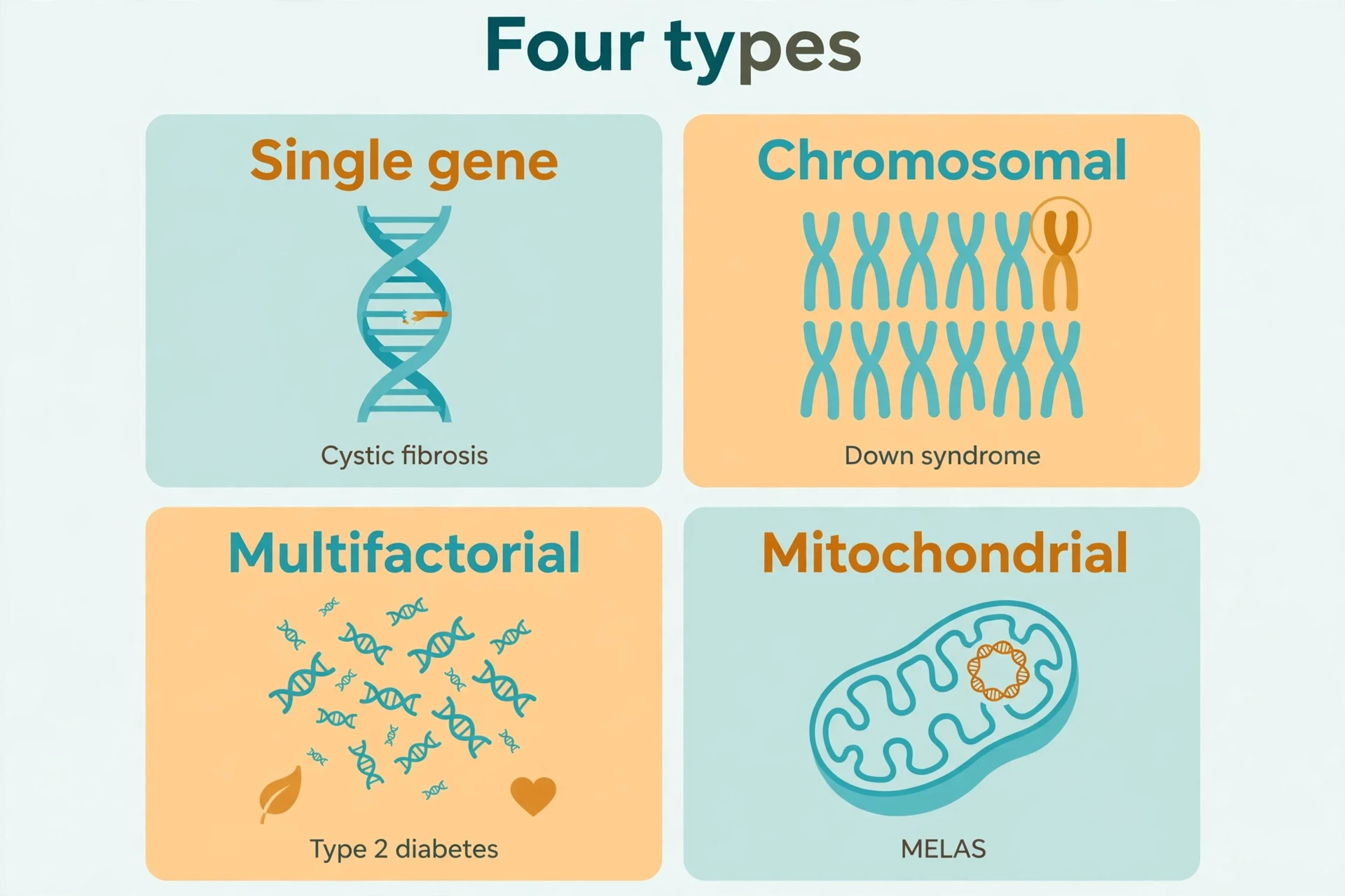
Every genetic disorder falls into one of four types. The types are not a trivia list. They are the first thing a clinician works out, because the type decides the questions that follow: Is one gene involved, or many? Is the chromosome count normal? Can the disorder be passed on, and to whom? The Cleveland Clinic's patient guide to genetic disorders uses the same four-type structure, and learning it here means the rest of the article falls into place.
3.1 Single-gene disorders (monogenic): one gene, one broken instruction
A single-gene disorder is caused by a pathogenic variant in one gene, and it follows one of the Mendelian inheritance patterns: autosomal dominant, autosomal recessive, or X-linked. One gene is enough. The protein that gene makes is broken, missing, or made in the wrong amount, and that single failure is enough to cause disease.
The canonical examples are cystic fibrosis (the CFTR gene), sickle cell disease (the HBB gene), Huntington disease (the HTT gene), Marfan syndrome (the FBN1 gene), Duchenne muscular dystrophy (the DMD gene), and fragile X syndrome (the FMR1 gene). Section 6 takes three of these — cystic fibrosis, sickle cell disease, and Huntington disease — and follows each one from the gene to the broken protein to daily life. The inheritance patterns themselves get their own section, section 5, because they are the part readers most often need to work through slowly.
3.2 Chromosomal disorders: the wrong number, or the wrong structure
A chromosomal disorder is caused by a change in the number or the structure of whole chromosomes. The two kinds of change behave differently. In aneuploidy, a cell has an extra copy of a chromosome, or is missing one entirely: Down syndrome is three copies of chromosome 21 instead of two, Turner syndrome is a single X chromosome instead of two, and Klinefelter syndrome is an extra X chromosome in a male. In structural changes, the total chromosome count is normal, but a piece is missing (a deletion), copied twice (a duplication), or moved to a different chromosome (a translocation). All three can cause disorders even when the count is exactly 46.
The examples in this group include Down syndrome (trisomy 21), Edwards syndrome (trisomy 18), Patau syndrome (trisomy 13), Turner syndrome (45,X), and Klinefelter syndrome (47,XXY). Section 7 goes deep on Down syndrome, the most common chromosomal condition, and pairs Turner and Klinefelter as the two sex-chromosome disorders most often encountered in clinics.
3.3 Multifactorial (complex) disorders: many genes plus environment
A multifactorial disorder is caused by a combination of variants in many genes plus environmental and lifestyle factors. No single variant causes the condition. Dozens or hundreds of variants each add a small amount of risk, and diet, exercise, smoking, infections, and other exposures add more. This is the largest group by number of people affected, and the hardest to predict for any one person.
Type 2 diabetes and heart disease belong here, along with many cancers, asthma, autism, and schizophrenia. The BRCA1 and BRCA2 genes appear in this section too, but with a careful distinction: a harmful BRCA variant sharply raises cancer risk, yet it does not cause cancer by itself, and most breast cancers are not inherited at all. Section 8 works through that distinction, because it is the one that most often gets flattened into a false certainty. Our dedicated articles cover the conditions themselves in full — How Cancer Works, Autism Explained, ADHD Explained, Alzheimer's Disease Explained, and Parkinson's Disease Explained — and this article points to them rather than repeating them. How environment shapes gene activity, and with it multifactorial risk, is the subject of Epigenetics Explained.
3.4 Mitochondrial disorders: the small genome with its own rules
Mitochondria, the energy-producing structures inside our cells, carry their own tiny DNA — just 37 genes, compared with roughly 20,000 in the nucleus. A mitochondrial disorder is caused by a variant in that small mitochondrial genome, or in a nuclear gene that supports mitochondria. Because mitochondria are inherited only from the mother, these disorders follow their own inheritance rules, covered in section 5.3.
Two features make mitochondrial disorders distinctive. The first is heteroplasmy: a person can carry a mixture of healthy and faulty mitochondria in the same body, and the proportion of faulty ones helps explain why severity varies so widely, even within one family. The second is which tissues suffer most. Mitochondrial disorders most often affect the tissues with the highest energy demand — brain, muscle, heart, and liver. MELAS, a disorder whose name spells out its features (mitochondrial encephalomyopathy, lactic acidosis, and stroke-like episodes), is most often caused by a variant in the MT-TL1 gene and affects about 1 in 4,000 people, though estimates vary widely. Leigh syndrome, a severe disorder that usually appears in infancy, can be caused by variants in either mitochondrial DNA or nuclear DNA, and more than 80 different genes have been implicated.
4. How Mutations Happen — and How They Break a Protein
A gene is a sentence written in four letters — A, T, C, and G — read three letters at a time. Each three-letter word, a codon, tells the cell which amino acid to add next to a growing protein. Change one letter and you may change one word. Delete one letter and every word after it shifts. That is the whole mechanics of mutation, and once you see it, you can work out for yourself why one single-letter change can be harmless and another can be fatal.
4.1 Point mutations: missense, nonsense, and silent
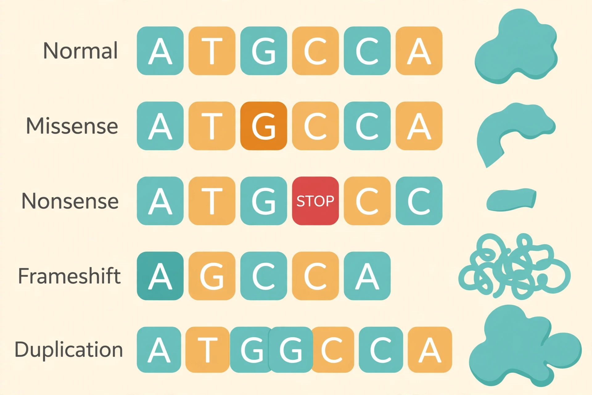
A point mutation changes a single DNA base. What it does to the protein depends on what the new three-letter word says. A missense variant swaps one amino acid for a different one. The protein may still work, work poorly, or fail entirely — it depends on where the swap happens and what the new amino acid does to the protein's shape. A nonsense variant creates a premature stop signal, so the protein is cut short and is usually non-functional. A silent, or synonymous, variant changes a DNA base but not the amino acid, and is usually harmless, though some can affect how the gene's message is spliced.
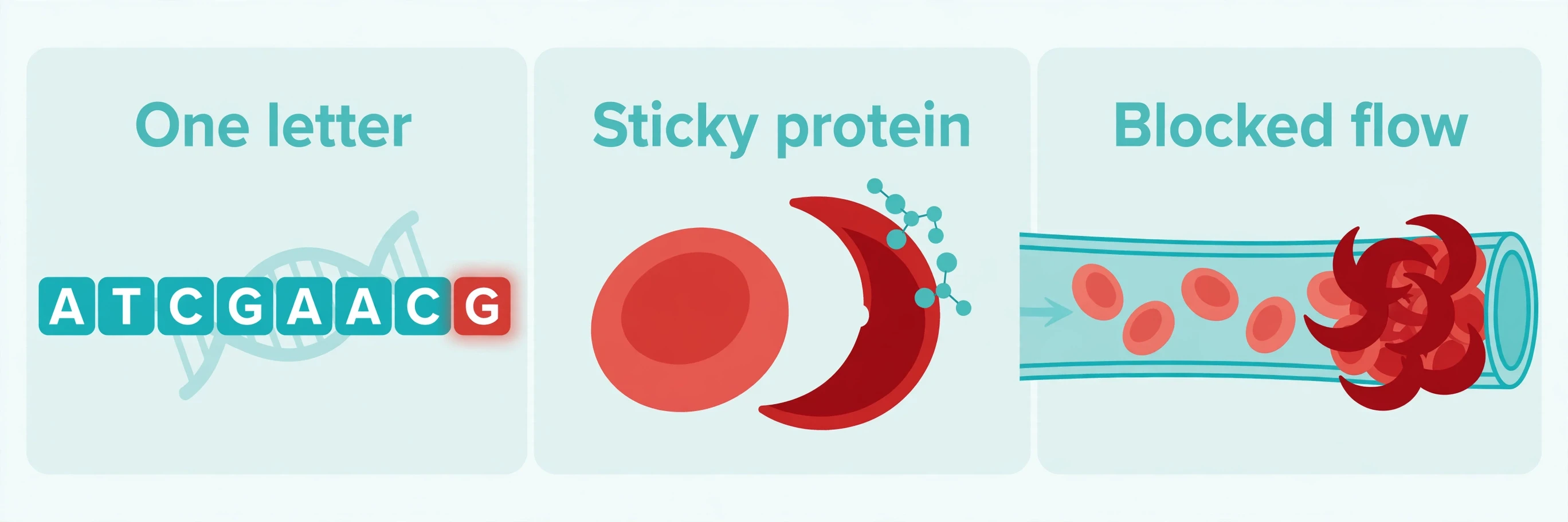
The worked example for missense is sickle cell disease. The sickle variant is a single missense change in the HBB gene: glutamic acid is replaced by valine at position 6 of the beta-globin protein. One amino acid, in one protein, in one gene. That is all it takes to make hemoglobin polymerize into stiff fibers, bend red blood cells into crescents, and block blood flow. One letter, a lifetime of consequences — and section 6.2 returns to this variant in full.
4.2 Insertions, deletions, and the frameshift disaster
An insertion adds DNA letters; a deletion removes them. The damage depends on how many. Because the code is read three letters at a time, an insertion or deletion that is not a multiple of three shifts the entire reading frame, scrambling every amino acid downstream and usually producing a non-functional protein. Geneticists call this a frameshift, and the word is apt: the frame itself has moved, so every word after the break is read wrong.
The common cystic fibrosis variant, F508del, is a useful contrast, because it is often misunderstood. F508del is a deletion of three DNA bases in CFTR that removes one amino acid, phenylalanine at position 508. Three is a multiple of three, so the reading frame does not shift. The protein is missing one amino acid, misfolds, and is degraded before it reaches the cell surface — which is serious enough — but it is not a frameshift. The distinction matters, and Figure 2 spells it out: the frameshift row shows a one-base deletion, not F508del.
4.3 Nondisjunction: when chromosomes fail to separate
Not every genetic error is a change in a single gene. Nondisjunction is the failure of chromosomes to separate properly during meiosis, the cell division that makes eggs and sperm. The result is a gamete with an extra chromosome (24 instead of 23) or one missing a chromosome (22 instead of 23). When a gamete with an extra chromosome 21 fuses with a normal gamete, the embryo has three copies of chromosome 21 in every cell. That is trisomy 21.
Most nondisjunction events happen in the egg, during maternal meiosis I, and the risk rises with maternal age. The stages of meiosis themselves are covered in our comparison article, Mitosis vs Meiosis; what matters here is the consequence. A single failed separation, in a single cell division, changes the chromosome count of every cell in the body that follows. Section 7 traces that consequence through Down syndrome.
4.4 Repeat expansions: the gene that grows longer each generation
Some genes contain a short three-base sequence repeated several times, like a stutter in the code. A trinucleotide repeat expansion is a mutation in which that sequence is repeated too many times. Above a threshold it causes disease, and the repeat can grow longer when it is passed to the next generation — a phenomenon called anticipation, in which the disorder appears earlier or more severely in successive generations.
In Huntington disease, the HTT gene normally has fewer than 35 CAG repeats. Thirty-six or more repeats cause the disease, and longer repeats are associated with earlier onset. In fragile X syndrome, more than 200 CGG repeats in the FMR1 gene — a "full mutation" — silence the gene by methylation, so no FMRP protein is made at all. The repeat is not a broken word. It is a word repeated until the sentence cannot be read.
Most of the variants in your genome are passengers, not drivers. The question is never whether you carry variants — you do — but whether any of them breaks a protein your body cannot do without.
That sentence is worth carrying into the next section. A genome is not a clean instruction manual. It is a working copy, full of small differences, and almost all of them are harmless. Disease begins only when a difference lands in the wrong place, in the wrong gene, in the wrong number of copies.
5. How Genetic Disorders Are Inherited — Reading the Family Tree
If section 4 is about how the code breaks, this section is about how the break travels. Inheritance is the part of genetics that families actually live with, and it is also the part most often misunderstood. The patterns have plain rules, and once you can read them, a family tree stops looking like a tangle of names and starts looking like a diagram with a logic to it. The basic mechanics — Mendel's laws, dominant and recessive alleles, Punnett squares — are explained in Genetics Explained. What follows here is what those mechanics mean when a disorder is at stake.
5.1 Autosomal dominant vs recessive: the comparison that matters
This is the comparison people search for most, and it is the one that decides what a family is told about risk. Autosomal means the gene sits on one of the 22 numbered chromosomes, not on a sex chromosome. Dominant and recessive describe how many faulty copies it takes to cause the disorder.
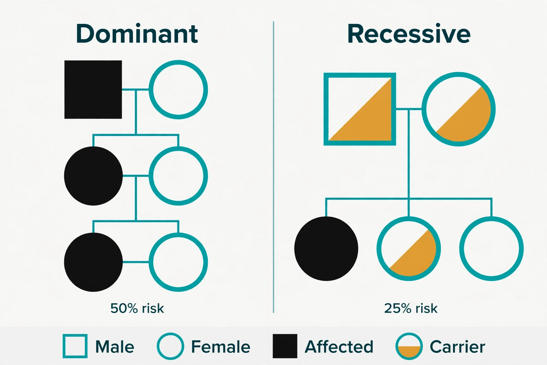
In autosomal dominant inheritance, one copy of the altered gene in each cell is enough to cause the disorder. An affected person has a 50 percent chance of passing the variant to each child, regardless of the child's sex. Dominant disorders typically appear in every generation of a family, and affected people usually have an affected parent. Huntington disease and Marfan syndrome are autosomal dominant.
In autosomal recessive inheritance, both copies of the gene must be altered for the disorder to appear. When both parents are carriers — each holding one faulty copy and one working copy, usually with no symptoms — each child has a 25 percent chance of being affected, a 50 percent chance of being a carrier, and a 25 percent chance of inheriting neither variant. Recessive disorders can appear to skip generations, because healthy carriers can pass the variant silently for years before two carriers have an affected child. Cystic fibrosis and sickle cell disease are autosomal recessive.
Feature | Autosomal dominant | Autosomal recessive |
|---|---|---|
Faulty copies needed to be affected | One | Two |
Who is affected | Anyone who inherits one faulty copy | Only people with two faulty copies; carriers are usually healthy |
Pattern in a family tree | Appears in every generation; an affected person usually has an affected parent | Can skip generations; healthy parents can have an affected child |
Risk per child | 50 percent if one parent is affected | 25 percent affected, 50 percent carrier, 25 percent neither — when both parents are carriers |
Canonical examples | Huntington disease, Marfan syndrome, achondroplasia | Cystic fibrosis, sickle cell disease, Tay-Sachs disease |
Table 2: Autosomal dominant versus autosomal recessive inheritance. The risk figures apply per child, per pregnancy — each child faces the same odds, whatever happened with a sibling.
One point in that table deserves emphasis, because families get it wrong in a way that causes real distress. A 25 percent risk per child does not mean that one child in four will be affected, as if the family were drawing lots from a fixed set. Each pregnancy is an independent event. A couple can have three affected children in a row, or four healthy children, and neither outcome changes the odds for the next pregnancy. The numbers describe chance, not a quota.
5.2 X-linked inheritance: why some disorders mostly affect males
Some genes sit on the X chromosome, and that placement changes the pattern. In X-linked recessive inheritance, males — who have one X chromosome — are usually affected when they inherit the variant, while females — who have two X chromosomes — are usually carriers, because their second X chromosome can compensate. A carrier mother has a 50 percent chance of passing the variant to each son, who would be affected, and a 50 percent chance of passing it to each daughter, who would be a carrier. An affected father cannot pass an X-linked variant to his sons, because sons receive his Y chromosome, but he passes it to all of his daughters, who become carriers.
Duchenne muscular dystrophy and hemophilia A follow this pattern. Duchenne is caused by variants in the DMD gene, the largest known human gene, spanning 79 exons and about 2.2 megabases; roughly 60 to 70 percent of cases are caused by large deletions of one or more exons. It affects about 1 in 3,500 to 5,000 male births. Hemophilia A, caused by variants in the F8 gene, affects about 1 in 5,000 male births.
Fragile X syndrome carries a classification nuance worth stating plainly rather than flattening. It is described as X-linked dominant in some sources and as X-linked with variable penetrance in females in others, because females with the full mutation can be affected, usually more mildly than males. The reason some carrier females show mild symptoms is X-inactivation, the process in which one X chromosome in each female cell is largely switched off. When inactivation is skewed, so that the working X is switched off in many cells, a carrier female can show features of the disorder. The biology is messier than the textbook categories, and the categories should bend to fit the biology, not the other way around.
5.3 Mitochondrial inheritance: passed down the maternal line only
Mitochondrial DNA is inherited only from the mother. A mother's mitochondrial variant can be passed to all of her children, while a father's cannot be passed to any of his children, because the mitochondria in a fertilized egg come from the egg, not the sperm. That single rule makes a mitochondrial pedigree easy to recognize: the disorder travels down the maternal line, through mothers, to children of both sexes, and stops at every father.
Heteroplasmy, introduced in section 3.4, is what makes the pattern hard to predict in practice. A mother passes a mixture of healthy and faulty mitochondria to each child, and the proportion differs from child to child. One child may inherit mostly healthy mitochondria and have mild symptoms or none; a sibling may inherit mostly faulty ones and be severely affected. Same mother, same variant, very different outcomes. Any risk estimate for a mitochondrial disorder has to carry that uncertainty with it.
5.4 How to read a pedigree: a worked family tree
A pedigree is the standard tool for tracing inheritance through a family, and its symbols are simple once you learn them. Squares represent males, circles represent females, filled shapes represent affected people, and a dot inside a shape marks a carrier. Partners are joined by a horizontal line, and a vertical line drops from that line to their children. A diagonal line through a shape means the person has died. That is nearly the whole alphabet.
Here is how a genetic counselor uses it, walking through the family from section 1. The parents are both healthy, so both are drawn as unfilled shapes — a square and a circle joined by a horizontal line. But we know from their daughter's diagnosis that both are carriers, so each shape carries a dot. A vertical line drops to their children: a filled circle for their daughter, who has cystic fibrosis. Now the counselor asks about the next pregnancy, and the pedigree does the arithmetic in plain sight. Each child has a 25 percent chance of inheriting two faulty copies and being affected, a 50 percent chance of inheriting one faulty copy and being a carrier like the parents, and a 25 percent chance of inheriting neither variant.
I have sat beside a family while a counselor drew exactly this tree on a piece of paper, and what struck me was how the drawing changed the conversation. Before the tree, the parents were trading guilt — whose side of the family had brought this in? After the tree, they could see it: both sides. Both of them carried one copy, silently, as about 1 in 25 people of European descent do for CFTR. The tree did not take away the diagnosis. It took away the blame, and it replaced a vague fear about future children with three numbers the couple could actually plan around.
Square: a male. Circle: a female.
Filled shape: an affected person. Unfilled shape: an unaffected person.
Dot inside a shape: a carrier — one faulty copy, usually no symptoms.
Horizontal line between two shapes: partners. Vertical line down from it: their children.
A disorder that skips a generation and appears in children of healthy parents points toward recessive inheritance; a disorder in every generation points toward dominant inheritance.
Two ideas from the pedigree deserve a line each, because they explain why the same variant can look different in different people. Penetrance is the proportion of people with a pathogenic variant who actually develop the condition; some variants have incomplete penetrance, so a person can carry the variant and never become ill. Variable expressivity means that people with the same pathogenic variant can have very different symptoms or severity — Marfan syndrome, for instance, varies widely even within one family. A pedigree shows who inherited a variant. It cannot, by itself, show how severely that variant will affect them.
6. Single-Gene Disorders Up Close
Three genetic disorders, three genes, three very different lives. Each case study follows the same arc: the gene, the broken protein, what goes wrong in the body, how common the disorder is, and what life and treatment look like now.
6.1 Cystic fibrosis: when mucus turns against the lungs (CFTR)
Cystic fibrosis is an autosomal recessive disorder caused by pathogenic variants in the CFTR gene on chromosome 7. CFTR makes a chloride channel that controls salt and water movement across epithelial cells — the cells that line the airways, the gut, and the pancreatic ducts. When the channel fails, the mucus those surfaces produce becomes thick and sticky. In the lungs, thick mucus traps bacteria and blocks airways. In the pancreas, it blocks the ducts that carry digestive enzymes, so food is poorly absorbed.
In populations of European descent, cystic fibrosis affects about 1 in 2,500 to 3,500 newborns, and about 1 in 25 people is a carrier of a CFTR variant. In the United States about 1 in 3,500 children is born with cystic fibrosis, and about 30,000 people in the US live with the condition. More than 1,900 CFTR variants have been described, and the F508del variant accounts for roughly 70 percent of CFTR variants in Northern European populations. Cystic fibrosis is the most common life-limiting autosomal recessive disease among people of European heritage.
The treatment story is the most hopeful in this section. CFTR modulator drugs — combinations such as elexacaftor, tezacaftor, and ivacaftor — target the faulty CFTR protein itself, helping it reach the cell surface and work better, and they have transformed cystic fibrosis care for people with responsive variants. A disorder that was once managed only by clearing mucus and fighting infections can now, for many people, be treated at the level of the protein. Not for everyone, and not a cure — but a change in kind, not just in degree.
6.2 Sickle cell disease: one letter, a lifetime of crises (HBB)
Sickle cell disease is a group of inherited red-blood-cell disorders caused by variants in the HBB gene. A child must inherit a sickle hemoglobin gene from both parents to have the disease. The mechanism is the single missense change from section 4.1: glutamic acid replaced by valine at position 6 of beta-globin. When oxygen levels fall, the altered hemoglobin polymerizes into stiff fibers, the red cell bends into a crescent, and the crescent jams in small blood vessels. Blood flow stops. Tissue is starved of oxygen. Pain follows, sometimes for days, and repeated blockages damage organs over a lifetime.
The numbers are stark. Sickle cell disease affects about 100,000 people in the United States and occurs in about 1 in every 365 Black or African American births and about 1 in every 16,300 Hispanic American births. About 1 in 13 Black or African American babies in the US is born with sickle cell trait — one copy of the variant, not the disease. Life expectancy for people with sickle cell disease in the US is more than 20 years shorter than the average. Sickle cell disease is the most common inherited red-blood-cell disorder in the United States.
And yet the variant persists, because carrying one copy protects against severe malaria. That is heterozygote advantage in its classic form: two copies cause disease, one copy confers resistance to a killer infection, and no copies leave a person exposed to malaria. In regions where malaria is endemic, the variant was favored by natural selection despite the cost. I find this the most unsettling fact in medical genetics — that evolution can keep a devastating variant in a population because, on balance, it saves more lives than it takes. It is also the fact that makes the gene-therapy story in section 11 feel like a kind of justice: a disease shaped by evolution may finally be answered by editing.
6.3 Huntington disease: the disorder that arrives in mid-life (HTT)
Huntington disease is caused by a CAG repeat expansion in the HTT gene on chromosome 4, and it is inherited in an autosomal dominant pattern. It affects around 1 in 10,000 to 20,000 people in the United States. Each child of a parent with Huntington disease has a 50 percent chance of inheriting the pathogenic variant, regardless of sex. The disease typically becomes noticeable in adulthood, between ages 30 and 50, with involuntary movements, cognitive decline, and psychiatric symptoms that worsen over time. Juvenile Huntington disease, with onset before age 20, accounts for about 5 to 10 percent of diagnosed cases.
What makes Huntington disease different from the other two case studies is not the mechanism. It is the timing, and what the timing does to a family. A person can grow up, build a career, have children, and only then learn that the disorder was in their future all along. Predictive testing can reveal the variant decades before symptoms appear, and the decision to test is one of the hardest in medicine: the result is certain, the onset is not yet treatable, and the knowledge affects not only the person tested but their children, their siblings, and their parents. That is why genetic counseling is recommended before and after predictive testing, and why the counseling in section 10 is described as a process, not a test. The science here is precise. The human decision is not, and it should not be rushed.
The other single-gene disorders in brief:
Marfan syndrome (FBN1, autosomal dominant): a connective-tissue disorder caused by variants in the fibrillin-1 gene; affects approximately 1 in 5,000 people worldwide, and at least 25 percent of cases arise from a new variant with no family history. The aorta needs regular monitoring.
Duchenne muscular dystrophy (DMD, X-linked recessive): variants in the dystrophin gene; affects about 1 in 3,500 to 5,000 male births, and about 30 percent of cases are caused by a spontaneous variant.
Fragile X syndrome (FMR1, X-linked): a CGG repeat expansion of more than 200 repeats silences the gene; the most common inherited cause of intellectual disability and the most common single-gene cause of autism, affecting about 1 in 4,000 males and 1 in 8,000 females.
Hemophilia A (F8, X-linked recessive): variants in the factor VIII gene; affects about 1 in 5,000 male births.
Achondroplasia (FGFR3, autosomal dominant): the most common form of short-limbed dwarfism, affecting about 1 in 15,000 to 40,000 newborns; about 80 percent of cases arise from a new variant.
Tay-Sachs disease (HEXA, autosomal recessive): carrier frequency about 1 in 27 in people of Ashkenazi Jewish ancestry, compared with about 1 in 250 in the general population, and routinely included in carrier-screening panels.
7. Chromosomal Disorders Up Close — Down Syndrome and Trisomy 21
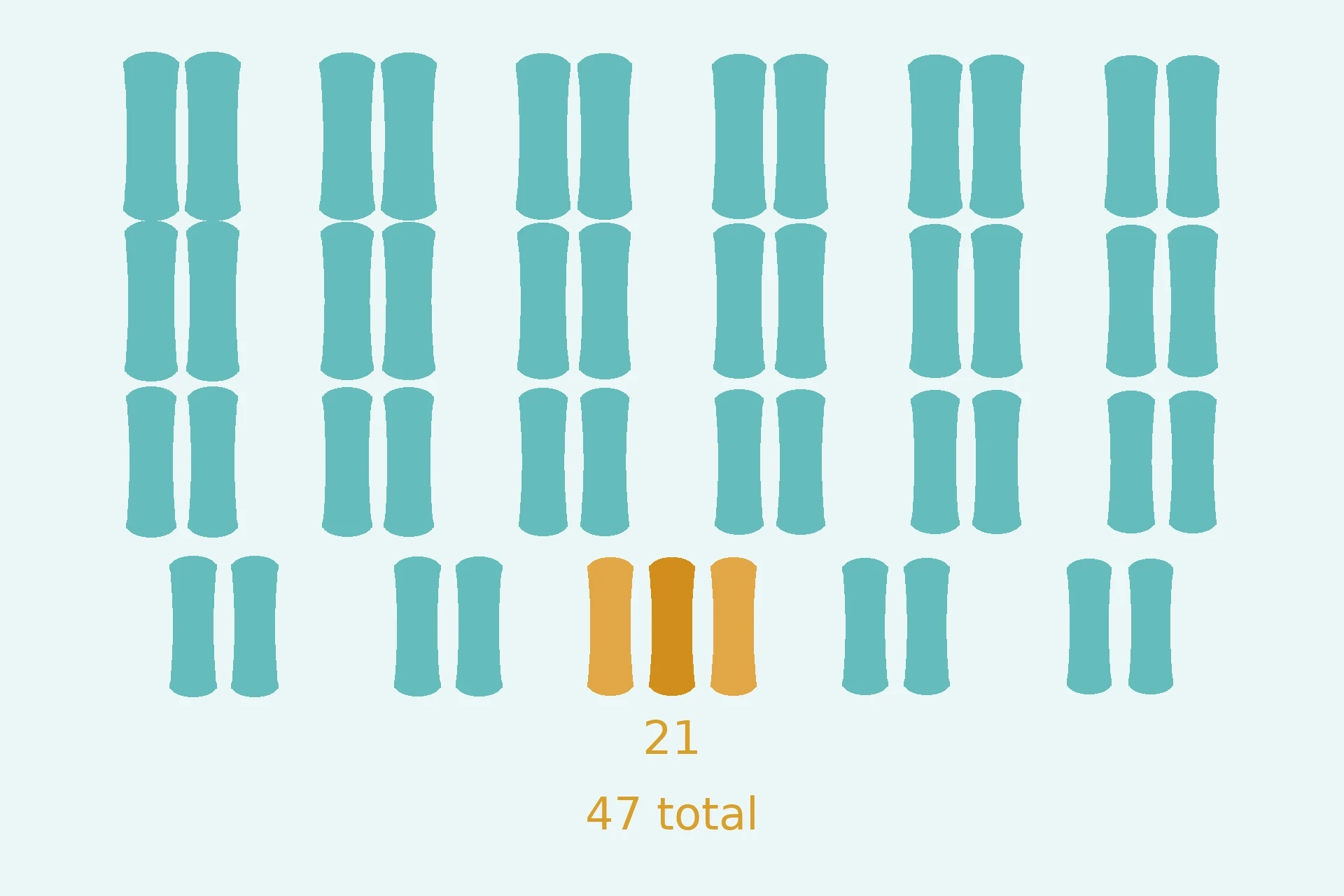
Down syndrome is caused by a full or partial extra copy of chromosome 21. A person with Down syndrome has 47 chromosomes instead of the usual 46. That is the answer to the question most people ask first, and it is worth stating before anything else: the cause is an extra chromosome, it arises from a random error in cell division, and no parental behavior or environmental factor is known to cause it. Down syndrome is not anyone's fault. It is the most common chromosomal condition diagnosed in the United States, and the rest of this section explains the three forms it takes, how common it is, and why age matters — in that order, and in plain language.
7.1 What causes Down syndrome?
The usual cause is nondisjunction, the failure of chromosomes to separate properly during meiosis that section 4.3 described. That failure usually happens in the egg, during maternal meiosis I, and the chance of it happening rises with maternal age. When an egg carrying an extra copy of chromosome 21 is fertilized by a normal sperm, the embryo has three copies of chromosome 21 in every cell. The US National Institute of Child Health and Human Development explains this mechanism directly: the extra copy usually comes from the mother, and the error is a random event in cell division.
7.2 The three types: trisomy 21 (~95%), translocation (~3–4%), mosaicism (~1–2%)
Trisomy 21, caused by nondisjunction, accounts for about 95 percent of Down syndrome cases. Every cell carries three copies of chromosome 21, and the total chromosome count is 47.
Translocation Down syndrome accounts for about 3 to 4 percent of cases. Here the total chromosome count remains 46, but an extra full or partial copy of chromosome 21 is attached to another chromosome, usually chromosome 14. Translocation is the one type that can be inherited: a parent can carry a balanced translocation, with no extra genetic material and no symptoms, and pass the unbalanced form to a child. The recurrence risk is about 3 percent if the father carries the translocation and 10 to 15 percent if the mother carries it.
Mosaic Down syndrome accounts for about 1 to 2 percent of cases. Only some of the body's cells carry the extra chromosome 21, because the chromosome error happened after fertilization, when the embryo was already dividing. Mosaicism is a reminder that "having Down syndrome" describes a range of genetic patterns, not a single one — and that the features associated with the condition vary from person to person.
After having one child with trisomy 21, the chance of having another baby with trisomy 21 is about 1 in 100 up to age 40, or about 1 percent above the age-related risk. That number is small, and it is also the number families ask about most. It deserves to be stated exactly, without rounding it away into reassurance.
7.3 How common is it, and why does age matter?
Down syndrome occurs in about 1 in every 640 babies born in the United States, or about 5,700 babies per year, according to the CDC's current figures. The risk rises with maternal age: about 1 in 1,250 at age 25, about 1 in 350 at age 35, about 1 in 100 at age 40, and about 1 in 30 at age 45. Those numbers look alarming at 40 and 45, and they should be read alongside the fact that puts them in context: most babies with Down syndrome are born to mothers under 35, because most babies overall are born to younger mothers. A higher risk per pregnancy multiplied by far fewer pregnancies still produces fewer babies than a lower risk multiplied by far more pregnancies. Both statements are true at once, and both are needed to understand the picture.
7.4 Turner and Klinefelter: the sex-chromosome disorders
Turner syndrome is caused by a complete or partial loss of one X chromosome, giving a karyotype of 45,X. It affects only females and occurs in about 1 in 2,500 female births. Women with Turner syndrome are almost universally infertile, and the condition is associated with short stature and heart defects, notably bicuspid aortic valve and coarctation of the aorta. Turner syndrome is the only viable monosomy in humans; an estimated 95 percent or more of 45,X conceptions do not survive to birth.
Klinefelter syndrome is caused by an extra X chromosome in a male, giving a karyotype of 47,XXY. It occurs in about 1 in 500 to 1,000 male births and is often undiagnosed until puberty or adulthood, when low testosterone, infertility, or learning differences prompt testing. Both Turner and Klinefelter usually arise from nondisjunction, not from inheritance, and both are milder in their effects than the autosomal trisomies — a pattern that holds more broadly: sex-chromosome abnormalities occur in about 1 in 400 male live births and 1 in 650 female live births, though most are clinically much milder than autosomal abnormalities.
For completeness, the other two autosomal trisomies: Edwards syndrome (trisomy 18) occurs in about 1 in 5,000 live births, and Patau syndrome (trisomy 13) in about 1 in 10,000 to 16,000 live births. Both have much lower survival in infancy than Down syndrome, which is why they are less often encountered outside specialist care.
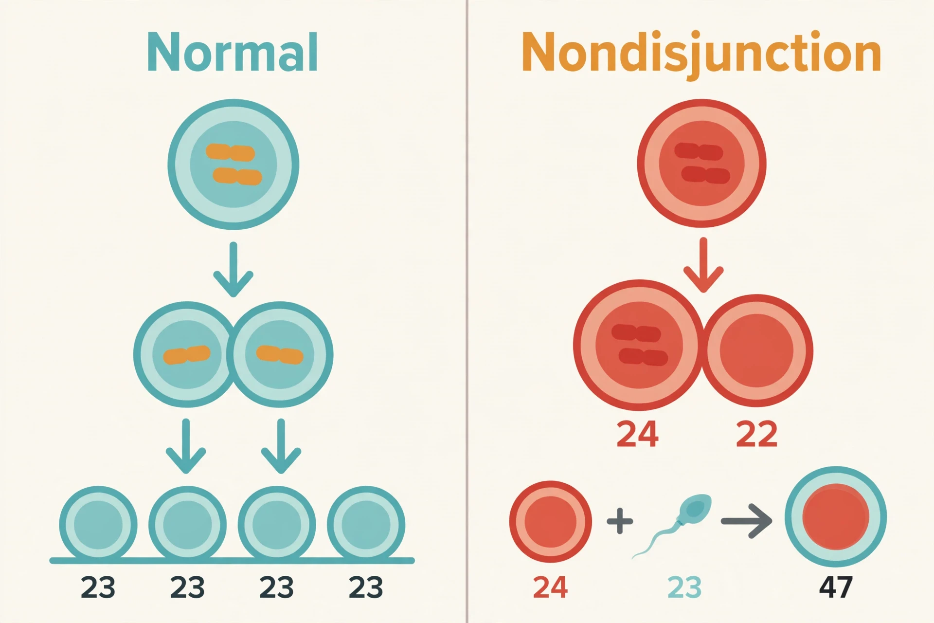
Down syndrome is caused by a random error in cell division. It is not caused by anything the parents did — not by anything they ate, drank, or failed to do, and not by anything in their family history.
That is the sentence to remember from this section, and it is worth repeating in plain words because it is the sentence families need most. A random error is not a judgment. It is a fact about how cell division works: a process that succeeds billions of times can still fail once, and when it fails in the wrong division, the result is a child with an extra chromosome.
8. Multifactorial Disorders: When Genes Load the Gun and Environment Pulls the Trigger
Not every genetic disorder follows the clean patterns of section 5. Multifactorial disorders — including heart disease, type 2 diabetes, many cancers, asthma, autism, and schizophrenia — result from variants in many genes combined with environmental and lifestyle factors. No one variant on its own causes the condition. Dozens of variants each add a little risk, and exposures such as diet, smoking, or infection add more. The phrase in the heading is a metaphor, and like all metaphors it needs handling with care: genes do not force an outcome, and environment does not act alone. The two work together, and the balance differs from person to person.
The clearest way to think about multifactorial risk is the polygenic risk score: a calculation that combines the small effects of many common variants into a single estimate of a person's genetic predisposition. A polygenic risk score is a probability, not a diagnosis. It cannot predict with certainty whether someone will develop a condition, and a high score does not mean the condition is inevitable, any more than a low score means it is impossible. Identical twins share essentially the same DNA, yet they can differ in multifactorial conditions — and can even differ for a genetic disorder when a new variant or mosaicism arises after the embryo splits. If genes alone decided everything, identical twins would always match. They do not, and that mismatch is the evidence that environment and chance matter.
BRCA1 and BRCA2 are the worked example, because they show how a single gene can matter enormously inside a multifactorial picture without deciding it. BRCA1 and BRCA2 are tumor-suppressor genes that help repair damaged DNA. A harmful variant disables that repair function and sharply raises cancer risk. The National Cancer Institute's BRCA fact sheet sets out the numbers: women with a harmful BRCA1 variant face a lifetime breast-cancer risk of roughly 55 to 72 percent, versus about 12 to 13 percent in the general population; for BRCA2 the figure is roughly 45 to 69 percent. Lifetime ovarian-cancer risk is roughly 39 to 44 percent for BRCA1 carriers and roughly 11 to 17 percent for BRCA2 carriers, versus about 1 to 2 percent in the general population. A child of a BRCA1 or BRCA2 carrier has a 50 percent chance of inheriting the variant, and testing of relatives — cascade testing — is standard practice. And the crucial context: about 5 to 10 percent of breast cancers and about 15 percent of ovarian cancers are linked to an inherited pathogenic variant, most commonly BRCA1 or BRCA2. The large majority are not inherited. A positive BRCA result means a sharply higher risk, not a certainty, and the difference between 55 to 72 percent and 100 percent is the difference between a risk to manage and a fate to accept. For the full biology of cancer, see How Cancer Works.
Alzheimer's disease is multifactorial too. The APOE epsilon 4 variant is the strongest common genetic risk factor for late-onset Alzheimer's, but carrying it does not mean a person will develop the disease. Most Parkinson's disease is multifactorial as well, though a small proportion — roughly 10 to 15 percent — has a clear single-gene cause, involving variants in genes such as LRRK2, SNCA, and PARKIN. Autism and ADHD, covered in full in Autism Explained and ADHD Explained, are also multifactorial: strongly heritable, yet shaped by many genes and by environment. The chemical marks on DNA that affect gene activity without changing the DNA sequence — epigenetic changes — are one of the links between environment and genes, and they are the subject of Epigenetics Explained.
Five multifactorial conditions and what is known about their genetic contribution:
Type 2 diabetes: many common variants plus diet, weight, and activity; family history raises risk but does not decide it.
Heart disease: polygenic risk plus cholesterol, blood pressure, smoking, and exercise; risk scores are probabilities, not verdicts.
Breast and ovarian cancer: mostly not inherited; BRCA1 and BRCA2 variants sharply raise risk in the minority who carry them.
Autism: strongly heritable and polygenic, with rare single-gene causes such as fragile X in a minority of cases.
Alzheimer's disease: APOE epsilon 4 is the strongest common risk factor for late-onset disease, yet most carriers never develop it.
9. Can Genetic Disorders Be Inherited? The Short Answer Is: Not Always
This is the question worried families actually ask, and it deserves its own short answer. Some genetic disorders are inherited. Some are not. And some sit in a third bucket that is neither.
Inherited disorders pass from parent to child in the patterns of section 5: cystic fibrosis, when both parents are carriers; Huntington disease, when one parent carries the variant; hemophilia, down the X chromosome. De novo disorders begin new in the child: roughly a quarter of Marfan cases, around 30 percent of Duchenne cases, and about four in five achondroplasia cases start with a variant neither parent carries. Chromosomal errors are neither inherited nor, strictly, new variants: Down syndrome usually comes from nondisjunction, a random error in cell division, and only the translocation type can be passed on.
The practical takeaway is simple, and it is the bridge to the next section. A family history matters — a counselor will always take one, and it can change what testing is offered. But the absence of a family history does not rule a genetic disorder out, and the presence of one does not mean a child will be affected. That gap, between what a family history can and cannot tell you, is exactly what testing is for.
10. Genetic Testing and Screening: Finding the Answer Before — or After — Birth
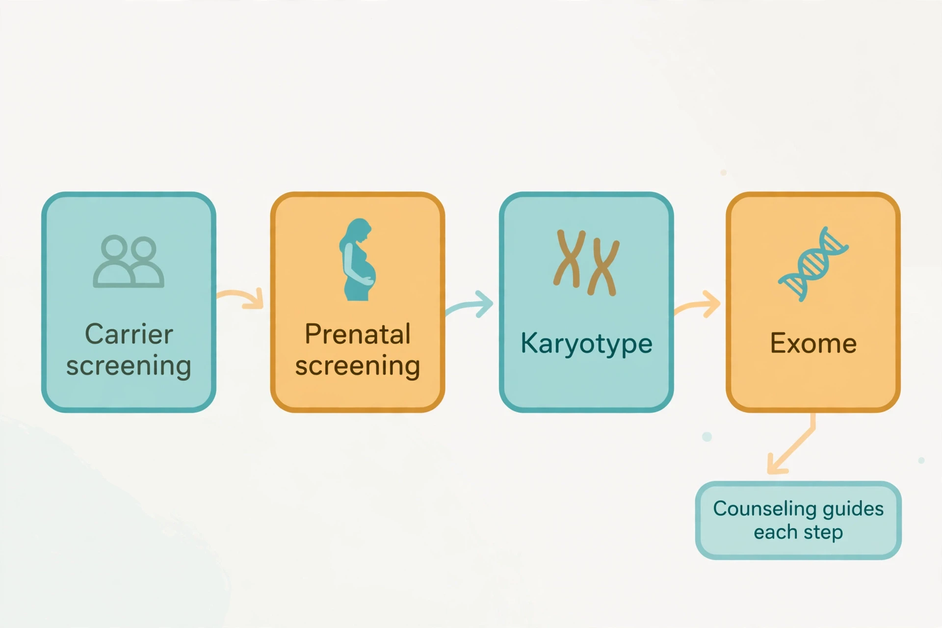
The first distinction in this section is the one readers confuse most often. Screening tests estimate risk. Diagnostic tests confirm — or rule out — a condition. A screening result is not a diagnosis, and a negative screening result does not rule everything out. Everything that follows is a variation on that distinction, and the counseling that surrounds testing exists largely to make sure nobody mistakes one kind of answer for the other. Nothing in this section is individual medical advice. It is a map of the tests that exist, what each one can say, and where each one falls short.
10.1 Carrier screening: knowing before pregnancy
Carrier screening tests healthy people for variants they could pass to a child, and it is most useful before or early in pregnancy, when both partners can be tested. The logic is the pedigree logic from section 5.4: if both partners carry the same recessive variant, each child has a 25 percent chance of being affected. Knowing that in advance does not change the odds. It changes what the couple can plan for — including the option of testing during pregnancy, or of considering other paths to parenthood.
Cystic fibrosis carrier screening is the flagship example. The American College of Obstetricians and Gynecologists recommends that it be offered to every woman who is considering or experiencing pregnancy. In 2023 the American College of Medical Genetics and Genomics updated its recommendation from a fixed 23-variant CFTR panel to a panel of 100 specific pathogenic CFTR variants — a quiet change that reflects how much more is now known about which variants matter, and a reminder that the panels themselves keep improving.
10.2 Prenatal screening and diagnosis: NIPT, amniocentesis, and what each can and cannot say
Non-invasive prenatal testing, or NIPT, analyzes cell-free fetal DNA in the mother's blood. It is a highly sensitive screen for trisomy 21, 18, and 13 — but a positive NIPT result must be confirmed by a diagnostic test. The blood draw is simple; the interpretation is not. A screen that is 99 percent sensitive still produces false positives, because the condition it screens for is uncommon, and that is why a positive screen is the start of a conversation, not the end of one.
Amniocentesis and chorionic villus sampling, or CVS, are diagnostic prenatal tests that analyze fetal cells directly. They can give a definite answer about chromosome number and many single-gene disorders. They are invasive, and they carry a small risk of miscarriage — a risk that varies by source and by era, and that a clinician should quote from current guidance rather than from a textbook figure. The trade-off is the point: the tests that can confirm a diagnosis are the tests that carry a risk, and the tests that carry no risk can only estimate one.
10.3 Diagnostic testing: karyotype, microarray, and exome sequencing
A karyotype is a picture of a person's chromosomes. It can detect extra or missing whole chromosomes — Down syndrome, Turner syndrome — and large structural changes, but not small deletions or single-gene variants. It is the oldest tool in this section, and still the right one when the question is about chromosome number.
Chromosomal microarray can detect deletions and duplications far smaller than a karyotype can see, including microdeletions such as 22q11.2 deletion syndrome, which affects about 1 in 4,000 births and is usually detected by microarray, not by standard karyotype. Cri du chat syndrome, caused by a deletion on the short arm of chromosome 5, affects about 1 in 25,000 to 50,000 newborns and is another condition microarray can find.
Whole-exome sequencing reads the protein-coding regions of all roughly 20,000 genes at once. The exome is only about 1 to 2 percent of the genome, yet it harbors roughly 85 percent of known disease-causing pathogenic variants — which is why reading it is so productive. Exome sequencing is now recommended as a first-tier or second-tier test for children with suspected single-gene disorders, intellectual disability, or developmental delay. Reported diagnostic yields are roughly 25 to 40 percent in children with suspected single-gene disorders, and 30 to 50 percent in neurodevelopmental delay, intellectual disability, or epilepsy. The landmark study by Yang and colleagues, published in the New England Journal of Medicine in 2013, showed that clinical whole-exome sequencing could diagnose Mendelian disorders that had resisted years of conventional testing.
Why can a test still come back with no answer? Because a yield of 25 to 40 percent is also a failure rate of 60 to 75 percent. Some disorders are caused by variants in parts of the genome the exome does not cover. Some are caused by variants science has not yet linked to disease. And some results come back as a variant of uncertain significance — a change whose effect is not yet known, which is not a positive result, and whose classification can change as evidence accumulates. Tests are available for more than 2,000 genetic conditions, and one 2018 study estimated that more than 68,000 genetic tests were on the market. The catalog is enormous. Certainty is not.
10.4 Newborn screening and the diagnostic odyssey
Newborn screening in the US — the heel-prick test — screens for dozens of conditions, including cystic fibrosis and sickle cell disease, in the first days of life. It is a population-wide safety net, and it catches disorders early enough for treatment to change the outcome. But it screens for a defined list, not for everything, and a normal newborn screen does not mean a child has no genetic disorder.
Many families, instead, face what clinicians call the diagnostic odyssey: the years spent seeking a diagnosis for a rare disease. About 25 percent of people with a rare disease wait 5 to 30 years for a confirmatory diagnosis, and about 40 percent receive a misdiagnosis along the way. I have heard a parent describe that period as living in a waiting room with no door — appointment after appointment, each one ruling something out and nothing in. A diagnosis, even without a cure, ends that waiting. It gives the condition a name, connects the family with others living with it, and opens the door to research and support. That is why diagnosis matters even when treatment does not yet exist.
Genetic counseling is the human guide through all of the above. A genetic counselor is a trained professional who helps people understand their family history, their test options and results, and the risks for themselves and their children. Counseling is a process, not a test. It is recommended before and after testing, especially for predictive testing such as Huntington disease, because results affect the whole family and can have psychological, insurance, and reproductive consequences. A good counselor does not tell a family what to decide. A good counselor makes sure the family understands what they are deciding about.
11. Treatment and the Gene-Therapy Era: From Managing Symptoms to Fixing the Code
Here is the honest picture, stated plainly before the hopeful part. Most genetic disorders have no cure. Treatment usually means managing symptoms, monitoring for complications, and intervening early — for example, regular aortic monitoring in Marfan syndrome. About 95 percent of rare diseases lack an FDA-approved treatment. That is the baseline, and any article that hides it to make the hopeful part sound bigger is doing its readers a disservice.
Against that baseline, a small number of disorders now have genuine one-time genetic treatments, and the change in the last few years has been fast. The mechanics of CRISPR editing are covered in CRISPR Gene Editing Explained; this section is about what the editing has actually delivered, and what it has not.
11.1 What treatment looks like today for most disorders
For most disorders, treatment is practical and specific rather than genetic. CFTR modulators for cystic fibrosis, described in section 6.1, target the faulty protein itself. For sickle cell disease, hydroxyurea reduces the frequency of pain crises, and blood transfusions manage severe anemia and prevent strokes. For Marfan syndrome, surveillance — regular imaging of the aorta — catches the complication that kills, before it kills. Early intervention, physiotherapy, and special education support children with Down syndrome and fragile X syndrome. None of these treatments fixes the underlying gene. All of them change lives, and the gap between those two sentences is the whole history of medical genetics until very recently.
11.2 Casgevy and Lyfgenia: the first CRISPR gene therapy (December 2023)
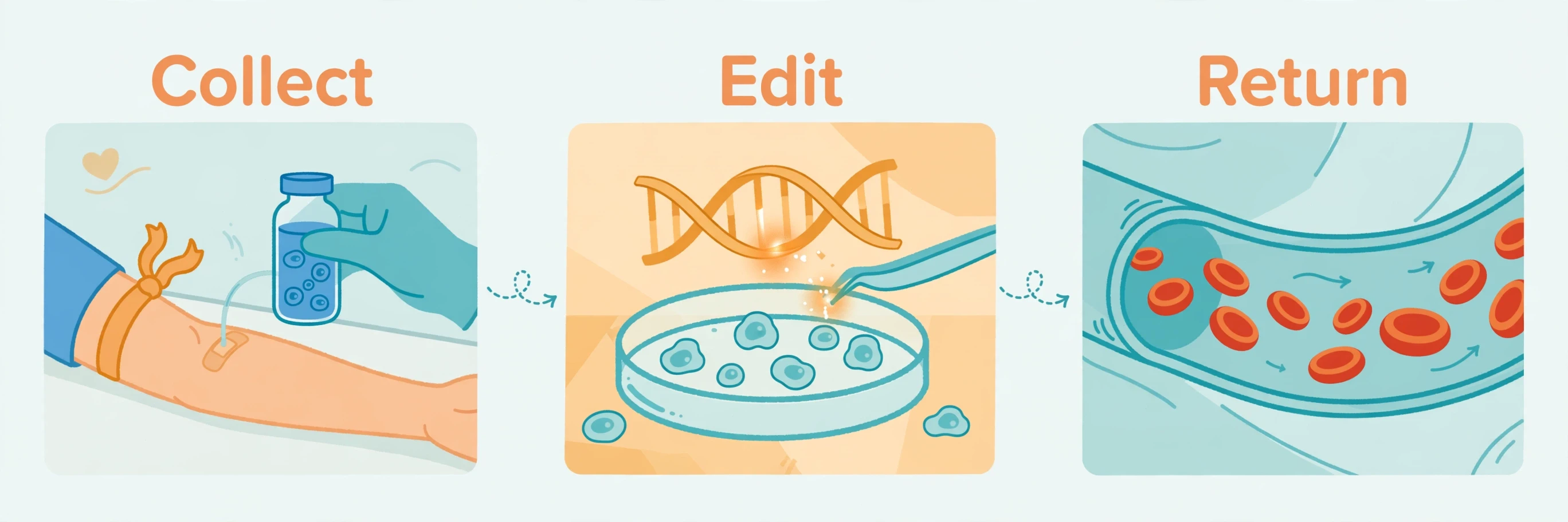
On 8 December 2023, the US Food and Drug Administration approved two gene therapies for sickle cell disease on the same day: Casgevy and Lyfgenia. Casgevy was the first FDA-approved treatment to use CRISPR/Cas9 genome editing. It edits the patient's own blood stem cells, outside the body, to re-switch on fetal hemoglobin — the form of hemoglobin babies use before birth, which does not sickle — and then returns the edited cells after chemotherapy conditioning. Lyfgenia uses a lentiviral vector to add a modified hemoglobin gene to the patient's own stem cells, and it carries an FDA boxed warning for a risk of blood cancer seen in trials.
Casgevy was originally approved for people aged 12 and older with sickle cell disease and recurrent vaso-occlusive crises. On 1 July 2026 the FDA extended the US approval to children as young as 2, covering sickle cell disease with recurrent vaso-occlusive crises and transfusion-dependent beta thalassemia. The expansion rested on trial data in children aged 5 to under 12; the ages 2 to 4 were included by extrapolation from that data, not from direct trial evidence in that age group. In the pivotal adult and adolescent sickle-cell trial, 29 of 31 evaluable participants — 93.5 percent — had no severe vaso-occlusive episodes for at least 12 consecutive months. Those are remarkable numbers for a disorder that had, for decades, only symptom management to offer.
Then there is the price, and it cannot be left out of an honest account. Casgevy's US list price is about two point two million dollars per patient, and Lyfgenia's is about three point one million dollars. Both are one-time treatments given in specialist transplant centers. A one-time treatment that costs more than most houses is a medical breakthrough and an access problem at the same time, and the access problem is not a footnote. It decides who actually benefits.
11.3 Zolgensma and the other approved gene therapies
Zolgensma was approved by the FDA in May 2019 for children under 2 with spinal muscular atrophy caused by SMN1 variants. It is a one-time intravenous infusion delivering a functional copy of the SMN1 gene, and its US list price at approval was about two point one million dollars — the most expensive drug in the world at the time. Luxturna, approved in 2017, was the first gene therapy approved in the US for an inherited disease: a form of inherited blindness caused by RPE65 variants, at a list price of about eight hundred and fifty thousand dollars. Elevidys, the first gene therapy for Duchenne muscular dystrophy, was approved in 2023.
Honesty requires reporting the safety story too. On 5 November 2026, Novartis disclosed nine reports of tumors in patients previously treated with Zolgensma. The next day, the FDA confirmed that in two of the three tumor cases involving the GLI1 oncogene, molecular analysis of tumor tissue showed the therapy's vector DNA had integrated into the genome near GLI1 — product-related insertional oncogenesis, confirmed in those two cases, not merely suspected. How GLI1 was overexpressed in the third case remains under investigation, as does the overall frequency of this risk. The FDA determined the risk applies to Itvisma as well — a related therapy using the same vector and construct, approved on 24 November 2025 for patients aged 2 and older — even though no tumors have been reported after Itvisma to date. No recall or withdrawal has been issued, and the FDA states the benefits of both therapies continue to outweigh their risks for their approved uses. The story is still developing, but the mechanism in those two cases is no longer an open question. It belongs in this article because gene therapy's promise and its risks are the same story, and a reader who hears only the promise has been misled.
11.4 What gene therapy cannot do (yet)
Gene therapy exists for only a tiny fraction of the 6,000-plus known genetic disorders. Delivery to the brain remains hard, which is why so many neurological disorders are still out of reach. Long-term durability is still being measured — the first patients are still, in effect, part of a long experiment whose results arrive year by year. And germline editing, which would change the DNA passed to future generations, is not an approved treatment anywhere; it is prohibited or subject to a moratorium in most countries. Every approved gene therapy edits only the treated person's own somatic cells. The line between treating a patient and changing a bloodline is one medicine has chosen, deliberately, not to cross.
The approved gene therapies at a glance:
Luxturna (2017): RPE65-related inherited blindness; the first US-approved gene therapy for an inherited disease.
Zolgensma (2019): spinal muscular atrophy, SMN1 variants, children under 2; one-time intravenous infusion.
Casgevy (2023): sickle cell disease; the first FDA-approved CRISPR therapy; edits the patient's own stem cells outside the body.
Lyfgenia (2023): sickle cell disease; lentiviral addition of a modified hemoglobin gene; carries a boxed warning for blood-cancer risk.
Elevidys (2023): Duchenne muscular dystrophy; the first gene therapy for the disorder.
12. Myths and Mistakes About Genetic Disorders
Myths about genetic disorders are unusually sticky, because they sound like common sense and they attach to decisions people make about children, pregnancy, and testing. Each one below gets a short correction. Every correction traces to a fact in this article.
"Genetic disorders always run in families." They do not. About 25 percent of Marfan cases, about 30 percent of Duchenne cases, and about 80 percent of achondroplasia cases arise from new variants with no family history, and most Down syndrome comes from a random error in cell division.
"If no one in my family has it, my children are safe." Healthy carriers are invisible. About 1 in 25 people of European descent carries a CFTR variant without knowing it, and two carriers can have an affected child.
"A carrier is sick, or will develop the disorder." A carrier of a recessive disorder has one faulty copy and one working copy, and usually has no symptoms. The small print: carriers of some X-linked disorders can show mild symptoms, and sickle cell trait has its own health considerations — but a carrier is not a patient with the disorder.
"Down syndrome is caused by something the mother did." No parental behavior or environmental factor is known to cause Down syndrome. It results from a random error in cell division, usually nondisjunction in the egg.
"Genetic testing gives a yes-or-no answer." Screening estimates risk; only diagnosis confirms. A variant of uncertain significance is not a positive result, and a negative result does not rule everything out — exome sequencing still leaves 60 to 75 percent of suspected single-gene cases undiagnosed.
"Gene therapy can cure any genetic disorder now." Only a handful of gene therapies are approved, for a handful of disorders. The great majority of the more than 6,000 known genetic disorders have no gene therapy, and about 95 percent of rare diseases lack an FDA-approved treatment of any kind.
"If a disorder is genetic, nothing can be done." CFTR modulators target the faulty protein in cystic fibrosis. Aortic monitoring saves lives in Marfan syndrome. Early intervention changes developmental outcomes for children with Down syndrome. A genetic cause is not a shrug.
"Identical twins always share genetic disorders." Twins who start life from one embryo share essentially the same DNA, yet one twin can develop a multifactorial condition the other never gets — and a new variant or mosaicism arising after the split can even leave them different for a single-gene disorder.
"A positive BRCA result means you will get cancer." It means a sharply higher risk, not a certainty. A BRCA1 carrier's lifetime breast-cancer risk sits roughly between 55 and 72 percent, against about 12 to 13 percent in the general population. Risk is a number to manage, not a verdict.
"Genetic disorders are always present at birth." Huntington disease typically appears between ages 30 and 50. Some mitochondrial disorders appear in adulthood. A gene can be present from conception and silent for decades.
13. Living With a Genetic Disorder: What the Science Means for Families
A diagnosis is a piece of information. A life is what a family builds around it. The families who live with genetic disorders will tell you — and the patient communities, the support groups, and the advocacy organizations built by those families will confirm — that the science in this article matters most at three moments: when a diagnosis arrives, when a decision has to be made, and when a child grows up and starts asking questions of their own.
Early intervention matters, often more than any drug. Children with Down syndrome benefit from physiotherapy, speech therapy, and early education support. Children with fragile X syndrome benefit from structured learning and behavioral support. Adults with Marfan syndrome benefit from a cardiologist who images the aorta on schedule, every year, without fail. None of this is dramatic. All of it is the difference between a condition that is managed and a condition that is left to surprise.
Patient communities matter too. A rare disease can be isolating in a way that a common one is not: a family may be the only one in their town, their school, or their clinic with a given diagnosis. The organizations built around these conditions — for cystic fibrosis, for Huntington disease, for Down syndrome, for sickle cell disease — do what no textbook can: they connect a new family with families who have already walked the road, and they turn a private worry into shared knowledge. Research participation matters in the same way. For disorders with no approved treatment, a family's willingness to join a registry or a trial is often what moves the science forward for the next family.
And the diagnosis itself has value, even when there is no cure. I think back to the couple from section 1, the ones who learned they were both carriers only after their daughter's diagnosis. What changed for them was not their daughter's lungs. What changed was the shape of their ignorance: they went from not knowing why their daughter coughed to knowing the gene, the pattern, the odds for a next child, and the community of families living with the same answer. Understanding the pedigree did not cure anything. It gave them decisions they could make with their eyes open — about testing, about a next pregnancy, about who in the wider family should be told they might be a carrier too. That is what genetic knowledge is for. Not certainty. Not a cure. A clearer view of the road, and the chance to walk it deliberately.
Person-first language is part of that clarity. People live with genetic disorders; they are not defined by them. A child with Down syndrome is a child first. A person with Huntington disease is a person first, and a person with a future that deserves planning, not pity. The language in this article has tried to hold that line throughout, and it is worth stating once, plainly, at the end: no one chooses their genome, no one is to blame for it, and no diagnosis is a verdict on a person's worth.
14. Conclusion: Your Genome Is a Family Story
Four types of genetic disorders cover the field: a fault in one gene, a fault in a whole chromosome, many small faults plus environment, or a fault in the mitochondria's small genome. The inheritance rules are few and plain: one faulty copy is enough for a dominant disorder, two are needed for a recessive one, the X chromosome has its own pattern, and mitochondria travel down the maternal line only. A family tree, read with those rules, can tell a family more about its future than any single test.
Testing has grown powerful and stayed imperfect. Screening estimates risk, diagnosis confirms, and exome sequencing still leaves most suspected single-gene cases without an answer — while returning, for the lucky minority, a name for a condition that had resisted years of searching. Treatment is still, for most disorders, the careful management of symptoms. For a small and growing number, it is now something more: a one-time edit to the code itself, priced at a level that raises as many questions as it answers.
Every pedigree is a story written in DNA — a story of carriers who never knew, of errors no one caused, of risks that can be counted and choices that cannot be made by counting alone. Understanding that story is the first step to acting on it, and acting on it is the whole point. The genome is not a fate. It is a family history, written in a language we are finally learning to read.
15. Frequently Asked Questions (FAQ)
What is a genetic disorder?
Genetic disorders are health problems that start with a change in the genome — one faulty gene, several genes acting together, or an extra or missing chromosome. More than 6,000 such disorders are known, and they fall into four types — single-gene, chromosomal, multifactorial, and mitochondrial. Carrying a variant is not the same as having a disorder: most variants are harmless, and only pathogenic variants — changes that disrupt a protein enough to cause illness — lead to a disorder.
What are the types of genetic disorders?
There are four types. Single-gene (monogenic) disorders are caused by a pathogenic variant in one gene, such as cystic fibrosis or Huntington disease. Chromosomal disorders are caused by changes in the number or structure of whole chromosomes, such as Down syndrome. Multifactorial (complex) disorders are caused by variants in many genes plus environmental and lifestyle factors, such as type 2 diabetes and heart disease. Mitochondrial disorders are caused by variants in the mitochondria's own small genome of 37 genes, such as MELAS and Leigh syndrome.
What is the difference between autosomal dominant and autosomal recessive inheritance?
In autosomal dominant inheritance, one faulty copy of a gene is enough to cause the disorder, the disorder usually appears in every generation, and each child of an affected parent has a 50 percent chance of inheriting the variant — Huntington disease is the classic example. In autosomal recessive inheritance, both copies must be faulty for the disorder to appear, healthy carriers can pass the variant silently so the disorder can skip generations, and when both parents are carriers, each child faces a one-in-four chance of being affected, a one-in-two chance of being a carrier, and a one-in-four chance of inheriting neither variant — cystic fibrosis and sickle cell disease follow this pattern.
Can a genetic disorder be inherited?
Not always. Some are inherited from a parent, following dominant, recessive, X-linked, or mitochondrial patterns. Others begin new in the child: Marfan syndrome starts this way in about a quarter of cases, Duchenne in about 30 percent, and achondroplasia in about 80 percent — a new variant neither parent carries. Most Down syndrome comes from nondisjunction — a random error in cell division that is neither parent's fault — and only the translocation type can be passed on. A family history matters, but its absence does not rule a genetic disorder out.
What causes Down syndrome?
Down syndrome comes from a full or partial extra copy of chromosome 21. In about 95 percent of cases, the cause is trisomy 21: nondisjunction, a failure of chromosomes to separate properly during meiosis, usually in the egg, leaves the embryo with three copies of chromosome 21 and 47 chromosomes in total. About 3 to 4 percent of cases are translocation Down syndrome, the one type that can be inherited, and about 1 to 2 percent are mosaic Down syndrome, in which only some cells carry the extra chromosome. No parental behavior or environmental factor is known to cause it.
What is the most common genetic disorder?
The honest answer depends on which group you mean. The most common single-gene disorder group is led, in many populations, by cystic fibrosis among people of European descent and sickle cell disease among Black Americans — sickle cell disease is the leading inherited red-blood-cell disorder in the United States. The most common chromosomal condition is Down syndrome, affecting about 1 in 640 babies born in the United States. The most common genetic disorders overall are the multifactorial ones — type 2 diabetes, heart disease, and many cancers — because they are shaped by many genes plus environment and affect large numbers of people. There is no single unqualified answer, and any source that gives one is oversimplifying.
16. Sources
Centers for Disease Control and Prevention — Genetic Disorders: Genomics and Your Health
Cleveland Clinic — Genetic Disorders: What Are They, Types, Symptoms and Causes
Centers for Disease Control and Prevention — Data and Statistics on Sickle Cell Disease
National Institute of Child Health and Human Development — What causes Down syndrome?
National Cancer Institute — BRCA Gene Changes: Cancer Risk and Genetic Testing
MedlinePlus Genetics — What is a gene variant and how do variants occur?
MedlinePlus Genetics — Duchenne and Becker muscular dystrophy
MedlinePlus Medical Encyclopedia — Genetics (including mitochondrial inheritance)