
1. The Two Pains in a Single Second — A Stubbed Toe Teaches the Science of Pain
It was past midnight, the house was dark, and I was barefoot. My little toe found the corner of a bed frame with the kind of precision that feels almost deliberate. The first pain arrived instantly: a sharp, bright sting, so well located I could have pointed to the exact millimeter of skin it came from. Then, a second or so later, a different pain rolled in underneath it — duller, deeper, aching, spreading across the whole toe and the front of the foot. Two pains, one injury, a second apart. If you have ever stubbed a toe, touched a hot pan, or cut a finger while cooking, you have felt exactly this. The science of pain starts with that ordinary observation, because the double pain is not a quirk. It is a clue to how the whole system is built.
Here is the puzzle. The toe hit the bed frame once — one impact, one moment of damage — yet the nervous system delivered two separate reports, at two speeds, with two different characters. The first was fast, sharp, and precise. The second was slow, dull, and vague. Why would one event produce two experiences? The answer involves two different kinds of nerve fiber, traveling at very different speeds, carrying two different kinds of message to the spinal cord. By the end of this article, you will know why there are two pains, why rubbing a bumped knee genuinely helps, and why pain sometimes refuses to switch off long after the injury has healed.
I have spent years explaining this system to students, and the stubbed toe is always where I start, because it gives the reader a memory to attach the mechanism to. I once asked a room full of students to recall the last time they burned a fingertip on a pan. Nearly every hand went up, and nearly everyone described the same sequence without knowing why: the instant sting, the withdrawal, then the throbbing ache that arrived a beat later and stayed. Nobody in that room had been taught the names of the fibers involved. Everyone had felt them working. That gap — between what we all feel and what we are told about it — is what the science of pain exists to close.
This article follows the signal: the definition of pain, the four stages of the journey from skin to brain, the detectors and the two fiber types, gate control theory, the brain's own painkillers, and the two ways the alarm gets stuck — first in the injured tissue, then in the spinal cord itself — until pain outlasts healing. Along the way it covers phantom limb pain, the placebo and nocebo effects, and the myths that still mislead people living with chronic pain. The science of pain is a large subject, and this article takes it one step at a time, in the order the signal itself travels.
2. What Is Pain? The 2020 Definition That Changed Everything
How does pain work? In the plainest terms, pain is what the brain builds when it decides that the body may be in danger — an experience assembled from signals, memory, emotion, and context, not a direct readout from a sensor. That is the short answer, and the rest of this article is the long version of it. But before the mechanism, the definition matters, because for forty years the official definition of pain had a flaw that quietly excluded people whose pain was real but invisible. The science of pain changed in 2020, when the definition was rewritten.
The International Association for the Study of Pain, known as IASP, defines pain as "an unpleasant sensory and emotional experience associated with, or resembling that associated with, actual or potential tissue damage." That single sentence is the most important sentence in the science of pain. It was adopted in 2020, replacing a definition that had stood since 1979. The change looks small on the page. It is anything but small in practice. The old definition described pain as an experience "associated with actual or potential tissue damage, or described in terms of such damage." The new one replaces "described in terms of" with "resembling that associated with." One word — resembling — carries the whole revision. Under the old wording, pain was defined by reference to tissue damage, and people whose pain could not be described in tissue-damage terms sat outside the definition. Under the new wording, pain is recognized even when a person cannot describe it that way, and even when no damage can be found. The IASP revised definition and its history are set out on the association's own pages.
The 2020 definition comes with six key notes, and three of them do most of the work. First, pain is always a personal experience, influenced by biological, psychological, and social factors. Second, pain and nociception are different phenomena, and pain cannot be inferred solely from activity in sensory neurons. Third, a person's report of an experience as pain should be respected. The revision was produced by a multinational, multidisciplinary task force chaired by Srinivasa N. Raja, after a two-year process including public consultation that received over 800 responses from 46 countries, and it was published online in July 2020 in the journal Pain. Those details matter because they show the definition was not one committee's opinion: pain is real when the person experiencing it says it is real, whether or not a scan can show why.
Most people assume pain is a simple measurement — that the amount of pain matches the amount of damage, the way a thermometer matches temperature. What the data actually show is something else entirely. The same injury can produce very different pain in two people, and the same person can experience the same injury very differently on two different days. That is not a failure of honesty. It is the system working as designed, because pain is built from more than the injury. The science of pain is, at its core, the study of how that building happens, and why it sometimes goes wrong.
2.1 Pain is not the same as nociception
Nociception is the neural process of encoding a noxious stimulus — the detection of something potentially damaging and the conversion of that event into an electrical signal. Pain is the conscious experience the brain constructs from that signal, mixed with emotion, memory, attention, and expectation. The two are related, but they are not the same thing, and the distinction is the single most important idea in the science of pain. Nociceptive activity does not always result in pain, and pain can occur without any nociceptor activation at all. A soldier in combat or an athlete mid-game can sustain a serious injury and feel little pain until later, because the brain's descending systems have turned the volume down. A person with phantom limb pain feels pain in a limb that no longer exists, where no nociceptor could possibly be firing. Both cases break the simple picture of pain as a direct wire from injury to feeling, and both are explained once you accept that nociception is input and pain is output — and the brain sits between them, editing.
Think of nociception as a microphone and pain as what comes out of the speaker after the signal has passed through a mixing desk — turned up, turned down, filtered. The microphone matters, but it does not decide what the audience hears. The science of pain is largely the study of that mixing desk: the spinal cord, the brainstem, and the brain regions that shape the signal before it becomes experience.
2.2 The three kinds of pain, in one paragraph each
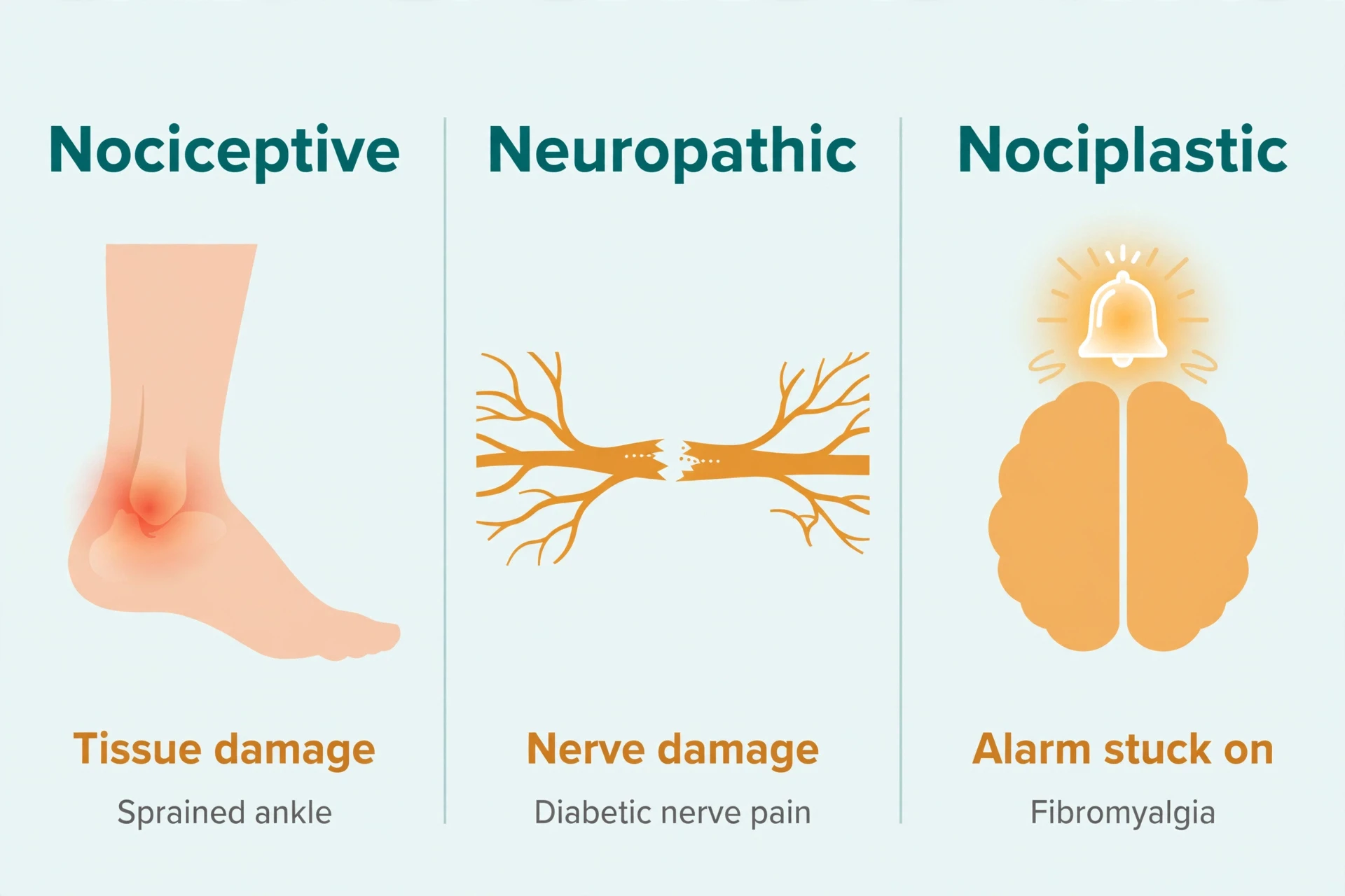
The IASP recognizes three mechanistic descriptors of pain. They describe mechanisms, not diagnoses, and using one does not mean a specific mechanism has been directly measured in an individual person. Still, the three-way split is the map the science of pain uses in the rest of this article.
Nociceptive pain arises from actual or threatened damage to non-neural tissue and is due to the activation of nociceptors — the pain of a sprained ankle, a burn, or an inflamed joint.
Neuropathic pain is caused by a lesion or disease of the somatosensory nervous system itself — the pain of diabetic nerve damage, shingles, or multiple sclerosis.
Nociplastic pain arises from altered nociception despite no clear evidence of actual or threatened tissue damage, or of disease or lesion of the somatosensory system — the pain of fibromyalgia and chronic widespread pain, where the alarm system itself has changed.
Each kind gets its full treatment in section 13, where the science of pain takes up the taxonomy in full. For now, notice what the three-way split already tells you: pain can come from damaged tissue, from a damaged nervous system, or from a nervous system that has changed how it processes signals. Only the first of those involves an injury you can point to. The other two are why the 2020 definition had to change, and why this article exists.
Chronic pain is far more common than most people realize. The numbers below come from the US Centers for Disease Control and Prevention, based on the National Health Interview Survey, and they are the latest official figures available.
Measure | Figure (2023, US adults) | What it means |
|---|---|---|
Chronic pain | 24.3% | Pain on most days or every day in the past 3 months — nearly one adult in four |
High-impact chronic pain | 8.5% | Pain that frequently limits life or work activities |
Women vs men | 25.4% vs 23.2% | Women were more likely than men to report chronic pain |
High-impact, women vs men | 9.6% vs 7.3% | The same gap appears for high-impact chronic pain |
Youngest vs oldest adults | 12.3% (ages 18–29) rising to 36.0% (age 65+) | Prevalence rises steeply with age |
Global estimate | About 20% of adults | About one adult in five worldwide, with some reviews suggesting nearly 1.6 billion people as a derived estimate |
The trend is worth noting too. In 2019, 20.4% of US adults had chronic pain and 7.4% had high-impact chronic pain. By 2023, both figures had risen — to 24.3% and 8.5%. Those figures come from the CDC's National Center for Health Statistics Data Brief No. 518, published in November 2024. A separate 2026 meta-analysis of 52 studies reported a pooled global prevalence of 26.99% — a single recent analysis rather than a consensus figure. Chronic pain is not rare, not marginal, and not a small problem at the edge of medicine. It touches roughly a quarter of adults, and understanding how it works is one of the most practical things the science of pain has to offer.
3. The Science of Pain in Four Steps: From Skin to Brain
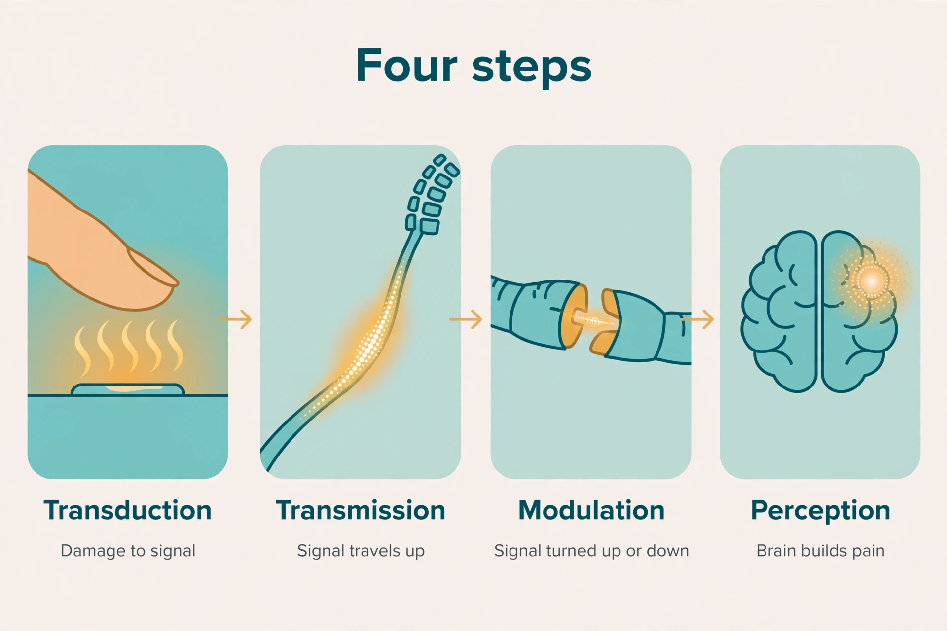
The science of pain has a spine: a four-step journey. A noxious stimulus — heat, pressure, a chemical irritant — is first converted into an electrical signal. That signal travels to the spinal cord and up to the brain. Along the way it is amplified or dampened. Finally, the brain constructs the conscious experience we call pain. Transduction, transmission, modulation, perception. Every section that follows is one of those stages in detail: transduction in section 4, transmission in sections 5 and 6, modulation in sections 7 through 10, perception in section 6, revisited in sections 15 and 16.
Each stage can go wrong in its own way: a detector that fires too easily, a spinal relay that amplifies, a descending brake that fails. Keeping the four stages apart is what lets a reader — and a clinician — work out where in the chain a particular pain is coming from, and the science of pain studies the four stages as a chain, not as separate facts. The hardware of this journey — neurons, action potentials, the spinal cord, the brain regions involved — is covered in depth in our article on How the Nervous System Works. This article spends its words on pain, not on re-teaching neuroscience.
4. Nociceptors: The Detectors That Only Fire When It Matters
What are nociceptors? They are high-threshold sensory receptors of the peripheral nervous system that detect stimuli with the potential to cause damage. They are free, bare nerve endings — not specialized encapsulated endings like the touch receptors that let you feel texture and vibration. That plain description hides an elegant piece of design. A nociceptor is excited only when stimulus intensity reaches the noxious range. Ordinary touch, ordinary warmth, ordinary pressure — none of it reaches the threshold, so none of it fires the alarm. The high threshold is the point. It is what lets the system selectively detect potentially injurious stimuli rather than reporting every contact with the world as pain. The science of pain begins here, with a detector that ignores almost everything and fires only when it matters.
The cell bodies of nociceptors sit in the dorsal root ganglia for the body and in the trigeminal ganglion for the face. Each neuron has a peripheral branch reaching into the target tissue — skin, muscle, joint, or organ — and a central branch entering the spinal cord. There are no nociceptors in the central nervous system itself. That fact will matter in the section on phantom limb pain.
I remember the first time I watched a student test this threshold for herself in a teaching session, pressing a warm metal rod against her own forearm and raising the temperature step by step. Nothing, nothing, nothing — then, at a particular point, a clear sting. She laughed, surprised by how sharp the boundary was. It felt like a switch, not a slope. That is the nociceptor's high threshold in action, and it is why you can rest your hand on a warm mug without pain and pull it away from a hot stove without thinking. The same receptor, the same skin, two different intensities, two different outcomes — and the line between them is the threshold.
4.1 How a nociceptor turns damage into electricity
Transduction is the conversion of a physical event into an electrical signal, and nociceptors do it with specialized ion channels. The transient receptor potential, or TRP, channel family is central. TRPV1 is activated by noxious heat and by capsaicin, the chemical that makes chili peppers hot. TRPM8 is activated by cold and by menthol, which is why mint feels cool. When a TRP channel opens, ions flow across the membrane, the ending depolarizes, and if the signal is strong enough, a voltage-gated sodium channel fires an action potential that races toward the spinal cord. One channel in particular, NaV1.7, is present only in nociceptor fibers and helps generate the action potential. It is one reason local anesthetics work: lidocaine dulls pain by blocking voltage-gated sodium channels.
The TRPV1 heat threshold is a good example of honest science: one textbook gives above 45 degrees Celsius, another gives above 43 degrees. The fair statement is that TRPV1 opens at around 43 to 45 degrees Celsius. That imprecision reflects real variation between experimental preparations, and a reader told one exact number as settled fact has been misled — which is why this article reports ranges wherever the sources disagree.
There is also an honest gap in the story. Heat and chemical transduction are far better understood than mechanical transduction. The molecules that let a nociceptor detect a pinch or a cut at the molecular level remain stubbornly elusive. We know the channel for heat. We know the channel for cold. We do not yet fully know the channel for a squeeze. That gap is worth stating plainly, because it is the kind of thing that separates a real explanation from a polished one. A real explanation admits what it does not know.
4.2 The nociceptor family: polymodal, silent, peptidergic
Most nociceptors are polymodal, meaning a single fiber can respond to more than one kind of noxious stimulus — mechanical, thermal, and chemical. A fiber that fires when you press hard on a bruise may also fire when the same spot is heated, or when inflammatory chemicals wash over it. That flexibility is useful for an alarm system: it means fewer detectors can cover more kinds of danger. A-delta nociceptors subdivide further, into Type I fibers with a high mechanical threshold and a heat threshold above 50 degrees Celsius, and Type II fibers with a low heat threshold and a very high mechanical threshold, which mediate the first acute response to noxious heat.
A stranger member of the family is the silent nociceptor. Under normal conditions, these C fibers are mechanically insensitive — they ignore pressure entirely. Only in the setting of injury or inflammation do they develop mechanical sensitivity, waking up and adding new pain-signaling channels that were not active before. It is a neat piece of design: the system keeps reserve detectors in the dark until damage justifies switching them on. The trouble, as section 9 shows, is that once switched on they do not always switch off quietly.
Nociceptors also fall into two chemical classes: peptidergic nociceptors, which contain substance P and calcitonin gene-related peptide (CGRP) and depend on nerve growth factor signaling, and nonpeptidergic nociceptors, which bind the plant lectin IB4, express the ATP-gated ion channel P2X3, and depend on glial cell line-derived neurotrophic factor signaling. Two chemically distinct populations, with different messengers — a division that matters for how pain is processed in the spinal cord. When activated, nociceptors release glutamate, substance P, CGRP, and ATP onto output neurons in the superficial dorsal horn. They also release transmitters outward into the surrounding tissue, triggering mast-cell degranulation and histamine release — one reason an injury site swells and becomes tender. And the neurons are not working alone: epithelial cells such as keratinocytes and Merkel cells directly interact with peripheral axon terminals and help modulate sensory transduction. The skin itself participates in sensing.
4.3 There are no pain receptors in your brain
Here is a fact that surprises almost everyone I tell it to: there are no nociceptors in the brain itself. The brain has no pain receptors in its own tissue, which is why brain surgery can be performed on an awake patient. The surgeon can cut brain tissue, and the patient feels nothing from the brain itself — though the scalp and the membranes around the brain, which do have nociceptors, must be anaesthetised. The brain builds pain. It does not feel its own tissue damage directly.
That sounds like a curiosity, and it is a striking one, but it is also the logical foundation of everything that follows. If pain were simply a readout from damaged tissue, then a brain with no damage detectors could never produce pain on its own. But it can — in phantom limb pain, in migraine, in nociplastic pain — because pain is constructed by the brain from signals, memory, and expectation, not read off a sensor in the tissue. The absence of nociceptors in the brain is not a gap in the system. It is the clearest proof that pain is a brain output, not a tissue input. Keep that fact in mind when section 15 arrives. It will make the phantom limb story feel inevitable rather than impossible, and it is one of the foundations of the science of pain.
5. Two Speeds of Pain: Why One Injury Hurts Twice
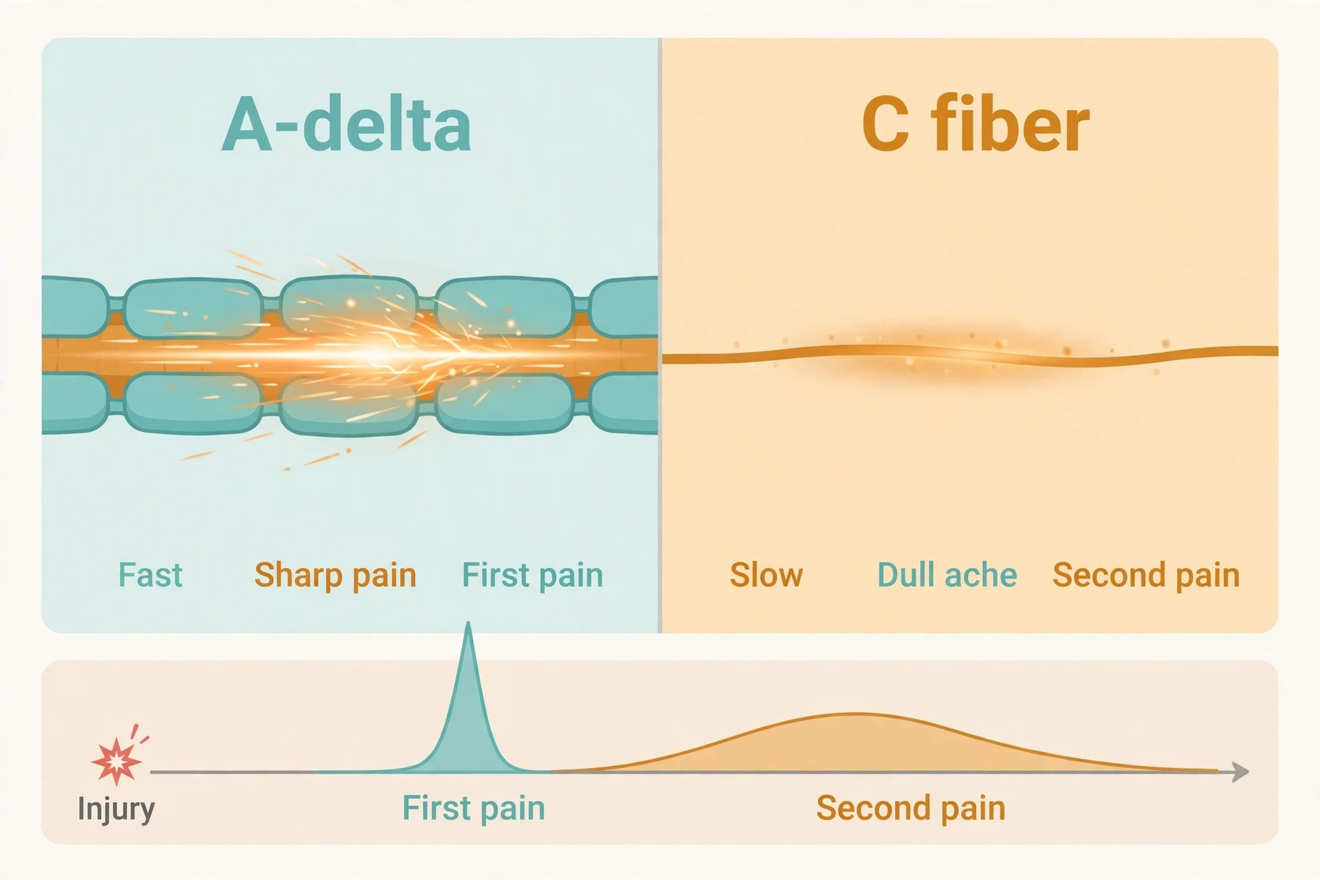
How does pain work at the level of a single injury? The science of pain answers that it works twice, at two speeds. The two major classes of nociceptor fibers are the reason. Medium-diameter, thinly myelinated A-delta fibers mediate acute, well-localised first pain — the sharp, pricking sting felt at the moment of injury. Small-diameter, unmyelinated C fibers convey poorly localised, second pain — the delayed, dull, aching, burning pain that follows. The two-pain sequence after a cut or a burn is the everyday demonstration of the two fiber types, and it is the same sequence from the stubbed toe in section 1. The science of pain explains it with a single variable: speed.
A-delta fibers conduct at roughly 5 to 30 meters per second. That is a wide range, and the width is honest: textbooks disagree. One standard text gives 5 to 15 meters per second, another gives 12 to 30, another 12 to 35, and a review figure gives 6 to 25. No single exact figure is settled, and this article will not pretend otherwise. C fibers are far more consistent across sources, conducting at 0.5 to 2 meters per second. The difference between the two is enormous — an order of magnitude or more — and it is the direct result of myelination. Myelin is insulation, and insulation is speed. A-delta fibers have a thin myelin sheath, so their signals travel fast. C fibers have none, so theirs crawl. C fibers are also smaller — 0.4 to 1.2 micrometers across, against 2 to 5 for A-delta fibers — and they are the majority of nociceptors, which is why the dull ache lingers long after the sharp sting has passed.
Return to the stubbed toe. The impact activates both fiber types at the same instant. The A-delta signal, traveling fast along its thinly myelinated fiber, reaches the spinal cord and the brain first: the sharp sting, precisely located, impossible to ignore. The C fiber signal, traveling slowly along an unmyelinated fiber, arrives a second or two later: the dull ache, poorly located, spreading, throbbing. One injury, two arrivals, two experiences — and the gap between them is simply the difference in conduction speed. Once you know this piece of the science of pain, you can never unfeel it.
Feature | A-delta fibers | C fibers |
|---|---|---|
Myelination | Thinly myelinated | Unmyelinated |
Diameter | 2–5 micrometers | 0.4–1.2 micrometers |
Conduction speed | About 5–30 meters per second (textbooks differ) | 0.5–2 meters per second |
Pain quality | Sharp, pricking, well localised | Dull, aching, burning, poorly localised |
Timing | First pain — immediate | Second pain — delayed by a second or two |
Role | Fast warning and precise location | Sustained ache that drives protection and rest |
The three fiber types at a glance:
A-delta fibers — thinly myelinated, fast, sharp first pain, precisely located.
C fibers — unmyelinated, slow, dull second pain, poorly located, and the majority of nociceptors.
A-beta fibers — large, myelinated, carrying innocuous touch and pressure, not pain at all. They will return in the next-but-one section, because their activation is what closes the spinal gate.
6. The Journey Up: Spinal Cord, Thalamus, and the Brain That Builds Pain
Transmission and perception are the middle and the end of the journey. The signal leaves the nociceptor, enters the spinal cord, and climbs to the brain — but it does not travel like a letter through the post, unchanged from sender to recipient. At every relay it is filtered, amplified, or suppressed. Understanding that filtering is the heart of the science of pain, and this section maps the route before the next section explains the filter.
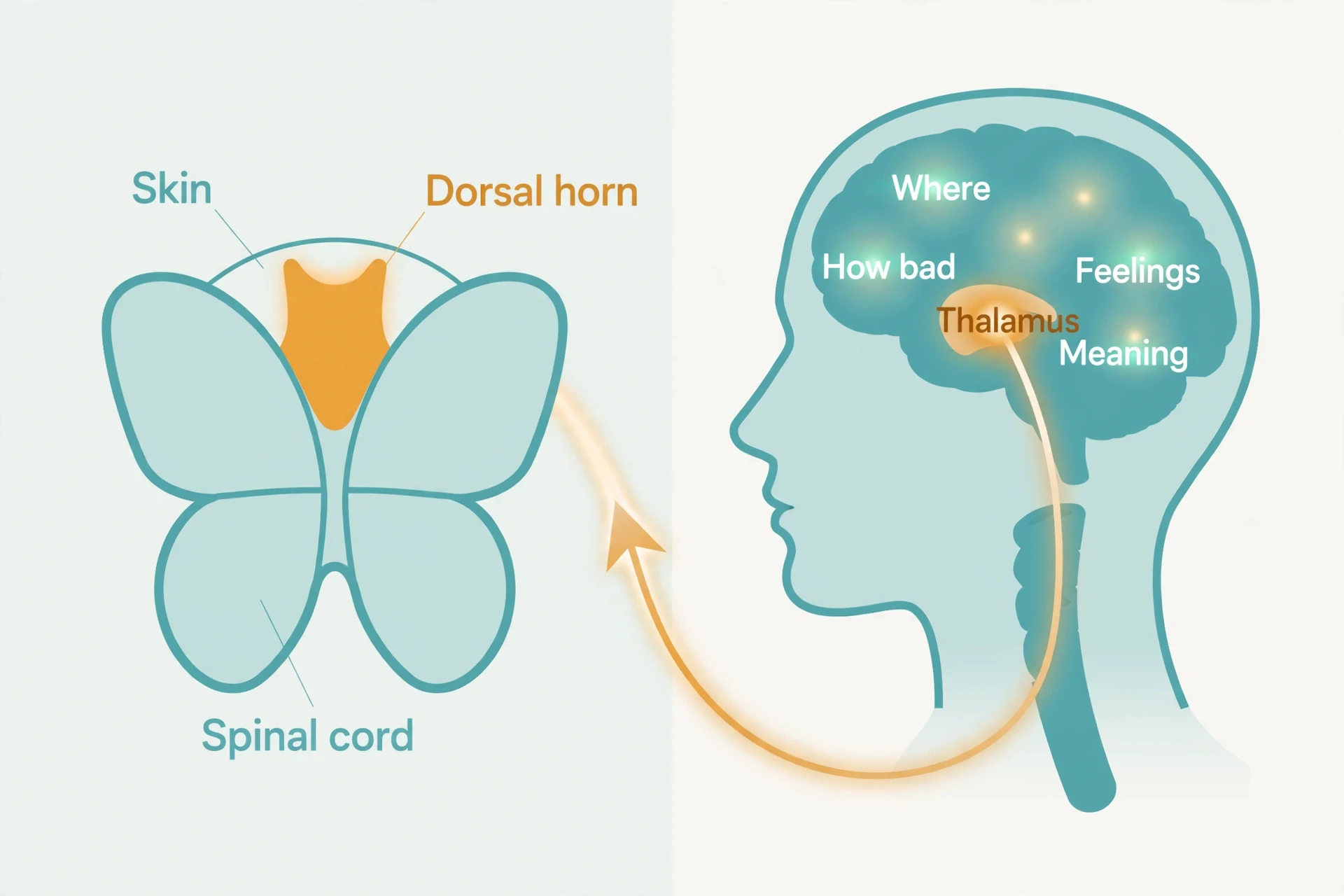
In the spinal cord, nociceptor central terminals synapse mainly in the superficial dorsal horn: A-delta and peptidergic C fibers in lamina I, nonpeptidergic C fibers in inner lamina II, A-delta fibers also in lamina V, while low-threshold touch fibers terminate deeper. That layered arrangement is the anatomy that makes the gate possible. From the dorsal horn, projection neurons carry the signal upward mainly through the spinothalamic tract to the thalamus, with the spinoreticular tract feeding brainstem centers and the spinomesencephalic tract feeding the periaqueductal gray. The thalamus relays onward to the somatosensory cortices, the insula, the anterior cingulate cortex, and the prefrontal cortex. The trigeminothalamic pathway carries the same kind of signal from the face, through the trigeminal ganglion.
Each brain destination does a different job, and the division of labor explains why pain has both a sensory and an emotional component. The somatosensory cortex handles the "where and how much" — location, intensity, quality. The insula and anterior cingulate cortex handle the emotional and unpleasant quality — how bad it feels, not just where it is. The prefrontal cortex handles evaluation, meaning, and decisions about what to do. Pain is a multidimensional experience with sensory-discriminative, affective-motivational, and cognitive-evaluative components, and there is no single designated pain center in the brain. Pain depends on distributed networks. That is why two people can have the same injury and describe their pain in completely different terms: one notices the location, the other the distress, because the networks weigh the signal differently. The anatomy is covered in full in our article on How the Nervous System Works, which this section builds on rather than repeats.
6.1 The spinal cord is not a telephone wire
The first synapse in the dorsal horn is already a decision point. The dorsal horn contains inhibitory interneurons that can suppress transmission-cell activity, and the substantia gelatinosa — lamina II — is the region where this gating circuitry is concentrated. Glutamate is the main fast excitatory transmitter at that first synapse, while substance P and CGRP are co-released and prolong and amplify the signal, especially with intense or repeated stimulation. The NMDA glutamate receptor is central to the activity-dependent strengthening of the dorsal-horn synapse — the mechanism behind central sensitization, which section 10 takes up in full.
The point to carry forward is simple, and it is the central insight of the science of pain: the signal does not travel straight from skin to conscious awareness like a telephone wire. At every relay, especially the dorsal horn, it is filtered, amplified, or suppressed before it reaches the brain. The same input can be experienced differently by different people, or by the same person at different times. Attention changes pain — focusing on it can increase its intensity, while distraction can decrease it — and that is a predicted consequence of descending modulation, not a sign that pain is imaginary.
7. Gate Control Theory: The 1965 Idea That Explains Why Rubbing Helps
What is gate control theory? In the science of pain, it is the idea that a "gate" in the dorsal horn of the spinal cord modulates pain signals before they reach the brain — and it is the single most famous theory in the science of pain. Proposed by Canadian psychologist Ronald Melzack and British neurophysiologist Patrick D. Wall in 1965, in the paper "Pain Mechanisms: A New Theory" published in Science, it did something no earlier theory had done: it gave a physiological explanation for how psychology could change pain. Attention, emotion, expectation, memory — all of them, the theory said, could reach down into the spinal cord and change what got through.
Start with the experience, not the wiring. You bump your knee on a table corner. Without thinking, you rub it. And the rubbing helps — not a little, and not only because it distracts you. It helps because touch and pressure input competes with pain input at the spinal cord. As the Cleveland Clinic puts it, gate control is why rubbing a bumped knee works. The same logic explains shaking a stubbed toe, holding a hot mug, or pressing on a bruise. Non-painful input carried by large A-beta fibers tends to close the gate. Pain input carried by small A-delta and C fibers tends to open it. The gate is the balance of excitatory and inhibitory signals at the transmission cells, and the balance shifts with every new input.
The wiring, as originally proposed, works like this. Both thin pain fibers and large touch fibers carry information to two destinations in the spinal cord: transmission cells that carry the pain signal up to the brain, and inhibitory interneurons in the substantia gelatinosa that impede transmission-cell activity. Large-fiber activity excites the inhibitory interneurons, closing the gate. Small-fiber activity inhibits them, opening it. The theory also proposed something bolder: that the brain sends signals back down to the spinal cord that can open or close the gate — descending control. That was the first physiological explanation for how a thought or a feeling could change a sensation, and it is the part of the theory that has proved most durable.
7.1 What came before: specificity vs pattern theory
Before 1965, two rival theories each explained only half the evidence. Specificity theory, traced to Descartes in the 17th century, held that a direct one-to-one line ran from injury to pain, like a wire from a sensor to a bell. Pattern theory, associated with Goldschneider in the late 19th century, held that pain came from patterned activity across fibers, with no dedicated pain pathway at all. Specificity theory could not explain why the same injury produced different pain in different people; pattern theory could not explain why some fibers were clearly specialized for noxious stimuli. Gate control theory combined elements of both and ended a century-old debate, keeping the specialized nociceptor from one and the patterned, modulated signal from the other, and adding the gate that let the two interact. That synthesis is why 1965 counts as a revolution in the science of pain rather than a small correction.
7.2 What the theory got wrong — and why it still matters
Honest science requires saying what happened next. The theory was met with considerable skepticism when first proposed, and its specific wiring diagram turned out to be wrong in detail. The nerve connections proposed in 1965 did not survive closer anatomical study. Patrick Wall himself published a re-examination and re-statement of the theory in 1978, revising details while keeping the core idea. Mouse experiments support a spinal gate for pressure-triggered pain, while human tests have given mixed results — the gate is supported as a concept, not proven in every detail in humans. The clinical technologies the theory inspired have a mixed record too: transcutaneous electrical nerve stimulation (TENS) and spinal cord stimulation both grew out of the idea that non-painful stimulation can close the gate, with Wall collaborating with neurosurgeon William Sweet on an early human trial in 1967. But the evidence for TENS is inconclusive in clinical trials, and spinal cord stimulation probably does not help low back pain. This article will not present either as proven effective, because the trial evidence does not support that claim.
So what survives? The basic conception remains unchanged, and the theory is still described as the one that most accurately accounts for both the physical and psychological aspects of pain perception. The spinal cord modulates pain. Psychology reaches the spinal cord. Those two claims, not the exact circuit diagram, are the lasting contribution. Melzack later extended the idea into neuromatrix theory, proposing that the brain contains a widespread network that generates the experience of pain — an idea that helps explain phantom limb pain, where pain is felt in a limb that no longer exists and no peripheral input is possible. The gate was the beginning, not the end, of the modern science of pain.
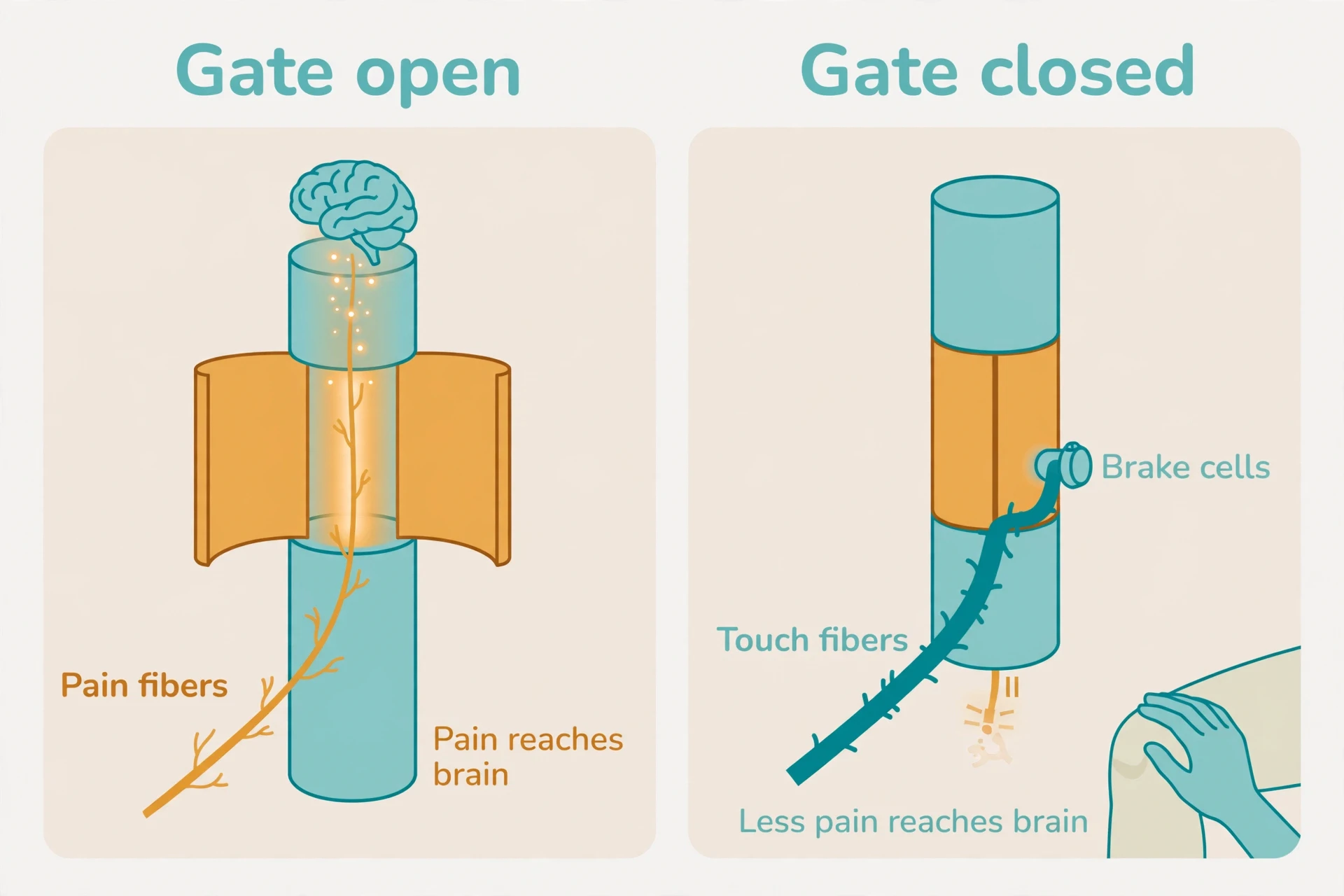
"Gate control is why rubbing a boo-boo works." — Cleveland Clinic, on gate control theory
8. The Brain Talks Back: Descending Modulation and Your Own Opioids
If the gate is the spinal cord's filter, descending modulation is the brain's hand on the filter. It explains the most mysterious observations in the field: why pain can vanish in a crisis, why expectation changes pain, and why the same injury hurts more on a bad day than on a good one. The brain does not only receive pain signals. It talks back, sending signals down to the spinal cord that can turn the volume up or down.
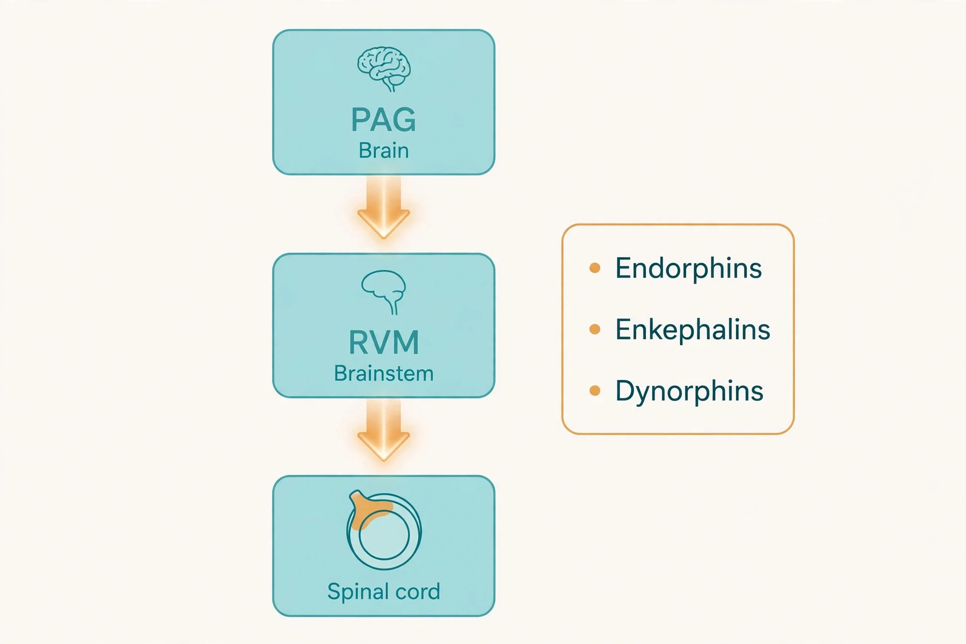
The descending pain modulatory system is a top-down pathway that starts in the periaqueductal gray, known as the PAG, a region of gray matter in the midbrain. The PAG relays to the rostral ventromedial medulla, or RVM, which includes the serotonin-rich nucleus raphe magnus. RVM neurons project down the spinal cord to the dorsal horn, where they modulate nociceptive transmission. The experimental proof dates from the 1960s onward: electrical stimulation of the PAG or the nucleus raphe magnus produces analgesia — stimulation-produced analgesia — showing that the brain has a built-in pain-suppression system. The system is driven by the body's own opioids. There are three classes of endogenous opioid peptides — beta-endorphins, which bind mainly mu receptors; enkephalins, which bind mainly delta receptors; and dynorphins, which bind mainly kappa receptors. Opioid receptors are found at high density in the PAG, the RVM, and the dorsal horn — the three stations of the descending system — as well as in the thalamus and on the peripheral terminals of nociceptors. Exogenous opioids such as morphine imitate endogenous opioids and produce analgesia by acting on those same stations.
How do endogenous opioids quiet a neuron? In two ways: they block neurotransmitter release by inhibiting calcium influx into the presynaptic terminal, and they open potassium channels, which hyperpolarises the neuron and inhibits firing. Picture a neuron as a room with a door that opens when calcium flows in. Opioids lock the door and open a window that lets potassium out, making the room harder to excite. The result is less transmitter released, less signal passed on, and less pain reaching the brain. Noradrenaline and serotonin are the other two main descending transmitters alongside opioids.
8.1 The brake and the accelerator
Here is the complication that makes the system interesting rather than simple: descending control is not inhibition only. Persistent nociception triggers both descending facilitation and descending inhibition, and both come mainly from the RVM. In inflammation models, inhibition usually predominates in circuits carrying input from the inflamed tissue, reducing primary hyperalgesia, while facilitation predominates in circuits carrying input from neighboring tissues, producing secondary hyperalgesia. RVM neurons include "off-cells", whose activity inhibits nociceptive transmission, and "on-cells", whose activity facilitates it. The balance between them sets the gain of the spinal gate. The same system can turn pain down or up, which is why context matters so much — and why the brain's influence on pain is not a simple story of relief.
Stress-induced analgesia is the vivid example: the endogenous opioid system can be activated by stressful states, and the effect can be reversed by naloxone, an opioid blocker. Soldiers and athletes who do not feel an injury until later are not ignoring pain out of toughness. Their descending system has turned the volume down while survival or performance takes priority, and the pain arrives when the crisis passes. I have watched this in a sporting context more than once: a player takes a hard knock, waves off attention, finishes the game, and only then discovers the bruise that has been there all along — a classic demonstration of the science of pain in action.
8.2 Why attention, emotion, and expectation change pain
The descending system is linked to higher brain areas — cingulofrontal regions, the amygdala, and the hypothalamus — which is how emotion, attention, and expectation reach down to the spinal cord. Focusing on pain can increase its intensity, while distraction can decrease it. The PAG–RVM–spinal cord pathway is the same route engaged by placebo analgesia: expectation of relief activates prefrontal and cingulate regions, which drive the PAG and RVM to suppress nociceptive transmission. That is physiology, not imagination, and it is the bridge to section 16, where expectation takes center stage. For now, the lesson is plain: what you feel, what you expect, and what you attend to are not separate from pain. They are inputs to the same circuit that carries the signal from your skin. The science of pain treats emotion as a mechanism, not as a footnote.
9. When the Alarm Gets Stuck: Peripheral Sensitization and the Inflammatory Soup
So far the system has behaved like a well-designed alarm: sensitive to real danger, quiet the rest of the time, adjustable from above. Now comes the first way it gets stuck. After tissue damage, the injured area does not simply wait to heal. It changes the detectors themselves, making them more excitable, lowering their thresholds, and increasing their firing. This is peripheral sensitization, and it is the first half of why pain outlasts injury. The science of pain treats it as a chemical story, because that is what it is: a soup of mediators, released by damaged tissue and inflammatory cells, that rewires the nociceptor from the outside in.
The cause is the inflammatory soup: damaged tissue, inflammatory cells, and tumor cells release prostaglandin E2 (PGE2), bradykinin, ATP, protons, histamine, serotonin, nerve growth factor (NGF), and pro-inflammatory cytokines such as tumor necrosis factor-alpha and interleukin-1beta. These mediators bind receptors on the nociceptor terminal and activate protein kinases A and C, which phosphorylate ion channels such as TRPV1 and the sodium channel NaV1.8. The result: the threshold of TRPV1 drops, the membrane becomes more excitable, and the nociceptor fires more easily and more often. The same heat that was harmless yesterday now hurts. The detector has not broken. It has been retuned, and the retuning is written in chemistry.
Two mediators deserve a closer look. Bradykinin binds B2 receptors on nociceptors, activating protein kinase C signaling that increases ion-channel activity, lowers the pain threshold, and activates TRPV1. Nerve growth factor is particularly potent: it promotes nociceptor sprouting and produces long-lasting sensitization. And the silent nociceptors from section 4 return here as a special case: C fibers that normally ignore mechanical stimuli gain mechanical sensitivity after injury, adding new pain-signaling channels that were not active before. The alarm does not only get louder. It gains new speakers.
The everyday example is sunburn. A sunburned shoulder hurts when a shirt brushes it — not because the shirt is dangerous, but because the nociceptors in the burned skin have been sensitized by the inflammatory soup and now fire at intensities that would normally be far below threshold. That is primary hyperalgesia: increased pain at the site of injury itself, where the tissue is actually damaged and inflamed. Secondary hyperalgesia — increased pain in the undamaged tissue around an injury — involves central mechanisms, not just peripheral ones, and it belongs to the next section. The chemistry of the soup is the same chemistry that drives fever and the redness, heat, and swelling of inflammation, and it is covered in depth in our article on Fever and Inflammation Explained. The inflammatory mediators that raise body temperature are also the chemicals that sensitize nociceptors. One soup, two jobs. Histamine plays a part here too, and its wider story — mast cells, allergic reactions, and the immune system's overreactions — is covered in our article on Allergies Explained. The blood that carries these mediators to and from the injury site is itself covered in our article on How Blood Works.
9.1 Why anti-inflammatory drugs help — and what they cannot do
Prostaglandins, especially PGE2, are made by cyclooxygenase enzymes: COX-1, which runs at baseline, and COX-2, which is induced by inflammation. Non-steroidal anti-inflammatory drugs, known as NSAIDs, work in part by blocking COX enzymes and so reducing PGE2 production. That is a general mechanism, stated here as information only: this article gives no individual advice, no dosage, and no recommendation about any specific treatment, and personal decisions about medication belong in a conversation with a clinician. The mechanism matters for understanding, not for prescribing. When PGE2 falls, the nociceptor's threshold rises back toward normal, and the sensitization eases — the clearest reason the science of pain describes anti-inflammatory drugs as turning down the soup, not switching off the alarm.
Then comes the limit, and it is the hinge of the whole article — and of the science of pain itself. Peripheral sensitization is normally reversible: as inflammation resolves and the mediator soup clears, nociceptor thresholds return toward normal. When it does not resolve — because inflammation persists or nerves are damaged — it feeds into central sensitization. When pain persists after the soup has cleared, the problem has moved centrally, into the spinal cord and the brain. The detectors are no longer the main problem. The amplifier is. That is the next section, and it is where acute pain becomes chronic pain.
10. Central Sensitization: The Volume Dial Turned Up
What is central sensitization? It is an amplification of neural signaling within the central nervous system that produces pain hypersensitivity — increased responsiveness of nociceptive neurons to normal or subthreshold input. That is the operational definition given by Clifford Woolf, who first described the phenomenon in 1983, showing that brief noxious inputs could trigger prolonged changes in spinal cord excitability. The analogy the science of pain uses is a good one: central sensitization is a volume dial turned up. The same input produces a larger output. The music has not changed. The amplifier has.
The mechanism is a strengthening of the first synapse in the dorsal horn. Sustained activation of primary sensory fibers induces long-lasting increases in synaptic strength at that relay, so the spinal cord amplifies signals it used to pass on quietly. Glutamate and NMDA receptor signaling are central to this strengthening. The relay becomes pathologically enhanced: a signal that once passed through at a whisper now comes through at a shout. And the change is not only amplification. Central sensitization reflects both increased activity of pain-facilitation pathways and malfunctioning descending inhibitory pathways — a failure of the brain's own analgesic control as well as an overactive amplifier.
10.1 The four signs: hyperalgesia, allodynia, wider fields, lingering pain
The science of pain describes four clinical manifestations of central sensitization — the signs clinicians look for, and worth knowing as a reader because they make an abstract mechanism concrete. They are not a self-diagnosis tool — only a clinician can assess them in a person — but as descriptions of what sensitization feels like, they are precise.
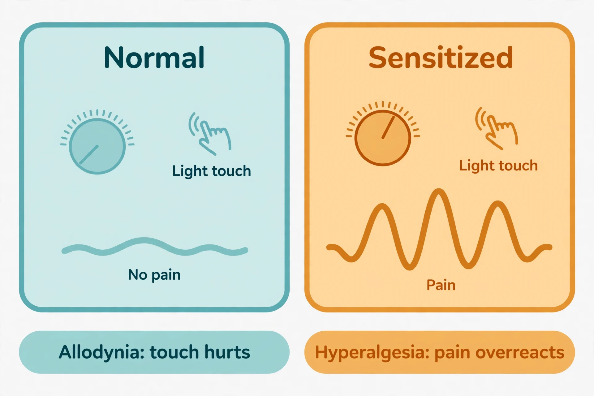
Hyperalgesia — increased pain from a mildly painful stimulus. The sunburned shoulder that hurts under a shirt.
Allodynia — pain from a normally non-painful stimulus. A light touch, or clothing contact, that should not hurt at all.
Expanded receptive fields — pain felt over a wider area than the injury, because the spinal cord's map of the body has blurred.
Persistent pain that continues after a transient stimulus ends — the signal keeps ringing after the bell has stopped.
Brain imaging in central sensitization shows overactivity in the insula, the anterior cingulate cortex, and the prefrontal cortex — the amplifier is not only in the spinal cord.
10.2 Fibromyalgia and the conditions where the dial is stuck
Central sensitization can arise from nerve injury and from neuropathies including diabetic peripheral neuropathy and postherpetic neuralgia — not only from ongoing tissue injury. It is increasingly recognized that it can also occur independently of peripheral injury or inflammation. Features of central sensitization have been identified in nearly all chronic pain conditions, and it is considered the primary underlying cause of pain in conditions such as fibromyalgia, characterized by widespread pain and multisite hyperalgesia and allodynia. The clinical grouping sometimes called central sensitization syndromes includes fibromyalgia, irritable bowel syndrome, and temporomandibular disorders — conditions where pain is widespread or out of proportion to any visible tissue damage.
Two honest limits belong here. First, there is no gold-standard test for central sensitization in a clinic. Classification criteria proposed in 2014 exist for research and clinical description, but there is no blood test for it. Second, central sensitization is a neurophysiological mechanism, not a diagnosis, and not a synonym for nociplastic pain. A person can have central sensitization as part of nociceptive or neuropathic pain too. The terms describe different things — a mechanism and a clinical descriptor — and the science of pain keeps them apart even when they travel together.
Peripheral sensitization and central sensitization, side by side:
Peripheral sensitization — happens in the tissue, at the nociceptor; the threshold drops because of the inflammatory soup; it feels like tenderness at the injury; it normally reverses as inflammation clears.
Central sensitization — happens in the spinal cord and brain; the synapse strengthens through NMDA signaling and the descending brake fails; it feels like pain spreading, lingering, and arriving from touches that should not hurt; it can persist long after the tissue has healed.
Both — amplify the signal, and both are physical mechanisms. Neither is imagination, and neither is a character flaw.
11. Allodynia and Hyperalgesia: When Touch Hurts and Pain Overreacts
What causes allodynia? The short answer is sensitization — peripheral, central, or both — lowering the threshold until a stimulus that should not hurt does hurt. But the question deserves a careful answer, because allodynia and hyperalgesia are two of the most commonly confused terms in the science of pain, and the confusion matters. They sound similar, they often appear together, and they describe different things — and sorting the two apart is a basic skill in the science of pain.
The IASP definitions, which the science of pain relies on, are precise. Allodynia is pain due to a stimulus that does not normally provoke pain. Hyperalgesia is increased pain from a stimulus that normally provokes pain. Read them again, because the difference is the whole point: allodynia is pain where there should be none, hyperalgesia is too much pain where there should be some. The shirt on a sunburned shoulder is hyperalgesia — the shirt presses, the pressure is real, and the pain is out of proportion. A light brush of a feather across skin that has become sensitized, or the contact of clothing on skin that is merely being touched, is allodynia — the stimulus is not painful at all in a healthy system, and yet it hurts. Both are clinical terms, and both are the visible signs of sensitization, not separate diseases. Neither by itself implies a mechanism: allodynia can come from a sensitized peripheral nociceptor or from a sensitized spinal cord, and working out which one is the clinician's job.
Allodynia is also a recognized feature of migraine attacks, where even brushing hair or wearing glasses can hurt during an episode — a detail covered in depth in our article on Migraine Explained. The mechanism involves central sensitization of the trigeminal system, the face's parallel pain pathway from section 6.
Feature | Allodynia | Hyperalgesia |
|---|---|---|
Definition | Pain from a stimulus that does not normally provoke pain | Increased pain from a stimulus that normally provokes pain |
Stimulus | Normally non-painful — light touch, clothing contact | Normally mildly painful — pressure, heat, a pinprick |
Response | Pain where there should be none | More pain than the stimulus warrants |
Everyday example | A feather brushing sensitized skin hurts | A shirt on a sunburned shoulder hurts far more than it should |
Mechanism link | Sensitized nociceptors or a sensitized spinal cord | Peripheral sensitization at the injury, or central amplification |
12. Acute vs Chronic Pain: The Three-Month Line
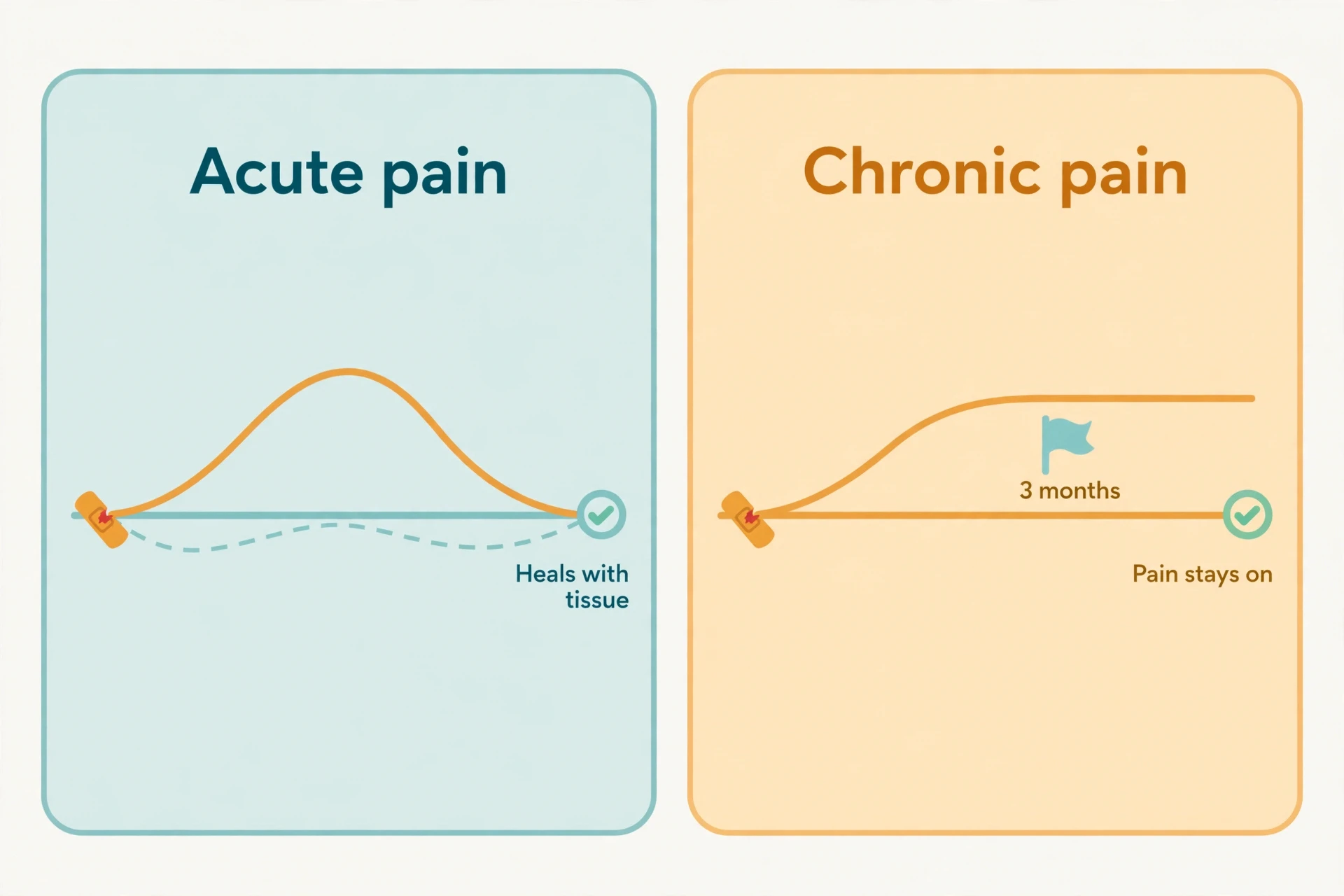
What is the difference between acute and chronic pain? Duration is the official answer, but it is not the real answer, and the science of pain is careful to say so. The IASP defines chronic pain as pain that persists or recurs for longer than three months. Acute pain is recent pain tied to injury or illness that resolves as tissues heal. Subacute pain is the intermediate stage, roughly one to three months. The three-month cut-off is a convention, not a biological switch: some sources use six months, and some researchers place the transition at twelve months. Nothing in the body changes on the ninety-first day, and duration alone does not describe the mechanisms behind a person's pain. The real distinction is functional. Acute pain protects: it warns of damage, triggers withdrawal, and enforces rest while tissues heal. Chronic pain has no such protective function. It persists past normal tissue healing time, and it is itself the disease, not a symptom. That is the difference that matters, and the chronic pain causes explored in section 14 are the mechanisms behind it.
The ICD-11, adopted by the World Health Organization in 2019 and in effect for mortality reporting from January 2022, contains a coding system for chronic pain for the first time: one code for chronic primary pain and six codes for chronic secondary pain syndromes. The seven categories are:
Chronic primary pain — pain in one or more anatomical regions that is not better explained by another condition, characterized by significant emotional distress or functional disability.
Chronic cancer-related pain
Chronic postsurgical or posttraumatic pain
Chronic neuropathic pain
Chronic secondary headache or orofacial pain
Chronic secondary visceral pain
Chronic secondary musculoskeletal pain
12.1 The ICD-11 map: one primary, six secondary
Chronic primary pain carries the biggest conceptual change in the classification. It is chronic pain in one or more anatomical regions, not better explained by another condition, characterized by significant emotional distress — anxiety, anger or frustration, or depressed mood — or functional disability, meaning interference in daily activities and reduced participation in social roles. It is multifactorial: biological, psychological, and social factors contribute. Fibromyalgia and chronic migraine are chronic primary pain conditions — health conditions in their own right, not symptoms of something else. Chronic secondary pain starts as the result of another condition, for example rheumatoid arthritis or inflammatory bowel disease, but reaches a stage where the pain itself requires treatment in its own right. The classification makes a point the science of pain has made since the 2020 definition: pain can be the disease, not merely the sign of one.
13. Three Types of Pain: Nociceptive, Neuropathic, Nociplastic
Types of pain — the taxonomy question — has a three-part answer, and this section gives each part its full treatment. The three mechanistic descriptors from section 2 return here as the article's classification, one H3 each, with mechanism, examples, and the feature that makes each one different.
13.1 Nociceptive pain
Nociceptive pain arises from actual or threatened damage to non-neural tissue and is due to the activation of nociceptors. A sprained ankle, a burn, an inflamed arthritic joint: in each case the tissue is damaged or threatened, the nociceptors fire, and the pain tracks the injury. This is the kind of pain the simple picture gets right. It is also the kind most responsive to the peripheral mechanisms of section 9: reduce the inflammatory soup, and the pain eases, because the pain was reporting the soup. Nociceptive pain is the alarm working as designed.
13.2 Neuropathic pain
Neuropathic pain is pain caused by a lesion or disease of the somatosensory nervous system itself. The damage is not in the tissue being reported on but in the reporting system. Diabetic peripheral neuropathy, postherpetic neuralgia after shingles, and the central pain of multiple sclerosis are the classic examples. The pain of multiple sclerosis comes from demyelinated pathways in the central nervous system, and it is covered in full in our article on Multiple Sclerosis Explained. Neuropathic pain often has a distinctive character — burning, shooting, electric-shock-like, or tingling — and it often comes with altered sensation in the affected area, because the same damaged nerves that produce pain also carry touch and temperature. Central sensitization can arise from nerve injury and from neuropathies, and it is a frequent companion of neuropathic pain, amplifying a signal that is already abnormal at its source.
13.3 Nociplastic pain
Nociplastic pain arises from altered nociception despite no clear evidence of tissue damage activating peripheral nociceptors, or of disease or lesion of the somatosensory system. The term was introduced by IASP in 2016 and formalised in 2017, for pain that cannot be adequately described as nociceptive or neuropathic. Fibromyalgia and chronic widespread pain are the leading examples. Central sensitization is probably the primary mechanism — but nociplastic pain should not be used interchangeably with central sensitization, because they describe different things: a clinical descriptor and a neurophysiological mechanism. Real patients often have more than one mechanism at once: a person with arthritis can have nociceptive pain from the joint and nociplastic pain from a sensitized system, and treating only one misses half the problem.
13.4 Why the labels matter — and where they fall short
The labels matter because they point to different mechanisms, and different mechanisms suggest different approaches. But the three-way split should not be presented as settled clinical fact, because researchers have raised a serious objection. In a recent editorial series, researchers Hoegh and Hodges argue that the three descriptors cannot yet be clearly discriminated in practice, and that their impact on treatment decisions is uncertain. The criticism is not that the mechanisms are imaginary. It is that, in a real person sitting in a clinic, the available tests cannot yet sort the mechanisms cleanly enough to make the label decisive. That is an honest limit, and it is worth stating plainly: the three kinds of pain are the best map the science of pain has, and the map is still being drawn. A reader who is told otherwise has been given a cleaner story than the science supports.
14. Why Chronic Pain Persists: Five Mechanisms, One Stuck System
Why does chronic pain persist? That is the question that brings most readers to the science of pain, and this section answers it as a synthesis. The chronic pain causes are not one cause but five mechanisms, converging and reinforcing each other until the system stays on after the reason for it has gone. Each was explained in its own section earlier. Here they come together.
The five chronic pain causes
Ongoing peripheral input and peripheral sensitization — the inflammatory soup keeps the detectors retuned, and the silent nociceptors stay awake (section 9).
Central sensitization — the amplifier stays on: NMDA-mediated strengthening of the dorsal-horn synapse means the same input produces a larger output (section 10).
Loss of descending inhibition — the brake fails: the PAG–RVM system's off-cells lose their grip on the spinal gate (sections 8 and 10).
Descending facilitation — the accelerator sticks: the RVM's on-cells keep amplifying the signal from above (section 8).
The biopsychosocial loop — fear, avoidance, poor sleep, and low mood feed back into the biology, and the biology feeds back into them (sections 2 and 12).
The transition from acute to chronic pain is not one event but a convergence of these five, and the science of pain studies that convergence rather than a single trigger. Chronic pain is multifactorial by definition in ICD-11: biological, psychological, and social factors contribute to the syndrome. Pain that lasts beyond three months is not automatically all in the head, and it is not automatically caused by ongoing tissue damage either. It can be driven by sensitized nerves, a sensitized spinal cord, or altered brain processing, all of which are physical mechanisms. That sentence is worth repeating to anyone who has been told their pain cannot be real because a scan came back normal. The scan shows tissue. The pain lives in the system that reports on tissue, and that system can be sensitized, amplified, and stuck, all while the tissue looks fine.
The biopsychosocial loop deserves one more paragraph, because it is the mechanism most often misunderstood as blame. Chronic pain and mental health travel together: the ICD-11 definition of chronic primary pain itself includes significant emotional distress as a defining feature, and the 2023 CDC data show high-impact chronic pain affecting 8.5% of US adults. The relationship runs both ways. Pain worsens mood and sleep, and low mood and poor sleep amplify pain — through the same descending pathways and the same sensitization mechanisms described earlier in this article. That is a general framing of a two-way relationship, not a causal claim about any individual, and not a suggestion that pain is caused by attitude or weakness. Fear and avoidance are biological inputs to a biological system: they change the gain of the spinal gate through the amygdala and the PAG. The science of pain treats them as mechanisms, and so should the reader.
"Injury increases the gain of the pain system, so the same input produces a larger output." — Woolf and Salter, Science, 2000, on neuronal plasticity and pain
15. Phantom Limb Pain: The Proof That the Brain Makes Pain
Phantom limb pain is the perception of pain in a limb that has been amputated. It is distinct from residual limb pain and from stump neuroma pain, which are pains in the remaining tissue. It is common: reported in roughly 50 to 80% of amputees, with a 2025 narrative review estimating global prevalence among amputees at approximately 64%, and a 2026 scoping review reporting about 82% of patients affected within the first year after amputation. Most patients who develop phantom limb pain report onset within the first month after amputation, with a secondary peak around one year, and phantom sensations — non-painful tingling, warmth, pressure — occur in as many as 80% of amputees. The science of pain treats phantom limb pain as its most powerful demonstration, because it breaks the simple picture beyond repair: there is no limb, no tissue, no nociceptor that could be reporting damage, and yet there is pain. If pain can exist without a limb, it can certainly exist without visible tissue damage.
The leading central explanation is maladaptive cortical reorganization: after amputation, the area of the primary somatosensory cortex that once represented the missing limb is invaded by representations of adjacent body parts — in upper-limb amputees, the neighboring face representation — and functional imaging shows the degree of reorganization correlates with phantom pain intensity. The face-to-hand remapping was first described by V.S. Ramachandran in the 1990s: touching an amputee's face could evoke sensations in the phantom hand, a finding later replicated in neuroimaging studies. It is one of the most cited findings in the science of pain, and it makes the cortical map feel less like a fixed chart and more like a living territory that redraws itself when part of it goes quiet.
But the mechanism is multifactorial, not cortical reorganization alone, and the exact cause is still unknown. That must be said plainly: the leading hypothesis is not a settled fact. Peripheral factors — ectopic discharges from the neuroma at the stump, nerve sprouting — spinal factors such as dorsal horn sensitization, thalamic reorganization, and psychosocial factors all contribute, and there is no consensus on a single mechanism. The four levels of phantom limb mechanisms are:
Peripheral — ectopic discharges from the neuroma at the stump, and nerve sprouting in the remaining tissue.
Spinal — dorsal horn sensitization, the same amplifier from section 10, now driven by a missing input.
Thalamic — thalamic neurons that did not normally respond begin responding, with enlarged maps.
Cortical — maladaptive reorganization of the somatosensory cortex, with neighboring representations invading the missing limb's territory.
Mirror therapy exploits the same plasticity in the other direction. The patient watches the reflection of their intact limb moving as if it were the missing one, and trials show consistent reductions in phantom limb pain accompanied by normalization of maladaptive sensorimotor activity. It is a first-line non-invasive approach, and its logic is the logic of the whole article: the brain builds pain from its maps and its expectations, so changing what the brain sees can change what it builds. Connect this back to section 2, and the circle closes. The 2020 definition says pain is recognized even when no tissue damage can be found. Phantom limb pain is the proof that the definition had to be written that way. The brain makes pain, and a brain that has lost a limb can keep making pain for a limb that is gone.
16. Placebo and Nocebo: Expectation Changes Pain — Really
The placebo effect is a genuine, measurable improvement in symptoms produced by a treatment with no specific therapeutic action — a sugar pill, a sham procedure — driven by expectation, belief, conditioning, and the context of care, not by any active ingredient. It is not imagination, and it is not merely reporting bias. The nocebo effect is the mirror image: the production or worsening of symptoms, including pain, by the expectation of harm. A warning, a frightening word, or a bad past experience can produce real pain, because the brain expects pain. The science of pain treats both as mechanisms, not mysteries.
Placebo analgesia recruits the brain's own opioid system: it can be partially blocked by naloxone, an opioid antagonist. It also engages dopamine and endocannabinoid systems, and prefrontal–cingulate circuits that drive descending inhibition through the PAG and RVM — the same descending pathway from section 8. Nocebo hyperalgesia runs on anticipatory anxiety and the signaling molecule cholecystokinin, known as CCK, which increases pain sensitivity and blocks mu-opioid receptor function — actively suppressing the body's own painkillers while amplifying transmission. CCK antagonists prevent nocebo hyperalgesia in experiments. Expectation changes activity in the brain regions that build pain: the anterior cingulate cortex, the insula, and the prefrontal cortex are the regions most consistently changed by expectation in human imaging studies. Direct evidence that pain's emotional and sensory parts can come apart comes from rodents, where lesions of the anterior cingulate cortex removed the learned aversion to a painful stimulus while leaving basic pain reflexes intact.
The modern framing is predictive processing: the brain treats pain as an inference, combining incoming sensory signals with top-down predictions. When a prior expectation of relief is strong, it dominates and drives descending inhibition. When a prior expectation of harm dominates, it biases perception toward threat and amplifies pain processing. The placebo effect was quantified in modern medicine by Henry K. Beecher's 1955 paper "The Powerful Placebo", and conditioning can drive placebo responses even in immune and endocrine function. But the limits matter as much as the power. Placebo effects are strongest for subjective, brain-mediated outcomes — pain, nausea, fatigue, mood, Parkinsonian symptoms, itch — and do not cure cancers, clear infections, or mend broken bones. They modulate experience and some physiology, not all disease. And a large share of reported drug side effects are nocebo responses, so the way risks are communicated can itself cause symptoms — a practical reason to discuss nocebo honestly, without blaming patients for their own expectations.
Placebo — trigger: expectation of relief; chemistry: endogenous opioids, dopamine, endocannabinoids; direction: pain decreases; it proves expectation can activate the brain's own analgesia.
Nocebo — trigger: expectation of harm; chemistry: anxiety and CCK blocking mu-opioid receptors; direction: pain increases; it proves expectation can suppress the body's own painkillers.
17. Myths and Mistakes About Pain
The science of pain has accumulated a set of stubborn myths, and most of them do their damage quietly — by telling people their pain cannot be real, or that it must mean something it does not. Each myth below gets a short correction, grounded in the mechanisms covered earlier in this article.
"Pain always means tissue damage." It does not. Nociplastic pain and phantom limb pain both occur without tissue damage, and the 2020 IASP definition recognizes pain even when no damage can be found.
"If the scan is normal, the pain is not real." A scan shows tissue. Pain lives in the system that reports on tissue, and a sensitized spinal cord or an altered brain network produces real pain with normal-looking tissue.
"Chronic pain is just acute pain that lasted too long." Different mechanisms, not just duration: central sensitization, loss of descending inhibition, and the biopsychosocial loop make chronic pain a different condition, not an extended one.
"Pain is all in your head." The brain makes pain — that is physiology, not imagination. Saying pain is constructed by the brain is a statement about mechanism, not a dismissal.
"Rubbing a bumped knee is just a distraction." Touch closes the spinal gate: A-beta fibers excite inhibitory interneurons in the substantia gelatinosa and reduce what reaches the brain. It is gate control, not distraction.
"Stronger pain means worse damage." Pain and tissue damage are not proportional. The same injury produces different pain in different people, and sensitized systems produce severe pain from minor input.
"Painkillers work by fixing the injury." They modulate the signaling system. NSAIDs, for example, reduce prostaglandin production by blocking COX enzymes; they do not repair the tissue.
"Complete rest is always the best treatment for a painful injury." Rest protects healing tissue, but prolonged avoidance feeds the fear-avoidance loop that reinforces chronic pain, and movement engages descending modulation. Recovery is a balance, decided with a clinician, not a rule.
"If you can still walk on it, it cannot be that bad." Function and pain intensity are different things. People can walk through severe pain, and people can be disabled by pain that looks minor from outside.
"Chronic pain means the body is still broken." The alarm system can stay on after healing. Central sensitization and a failed descending brake keep producing pain when the tissue has recovered.
18. Living With Chronic Pain: What the Science Means in Practice
Understanding the mechanisms changes what a person can do with their pain — not by curing it, but by changing the relationship with it. Movement engages descending modulation and challenges the fear-avoidance loop. Poor sleep amplifies sensitization, and sensitization worsens sleep. Stress reaches the spinal gate through the amygdala, the PAG, and the RVM. Expectations run on the same circuits that carry the pain signal. None of that is individual advice or a prescription. It is general information — the practical payoff of the science of pain — and personal decisions about treatment, activity, and care belong in a conversation with a clinician.
I think back to the stubbed toe that opened this article. The two pains — the sting and the ache — are still the same two pains. What changes, once you know the science of pain, is what they mean. The sting is the fast fiber doing its job. The ache is the slow fiber, and the inflammatory soup, and the spinal cord deciding how much of the signal to pass on. Knowing that does not make the toe hurt less. It makes the hurt understandable, and understanding is the first thing that turns pain from a mystery into a problem that can be worked on. That is what the science of pain offers the reader: not a cure, but a map.
19. Conclusion: Pain Is the Brain's Alarm — and Alarms Can Get Stuck
Pain begins at a nociceptor, crosses a threshold, and becomes an electrical signal. The signal travels up the spinal cord, through the gate, and into a distributed network of brain regions that build the experience. The gate can be closed by touch and opened by injury. The brain can turn the volume down with its own opioids, or turn it up through facilitation and expectation. The injured tissue can retune its own detectors, and the spinal cord can amplify the signal until the amplifier stays on. That is the whole journey, and the science of pain is the study of each step in it — and of what happens when any step gets stuck.
Pain is an alarm, not a damage meter. An alarm can be accurate, and it can be faulty. It can ring when there is a fire, and it can keep ringing after the fire is out, because the wiring has changed. Understanding the alarm — the detectors, the gate, the descending brake, the volume dial — is the first step to living with it. The science of pain does not promise that understanding will silence the alarm. It promises something more useful: that the person hearing it will know what it is, why it rings, and that the ringing is real, physical, and explicable, even when no scan can show the fire.
20. Frequently Asked Questions (FAQ)
How does pain work?
Pain works in four stages: transduction, transmission, modulation, and perception. Nociceptors convert a harmful stimulus into an electrical signal, the signal travels through the spinal cord to the brain, it is amplified or dampened along the way, and the brain constructs the conscious experience of pain. The science of pain studies each of these four stages and how they interact.
What are nociceptors?
Nociceptors are high-threshold sensory receptors — free nerve endings in the skin, muscles, joints, and organs — that detect stimuli with the potential to cause damage. They fire only when stimulus intensity reaches the noxious range, which is why everyday touch does not hurt. The science of pain begins with these detectors, because their high threshold is what makes pain selective rather than constant.
What is gate control theory?
Gate control theory, proposed by Melzack and Wall in 1965, says a gate in the dorsal horn of the spinal cord modulates pain signals before they reach the brain. Touch and pressure carried by large A-beta fibers tend to close the gate, pain input carried by small fibers tends to open it, and the brain can open or close the gate from above. The science of pain still uses this theory as its central explanation for why rubbing a bumped knee helps.
Why does chronic pain persist?
Chronic pain persists because five mechanisms converge: ongoing peripheral sensitization, central sensitization that keeps the spinal amplifier on, loss of descending inhibition, descending facilitation, and a biopsychosocial loop of fear, avoidance, poor sleep, and low mood feeding back into the biology. The science of pain treats chronic pain as a stuck system, not as a single cause, and not as a sign that the body is still damaged.
What is the difference between acute and chronic pain?
Acute pain is recent pain tied to injury or illness that resolves as tissues heal; chronic pain persists or recurs for longer than three months and becomes the disease itself rather than a symptom. The three-month line is a working convention, not a biological switch. The science of pain draws the real distinction functionally: acute pain protects, chronic pain does not.
What is central sensitization?
Central sensitization is an amplification of neural signaling within the central nervous system that produces pain hypersensitivity — the spinal cord and brain respond more strongly to normal or subthreshold input. It was first described by Clifford Woolf in 1983 and involves NMDA-mediated strengthening of the dorsal-horn synapse. The science of pain describes it as a volume dial turned up: the same input produces a larger output.
What causes allodynia?
Allodynia is caused by sensitization — peripheral, central, or both — lowering the threshold until a stimulus that should not hurt does hurt. A light touch or clothing contact becomes painful because sensitized nociceptors or a sensitized spinal cord treat ordinary input as dangerous. The science of pain counts allodynia among the clearest signs that the alarm system itself has changed.
Can you feel pain without an injury?
Yes. Pain can occur without nociceptor activation, because pain is built by the brain rather than read directly from tissue. Phantom limb pain, nociplastic pain, and migraine all involve pain without an injury to the painful area. The science of pain recognizes this in the 2020 IASP definition, which describes pain as an experience associated with, or resembling that associated with, actual or potential tissue damage.
Why does rubbing a bumped knee help?
Rubbing helps because touch and pressure input carried by large A-beta fibers excites inhibitory interneurons in the substantia gelatinosa and closes the spinal gate, reducing the pain signal that reaches the brain. It is gate control, not distraction. The science of pain has used this everyday observation as the theory's most familiar demonstration since 1965.
What is nociplastic pain?
Nociplastic pain arises from altered nociception despite no clear evidence of tissue damage or of a lesion or disease of the somatosensory system. Fibromyalgia and chronic widespread pain are the leading examples, and central sensitization is probably the primary mechanism. The science of pain keeps nociplastic pain distinct from central sensitization: one is a clinical descriptor, the other a neurophysiological mechanism.
21. Sources
International Association for the Study of Pain — Revised Definition of Pain (2020)
Raja SN et al. — The revised IASP definition of pain, Pain 2020;161(9):1976–1982
Melzack R, Wall PD — Pain mechanisms: a new theory, Science 1965;150(3699):971–979
Basbaum AI et al. — Cellular and molecular mechanisms of pain, Cell 2009;139(2):267–284
Basbaum AI et al. — Neural circuits for pain, Science 2016;354(6312):578–584
Braz J et al. — Transmitting pain and itch messages, Neuron 2014;82:522–536
Woolf CJ — Central sensitization, Pain 2011;152(3 Suppl):S2–S15
Woolf CJ, Salter MW — Neuronal plasticity: increasing the gain in pain, Science 2000;288:1765–1769
Treede RD et al. — IASP Classification of Chronic Pain for ICD-11, Pain 2019;160(1):19–27
University of Texas Neuroscience Online, Chapter 8 — Pain Modulation and Mechanisms
European Pain Federation — Types of pain: nociceptive, neuropathic, and nociplastic pain
Intracellular signaling in primary sensory neurons and persistent pain, Neurochem Res 2008
Advances in understanding nociception and neuropathic pain, PMC
Central Sensitization and CaV-alpha-2-delta Ligands in Chronic Pain Syndromes, J Pain
Pain without presence — phantom limb pain narrative review, PMC 2025
The Lancet Neurology — Public health approaches to chronic pain
The Lancet eClinicalMedicine — Paediatric chronic pain in low- and middle-income countries
Oxford Nuffield Department of Primary Care — IASP chronic pain definition summary