
1. The skeleton you thought you knew
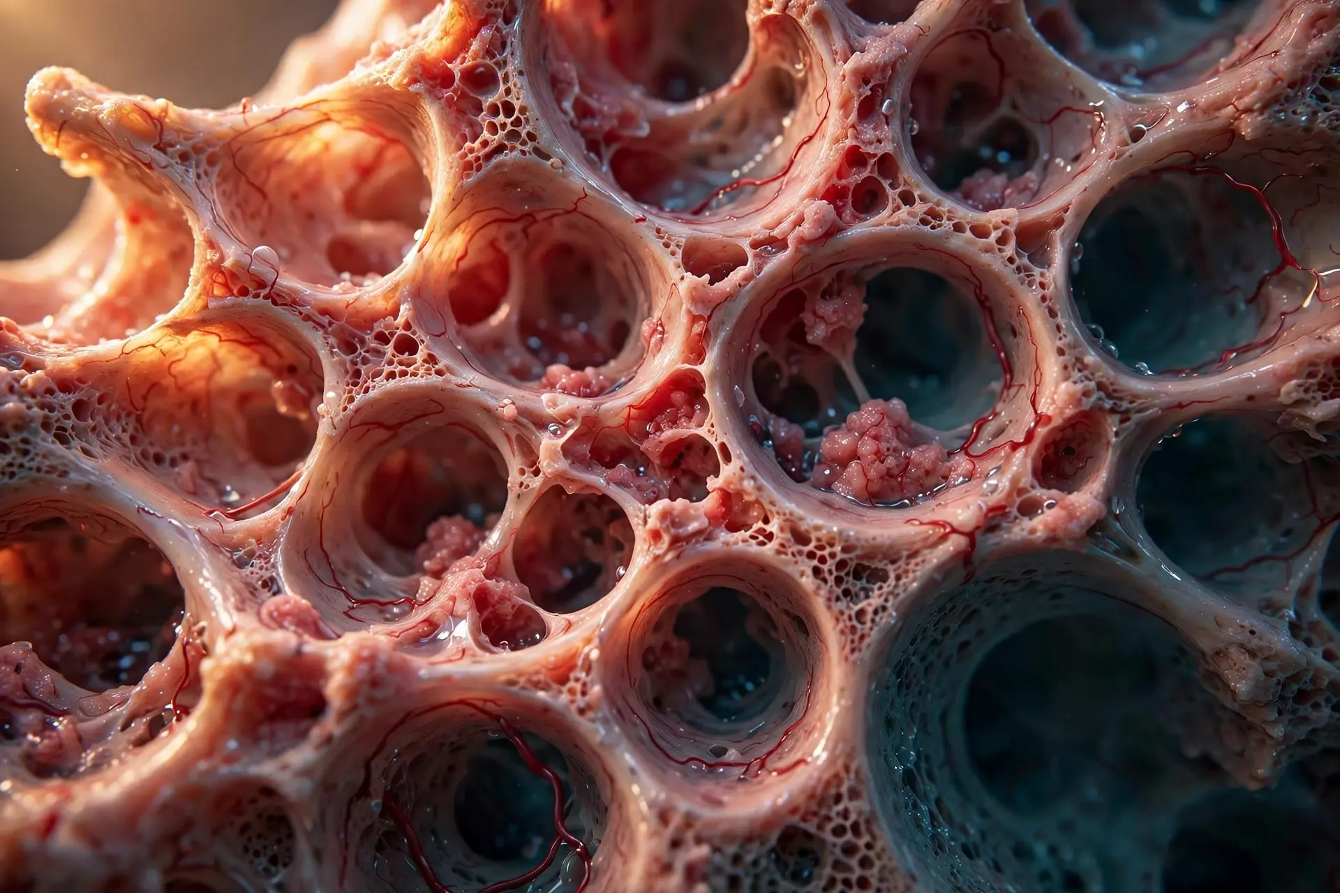
The first time I saw living bone, I did not believe what it told me about how bones work. It was not the white, dry thing from a museum display. It was pinkish and warm, wet with blood, and it bled when it was cut. I had spent years picturing the skeleton as a kind of scaffolding — inert, finished, silent. In front of me, the scaffolding was bleeding. That single image has stayed with me longer than any textbook diagram, because it overturned the quiet assumption most of us carry around: that our bones are dead objects we happen to live inside.
They are not. If you want to understand the living skeleton, the first thing to unlearn is the museum skeleton. A museum skeleton is bone with everything living removed — the cells scraped away, the blood vessels drained, the marrow gone. Living bone is a different object entirely. It is specialized connective tissue, and it is busy. Right now, as you read this, teams of cells are dissolving tiny patches of your skeleton and rebuilding them, a cycle that replaces roughly a tenth of your skeleton every year. Your skull, your ribs, your femur: none of them is the same bone you had a decade ago, at least not at the microscopic level. Some parts are older; some parts were made last month.
So how does something this hard rebuild itself? How does a broken arm knit back together without a scar on the bone? Why does the same skeleton that carries a teenager through a growth spurt turn fragile in old age? And what decides, cell by cell, whether bone is built or taken away?
This article answers those questions in order. You will meet the four cell types that run the skeleton, follow the five-step remodeling cycle they perform millions of times at once, learn the master switch that tells the demolition crew when to start, and see how load, hormones, calcium, and vitamin D all feed into one system. You will also see where the science is still unsettled — including a 2026 stem-cell trial that made headlines and deserves a careful look rather than a celebration. By the end, you will know why your skeleton is both a decade old and brand new, and what actually keeps it strong.
2. What bone actually is — and how bones work as a living composite
Bone is a composite material, and it is worth pausing on that word. A composite is two materials combined so that each covers the other's weakness, and bone is one of the best composites in nature. About 35 percent of its dry weight is organic material, mostly type I collagen — the same protein that gives tendons their pull strength — plus smaller amounts of proteoglycans and glycoproteins. The other roughly 65 percent is mineral: hydroxyapatite crystals, a form of calcium phosphate with the formula Ca10(PO4)6(OH)2. The collagen gives bone tensile strength and a measure of flexibility. The mineral gives it hardness and compressive strength. Take one away and the trick fails. Remove the mineral and bone turns flexible and rubbery, bending like a green twig. Remove the collagen and bone turns brittle like chalk, shattering on impact. Healthy bone needs both, in balance. Treat those percentages as approximate rather than exact — different textbooks put the mineral share at about 65 percent or about 70 percent — but the partnership itself is not in doubt.
That partnership serves six jobs. The skeleton supports the body against gravity, gives muscles leverage so we can move, and shields the brain, heart, and lungs behind hard walls. It makes blood cells in its marrow. It stores minerals — chiefly calcium and phosphate — and releases them when the blood needs them. And it acts as an endocrine organ, sending hormonal signals to the pancreas, fat tissue, and brain, a role science only recognized in the last two decades.
The headline number is the one that surprises people most. Under normal conditions, about 10 percent of the adult skeleton is replaced every year through remodeling, so most of the skeleton is renewed roughly every decade. That 10 percent is the standard textbook average; the honest range is 5 to 15 percent of total bone mass per year. In the first year of life, almost 100 percent of the skeleton is replaced — a baby is rebuilding at a pace an adult never matches again. The rates are not even across the skeleton. Dense outer bone turns over slowly, at about 2 to 3 percent a year. Spongy inner bone turns over at about 25 percent a year. Keep those two speeds in mind; they explain why some diseases show up in the spine years before they show up anywhere else.
Scale helps here too. An adult typically has 206 bones — 80 in the axial skeleton and 126 in the appendicular skeleton — though the count varies between people because of extra sesamoid bones and small accessory bones. Babies are born with far more, somewhere around 270 to about 300, and many fuse together as the child grows. The skull alone fuses from about 44 separate bones at birth into 22 in adulthood, and most fusion is complete by about age 25. Bone makes up about 14 percent of total body weight, roughly 10 to 11 kilograms for an average person, and reaches its maximum mass between the ages of 25 and 30.
None of this would be possible without blood. Bone is heavily vascularized: blood vessels run through every osteon, the cylindrical unit of compact bone, and the rich supply is exactly why fractures bleed and why bone can heal at all. A tissue with no blood supply cannot repair itself. Bone has plenty, and that is the first clue to everything that follows.
3. Anatomy: two tissues, one clever design
Cut a bone open and you find two tissues doing two different jobs. Compact bone, also called cortical bone, is dense and forms the hard outer shell of every bone and the shaft of long bones. Spongy bone, also called trabecular or cancellous bone, is a light honeycomb-like lattice of struts called trabeculae that fills the interior, especially at the ends of long bones. The lightness is the point. A solid bone would be enormously heavy; a hollow lattice is strong for its weight, which is why the skeleton can be both rigid and light enough to carry around all day.
3.1 Compact bone: the tree-trunk design
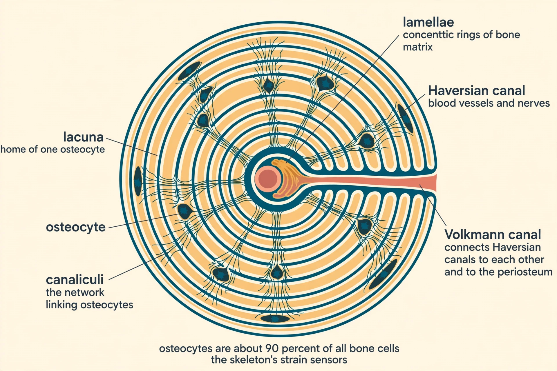
Compact bone is organized into cylindrical units called osteons, or Haversian systems. Each osteon is built like a tree trunk: concentric rings of bone matrix called lamellae surround a central Haversian canal carrying blood vessels and nerves. Volkmann canals run perpendicular to the Haversian canals and connect them with each other and with the periosteum, the membrane on the bone's surface, so blood reaches every osteon. It is a plumbing system as much as a structure, and it explains why even the densest part of the skeleton stays alive.
3.2 Spongy bone: the engineered lattice
Spongy bone has no osteons. Its trabeculae align along the lines of mechanical stress, which is why the internal pattern looks engineered: it puts material exactly where loads travel and leaves empty space everywhere else. That alignment is not an accident of growth. It is the skeleton responding to a lifetime of loading, laying struts along the paths that forces actually take. The lattice is mostly empty space, filled with marrow, and that emptiness is what makes the skeleton strong for its weight. Most people assume a bone is solid all the way through. What the data show is the opposite: only the outer shell is dense, and the interior is a carefully arranged void.
3.3 The map of a long bone
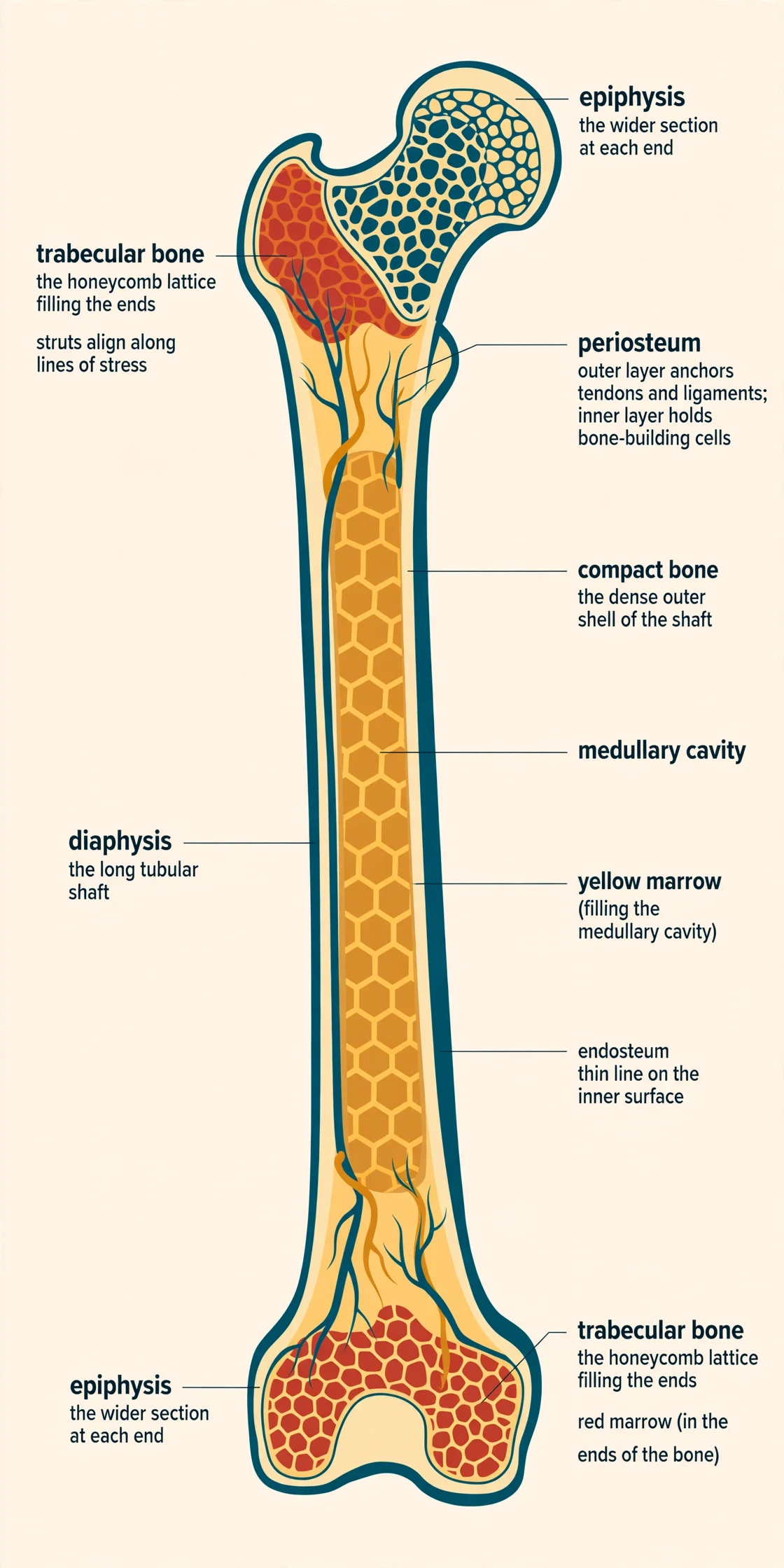
A long bone has a tubular shaft, the diaphysis, and a wider section at each end, the epiphysis. The hollow region inside the diaphysis is the medullary cavity, filled with yellow marrow in adults. The outer surface is covered by the periosteum, a fibrous membrane containing blood vessels, nerves, and lymphatic vessels. It covers the entire outer surface except where the epiphyses meet other bones to form joints. The periosteum has two layers: an outer fibrous layer that anchors tendons and ligaments, and an inner cellular layer rich in osteoprogenitor cells that can build new bone after injury. That inner layer is one reason a broken bone can rebuild itself — the repair crew is already stationed on the surface. The inner surfaces of bone, the medullary cavity and the trabeculae, are lined by the endosteum, a delicate membrane where bone growth, repair, and remodeling occur.
3.4 Red and yellow marrow: the blood factory and the fat store
Red bone marrow, found mainly in the pelvis, sternum, vertebrae, ribs, and the ends of long bones in adults, produces blood cells through hematopoiesis: red blood cells, white blood cells, and platelets, all from hematopoietic stem cells. Yellow bone marrow, found mainly in the medullary cavity of long bones in adults, is mostly fat cells and is inactive in blood cell production under normal conditions, though it can convert back to red marrow when the body needs more blood cells. The division is practical rather than fixed. In a crisis — severe blood loss, for example — the yellow marrow can be called back into service.
3.5 The five bone shapes
Bone is classified by shape into five types: long bones such as the femur and humerus; short bones such as those of the wrist and ankle; flat bones such as the skull and sternum; irregular bones such as the vertebrae and mandible; and sesamoid bones such as the kneecap, which forms inside a tendon. Flat bones such as the skull vault consist of two layers of compact bone with a layer of spongy bone sandwiched between them — a design that protects the brain with the least possible weight. The two tissues differ in ways that matter for disease, and the comparison is worth setting out plainly.
| Feature | Compact (cortical) bone | Spongy (trabecular) bone |
|---|---|---|
| Structure | Dense; organized into osteons with concentric lamellae around Haversian canals | Honeycomb lattice of trabecular struts; no osteons |
| Location | Outer shell of every bone; shaft of long bones | Interior, especially the ends of long bones and the inside of flat bones |
| Turnover rate | About 2 to 3 percent per year | About 25 percent per year |
| Surface-to-volume ratio | Roughly 2 to 5 square meters per liter | Roughly 20 to 30 square meters per liter |
| Main role | Strength, stiffness, protection | Lightweight support, shock absorption, mineral exchange |
| Disease pattern | Loses mass slowly; fractures at the hip and wrist in late disease | Shows metabolic bone disease first — for example, in the spine |
Table 1: Compact bone versus trabecular bone. The tenfold difference in surface area is why trabecular bone remodels so much faster — and why bone loss appears in the spine first.
That last row is the one to remember. Trabecular bone has a surface-to-volume ratio of roughly 20 to 30 square meters per liter, against 2 to 5 for cortical bone. Remodeling happens on surfaces, so the tissue with more surface remodels faster, loses mass faster when the balance tips, and shows disease first. When a doctor worries about a spine fracture in someone whose hip still looks reasonable, this table is the reason.
4. The cell cast: four cell types run the skeleton
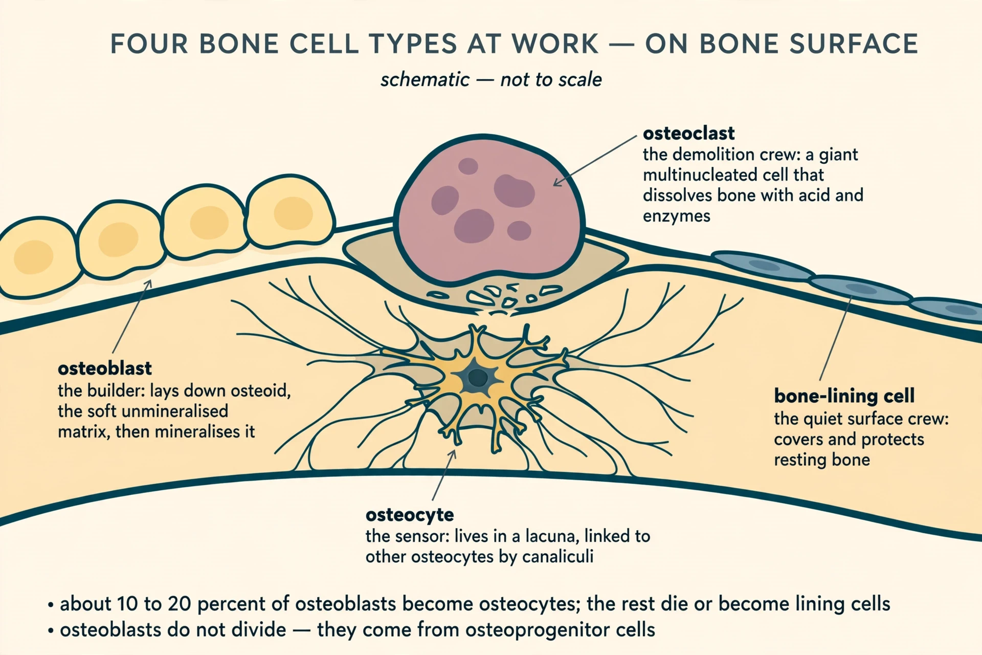
Bone looks simple from the outside. Inside, four cell types do all the work, and their division of labor is remarkably clean. Osteoprogenitor cells are the reserve workforce. Osteoblasts build. Osteocytes sense and supervise. Osteoclasts demolish. Everything else in bone biology — hormones, drugs, disease — works by changing what these four cells do.
4.1 Osteoblasts, the builders
Osteoblasts are the builders. They secrete type I collagen and other matrix proteins to form osteoid, the soft unmineralized organic matrix, and then drive its mineralization with calcium phosphate. They do not divide. They arise from osteoprogenitor cells — mesenchymal stem cells — under the control of the transcription factor Runx2, also called Cbfa1. Think of them as a construction crew that arrives, works for a fixed period, and then either settles into the building or leaves. When an osteoblast finishes building and becomes surrounded by the matrix it laid down, it becomes an osteocyte. Only about 10 to 20 percent of osteoblasts take this path; the rest die or become flat bone-lining cells on the surface.
4.2 Osteocytes, the sensors
Osteocytes are the most abundant bone cells by far, accounting for about 90 percent of all cells in mature bone, and they live embedded in tiny chambers called lacunae. They connect to each other through long, thin extensions running in tiny channels called canaliculi, forming the lacunocanalicular network, which occupies roughly 1.5 percent of total bone tissue volume. They are the skeleton's strain sensors. They sense mechanical loading and orchestrate remodeling, largely by adjusting how much sclerostin they release — a signal you will meet again in section 8, because it is the molecular link between exercise and bone strength. Osteocytes are roughly ten times as numerous as osteoblasts in bone tissue, which fits their role: a permanent resident network rather than a short-lived workforce.
4.3 Osteoclasts, the demolition crew
Osteoclasts are the demolition crew. They are very large, multinucleated cells formed by the fusion of monocyte and macrophage-lineage precursors, and they dissolve bone by secreting acid and enzymes into a sealed zone beneath the cell. A single osteoclast can have many nuclei, because it forms when several precursor cells fuse — which suits it for resorbing large areas of bone surface. The acid dissolves the mineral; the enzymes digest the collagen. Without osteoclasts, old bone would never be cleared away and new bone would have nowhere to go. They are not the enemy of the skeleton. They are half of it.
4.4 Bone-lining cells and osteoprogenitors
Bone-lining cells are flat, quiescent osteoblast descendants that cover bone surfaces, protect the matrix underneath, and help activate remodeling when needed. Osteoprogenitor cells are the reserve: stem cells waiting in the periosteum and marrow that can become osteoblasts on demand. Together they are the quiet infrastructure — the cells that do nothing dramatic until the moment something is needed, and then make everything else possible.
4.5 Coupling: removal matched by rebuilding
The balance between osteoblasts and osteoclasts is called coupling: in healthy adult bone, the amount removed at any one site is roughly matched by the amount rebuilt, so bone mass stays stable. Coupling is the whole story of bone health in one word. When coupling holds, the skeleton stays the same size for decades. When it breaks — when resorption outruns formation, or formation lags behind — bone thins, and that thinning is what the rest of this article is about.
| Cell type | Origin | Job | Fate | When it fails |
|---|---|---|---|---|
| Osteoblast | Osteoprogenitor (mesenchymal stem) cell, under Runx2 | Builds osteoid and mineralizes it | Becomes an osteocyte (10 to 20 percent), a lining cell, or dies | Too few or too weak: bone is not rebuilt after resorption |
| Osteocyte | An osteoblast trapped in its own matrix | Senses strain; controls remodeling through sclerostin | Lives for years embedded in lacunae; about 90 percent of bone cells | Signal lost: remodeling goes unregulated, microdamage accumulates |
| Osteoclast | Monocyte/macrophage-lineage precursors that fuse | Dissolves mineral with acid and digests collagen with enzymes | Large, multinucleated, short-lived; dies after resorbing | Overactive: bone is removed faster than it can be replaced |
| Bone-lining cell | Flat osteoblast descendant | Covers and protects resting surfaces; helps activate remodeling | Quiescent on the surface until called on | Surface left unguarded: remodeling starts at the wrong place or time |
Table 2: The four bone cell types — where each comes from, what it does, and what goes wrong when it fails.
5. How bones are built: ossification and growth
Bone formation, or ossification, begins in the embryo by the sixth or seventh week of embryonic life, and there are two pathways. Understanding both explains why a skull and a femur are built so differently, and why a child's bones can lengthen at all.
5.1 Two routes to bone
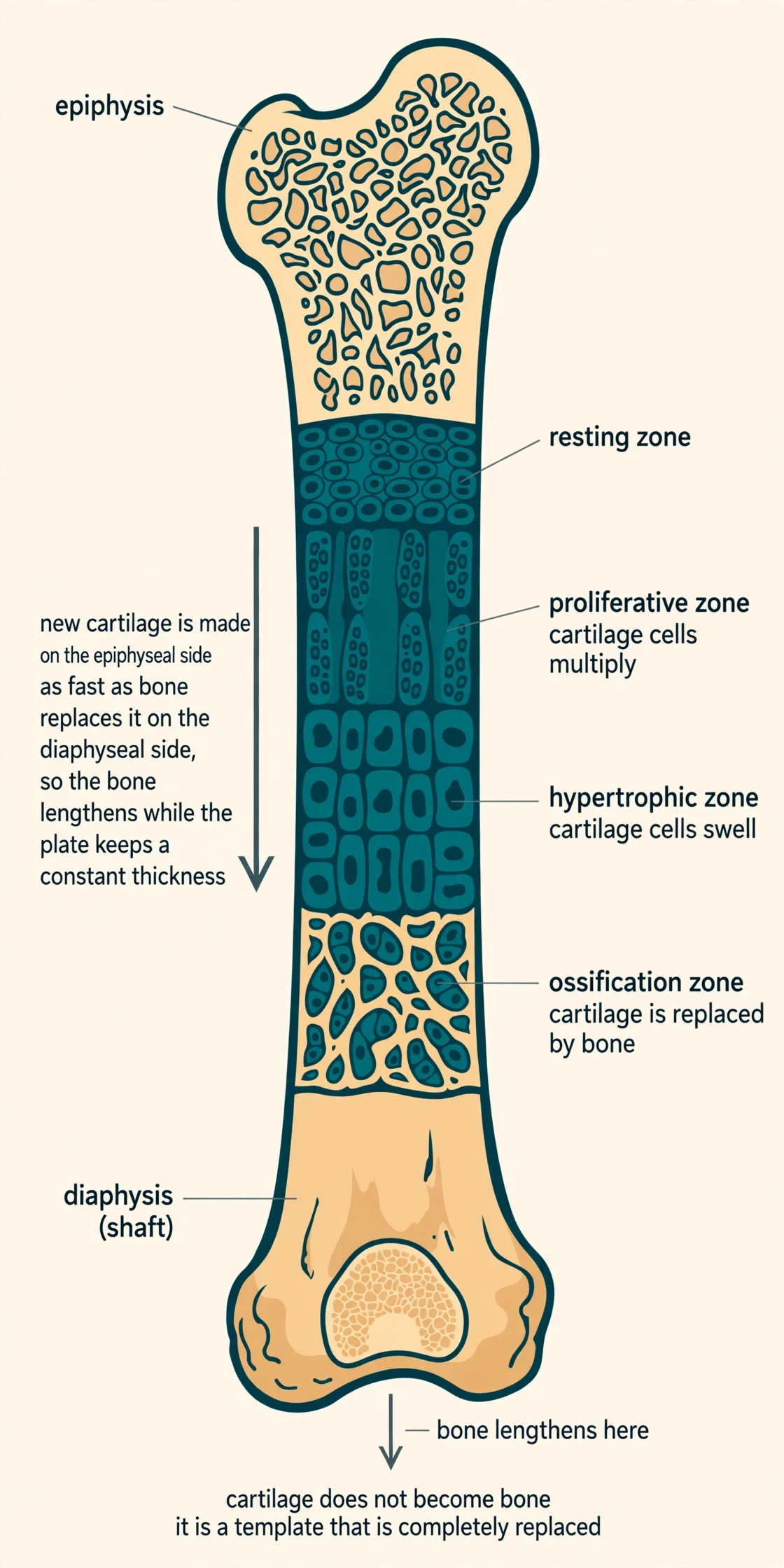
Intramembranous ossification forms bone directly from sheets of mesenchymal connective tissue, with no cartilage intermediate. It builds the flat bones of the face, most of the cranial bones, and the clavicles. Endochondral ossification builds most of the skeleton — long bones, vertebrae, ribs, and the base of the skull — by first sculpting a hyaline cartilage model and then replacing it with bone. Cartilage does not become bone; it serves as a template that is completely replaced. In endochondral ossification, a primary ossification center appears in the shaft, then secondary centers appear in the epiphyses after birth, leaving the growth plate between them.
5.2 The growth plate: a conveyor belt that lengthens bone
The epiphyseal growth plate is a layer of hyaline cartilage organized in zones: resting, proliferative, hypertrophic, and ossification zones. New cartilage is produced on the epiphyseal side as fast as bone replaces it on the diaphyseal side, so the bone lengthens while the plate keeps a constant thickness. It is a conveyor belt: cartilage cells multiply, swell, and are replaced by bone, and the plate itself never gets thicker. The lengthening happens at the plate and nowhere else, which is why a broken growth plate in a child is treated with such care — damage it and the limb may stop growing on that side.
5.3 The hormones of growth
Growth hormone from the pituitary drives overall growth, largely by stimulating the liver to release IGF-1, which promotes chondrocyte proliferation at the growth plates. At puberty, sex steroids — estrogen and testosterone — first accelerate growth, producing the growth spurt, then cause the epiphyseal plates to close: the plate ossifies completely and length growth ends, typically in the mid-to-late teens and earlier in females. The same hormones that make a teenager shoot up are the ones that eventually stop the growth. It is a tidy piece of design and a hard deadline: once the plates close, height is set.
5.4 After closure: wider, never longer
After the plates close, bones can still widen by appositional growth — new bone added at the periosteal surface — and can remodel, but they cannot lengthen. That is why an adult who breaks a leg heals at the same length, and why no exercise or supplement can add height after the plates have fused. Width, though, keeps changing for life, and that is the part loading can still influence.
5.5 Peak bone mass: the bone bank
About 90 percent of peak bone mass is acquired by age 18 in girls and by age 20 in boys, which makes childhood and adolescence the critical window for building the skeleton. Peak bone mass — the maximum amount of bone a person reaches — is typically achieved around age 30, then plateaus for about 10 years before a slow decline begins. Genetics plays a large role in determining peak bone mass, but nutrition, vitamin D, calcium intake, and weight-bearing exercise during youth all raise the peak a person reaches.
I think of it as a bank account. Childhood and adolescence are the years the deposits are made, and the peak around 30 is the balance. After that, everyone starts withdrawing, slowly at first. A person who built a large balance can afford decades of slow withdrawals. A person who built a small one runs short much sooner. I learned this the ordinary way: pencil marks on a doorframe, measuring a child who seemed to grow between one visit and the next. Each of those marks was the growth plate at work — cartilage made on one side, bone replacing it on the other, a millimeter at a time, too slow to see and too steady to stop. The marks look like a record of height. They are really a record of a bank account being filled, one deposit at a time.
6. The remodeling cycle, step by step
Remodeling is maintenance, not growth. Anyone asking how bones work eventually arrives here, at the cycle itself, because this is where the four cell types do their shared work. The same patch of bone is taken apart and rebuilt by a traveling team, and the patch ends up roughly the same size it started. The difference between modeling and remodeling matters here. Modeling is bone formation and resorption on separate surfaces, which changes the size and shape of a bone and produces net gain or loss. Remodeling is resorption followed by formation on the same surface, with no net change in mass when balanced. Growth is modeling. Adult maintenance is remodeling. This section is about the second.
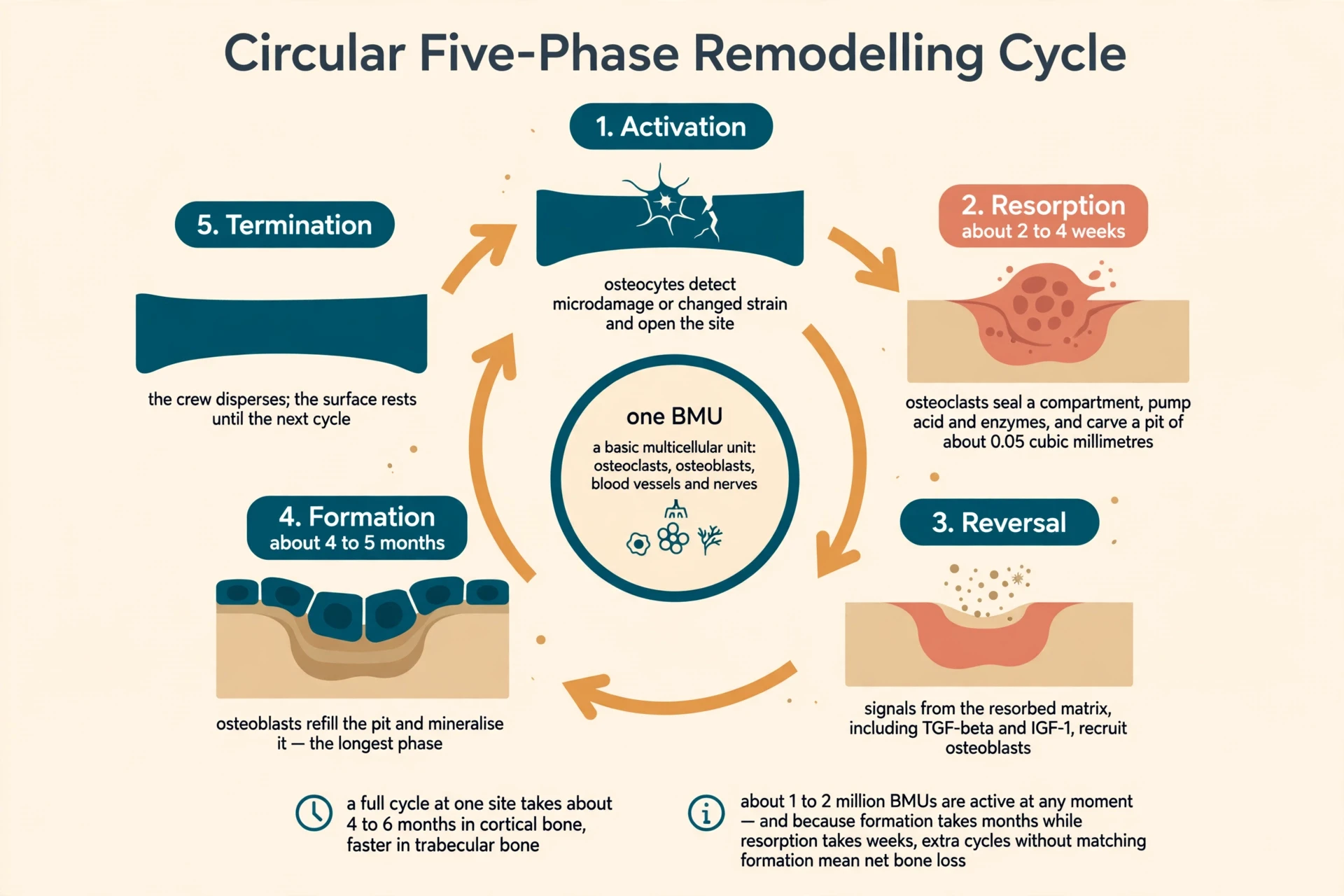
Remodeling happens in discrete patches of tissue called basic multicellular units, or BMUs — transient teams of osteoclasts, osteoblasts, blood vessels, and nerves organized in a specific spatial relationship. At any given time roughly 1 to 2 million BMUs are active throughout the adult skeleton. The full cycle at a single site takes approximately 4 to 6 months in cortical bone and is somewhat faster in trabecular bone. It follows five coordinated phases: activation, resorption, reversal, formation, and termination.
6.1 Activation: the call goes out
Osteocytes detect microdamage or a change in mechanical strain and signal bone-lining cells to withdraw, exposing the bone surface and recruiting osteoclast precursors. Microdamage is routine: everyday loading creates tiny cracks in bone, far too small to feel, and the osteocyte network is the system that notices them. Without this step, cracks would accumulate silently until a normal load broke the bone. Activation is the skeleton's inspection routine.
6.2 Resorption: the demolition crew moves in
Osteoclasts attach to the exposed surface, seal off a compartment beneath themselves, and pump acid and enzymes into it, dissolving mineral and digesting collagen. This phase lasts about 2 to 4 weeks and removes roughly 0.05 cubic millimeters of bone, leaving a pit called a Howship lacuna in trabecular bone or a cutting cone tunnel in cortical bone. The volume sounds tiny, and it is — but multiply it by a million active sites and the scale becomes clear. This is also the fast phase, and its speed is the source of most bone trouble, as you will see below.
6.3 Reversal: cleanup and recruitment
Osteoclasts leave or die, and the pit is cleaned and prepared by mononuclear cells. Signals released from the resorbed matrix, including TGF-beta and IGF-1, recruit osteoblast precursors to the site. The demolition crew leaves behind more than a hole. The matrix it dissolved was storing growth factors, and releasing them is what summons the builders. Resorption and formation are not two independent events. The first causes the second.
6.4 Formation: the long refill
Osteoblasts fill the pit with osteoid and mineralize it. This is the longest phase, taking about 4 to 5 months, which is why bone formation always lags behind resorption. That lag is the single most important timing fact in bone biology. A pit is emptied in weeks and refilled over months. Any process that starts extra cycles — menopause, for instance — creates a backlog of empty pits waiting for builders who work slowly. The skeleton can keep up with a normal pace of cycles. It cannot keep up when the pace doubles and the builders still take months per pit.
6.5 Termination: the crew disperses
Once the pit is refilled, osteoblasts either become osteocytes, become lining cells, or die, and the surface returns to a quiet, resting state until the next cycle. The patch looks the same as before, and at the microscopic level it is newer — fresh matrix, repaired cracks, a younger patch of tissue in an old bone.
6.6 Why it matters: three jobs in one cycle
Remodeling serves three purposes at once: it repairs microscopic cracks from daily wear before they can grow into fractures, it replaces old bone with fresh bone, and it releases calcium into the blood when the body needs it. And the trap is built into the timing. Because resorption takes weeks and formation takes months, anything that speeds up the start of new cycles without matching formation — such as the loss of estrogen at menopause — produces a net loss of bone with every cycle. Each individual cycle is balanced. A thousand cycles, each briefly in deficit while formation catches up, add up to a skeleton that is measurably thinner than it was a year ago. That is how a process designed to maintain bone becomes the mechanism of bone loss.
7. The master switch: RANK, RANKL, and OPG
The question this section answers is simple: who tells the demolition crew to start? Osteoclasts do not decide for themselves. They are summoned, and the summons runs through one molecular system. It is called the RANK/RANKL/OPG axis, and it is the master regulator of osteoclast formation and bone resorption. Its discovery in the mid-to-late 1990s transformed bone biology and led directly to new osteoporosis drugs — one of the rare cases where a laboratory discovery reached the clinic within a decade.
7.1 RANKL: the summons
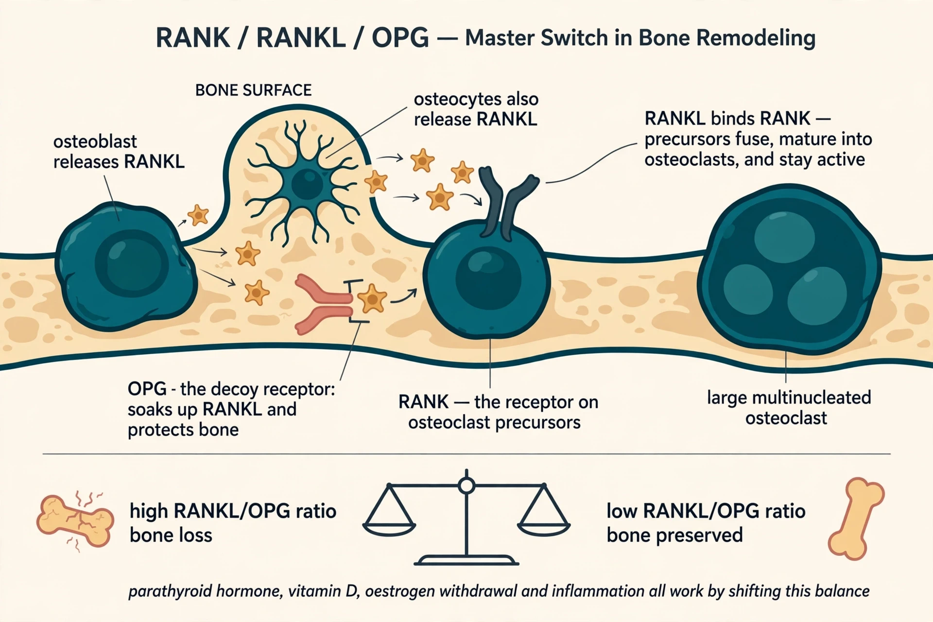
RANKL — receptor activator of nuclear factor kappa-B ligand — is a protein made mainly by osteoblasts and osteocytes. It binds to its receptor RANK on the surface of osteoclast precursors and drives them to fuse, mature into osteoclasts, and stay active. Notice who sends the signal: the builders summon the demolition crew. Osteoblasts, the cells that make bone, also control the cells that remove it. RANKL is a member of the tumor necrosis factor (TNF) superfamily. Before its discovery, researchers knew osteoblast-lineage cells controlled osteoclast formation but had not anticipated that TNF-family molecules would be the messengers — a surprise that reshaped the field's picture of how the two cell types talk.
7.2 OPG: the decoy receptor
OPG, osteoprotegerin, is a soluble decoy receptor secreted by osteoblasts. It binds RANKL and prevents it from reaching RANK, so it protects the skeleton from excessive resorption. Think of it as a sponge: RANKL molecules that OPG soaks up never reach an osteoclast precursor. The ratio of RANKL to OPG is therefore a major determinant of bone mass: a high RANKL/OPG ratio favors bone loss, while a low ratio favors bone preservation. Bone health, at the molecular level, is a ratio between a signal and its decoy.
7.3 The discovery story
OPG itself was discovered in 1997 by Simonet and colleagues, who found that mice engineered to overproduce OPG developed extremely dense bones (osteopetrosis), while mice lacking OPG developed severe early-onset osteoporosis (DOI: 10.1016/s0092-8674(00)80209-3). One gene, two opposite extremes: too much decoy and bone piled up; no decoy and bone vanished. It was the cleanest possible proof that the ratio, not either molecule alone, sets bone mass. The experiment is still cited because it settled an argument in a single stroke.
7.4 Why hormones work through this switch
The hormone story converges on RANKL. Parathyroid hormone, active vitamin D, estrogen withdrawal, and inflammatory signals all raise bone resorption largely by changing the RANKL/OPG balance, not by acting on osteoclasts directly. That is why such different conditions — menopause, overactive parathyroid glands, chronic inflammation — all end in the same place: more RANKL relative to OPG, more osteoclasts, more resorption. Different diseases, one switch.
7.5 Beyond bone
The discovery also explained a medical puzzle: the same RANKL/RANK signaling is required for lymph node formation and for mammary gland development during lactation, and OPG protects arteries from calcification. The system reaches far beyond bone. A molecule first found in the skeleton turned out to be part of the immune system and the reproductive system too, which is a reminder that the body rarely invents a new signal when an old one can be reused.
8. Wolff's law and the mechanostat: bone listens to load
If the RANKL/OPG axis is the switch, load is the hand on it. Bone adapts to the forces placed on it, and the history of that idea is longer and more contested than most textbooks admit.
8.1 Julius Wolff, 1892
Wolff's law, described by the German anatomist and surgeon Julius Wolff (1836–1902) in his 1892 book Das Gesetz der Transformation der Knochen (The Law of Bone Transformation), states that bone in a healthy animal adapts to the loads placed upon it: more loading makes bone stronger, less loading makes it weaker. Wolff observed that trabecular bone aligns along the lines of mechanical stress, and he argued — with a mathematical confidence later work softened — that trabecular architecture follows the principal stress trajectories in the bone.
"Every particle of mature bone is very active. Such activity must appear in the external shape of the bones." — Julius Wolff, 1892, Das Gesetz der Transformation der Knochen
Read that line again. In 1892, decades before Frost gave the process its modern name, remodeling, Wolff was saying that bone is active to its smallest particle and that its shape records its history of use. He was right about the activity and about the shape. He was too confident about the mathematics, and that overconfidence is part of the story.
8.2 The honest correction
The idea that bone senses and adapts to load predates Wolff: Galileo noted the mechanical implications of bone shapes in 1638, and the surgeon Charles Bell observed in 1827 that cancellous bone structure had reference to the forces acting on the bone. Modern research supports the core observation while qualifying the strict mathematics: bone tissue is anisotropic, with different mechanical properties in different directions, so Wolff's law is best understood today as the general principle of bone functional adaptation. The law survived as a principle, not as an equation. That is a common fate for a good nineteenth-century idea, and it is not a failure — the observation was sound even when the maths was not.
8.3 Harold Frost and the mechanostat
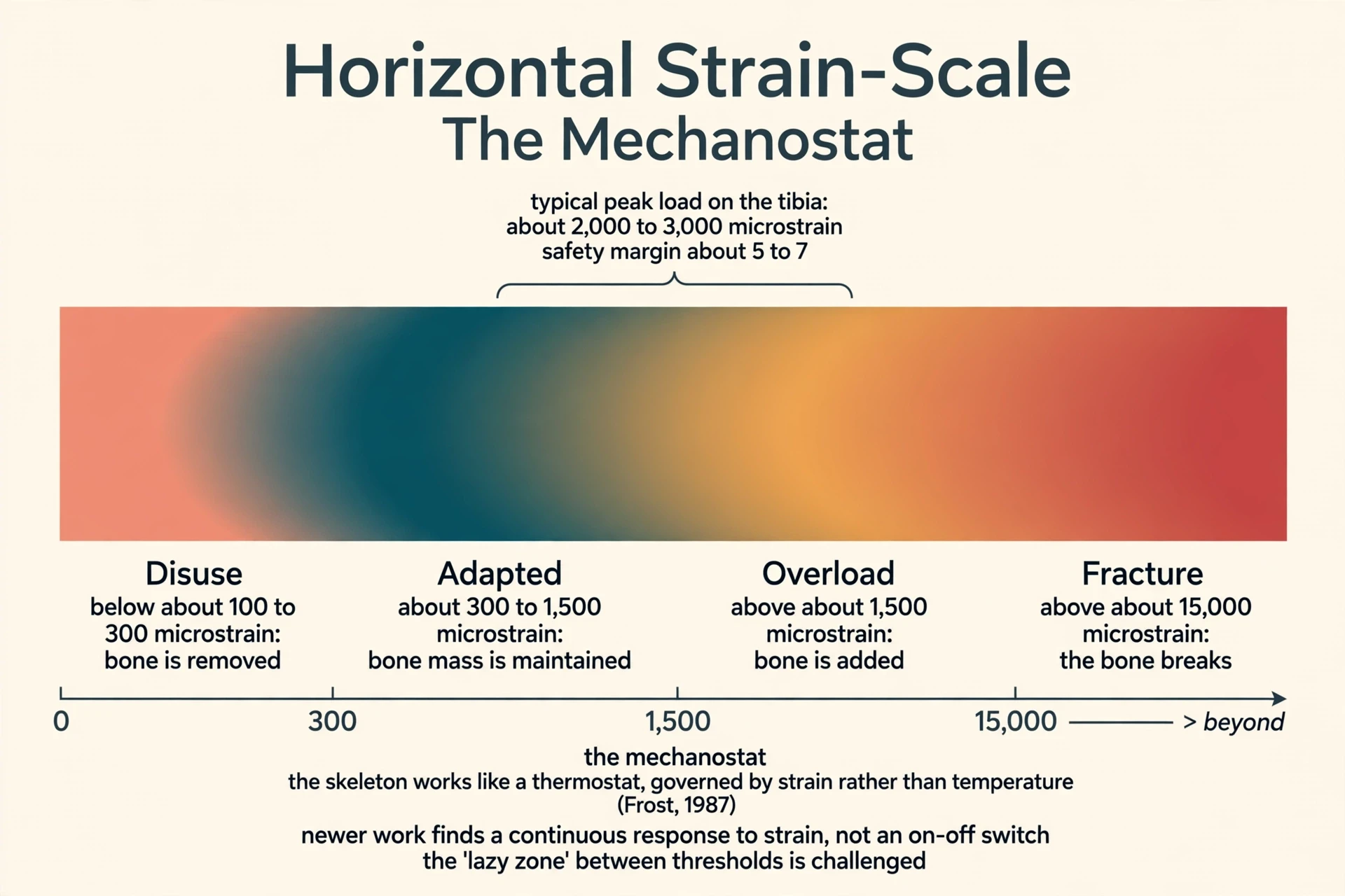
Harold Frost, an orthopedic surgeon and researcher, refined Wolff's law into the mechanostat theory, first proposed in 1987 and updated in 2003, likening the skeleton to a thermostat that is governed by mechanical strain rather than temperature. Frost's mechanostat defines strain thresholds: below roughly 100 to 300 microstrain, disuse-mode remodeling removes bone; between roughly 300 and 1,500 microstrain bone mass is maintained; above roughly 1,500 microstrain, modeling adds bone; and above roughly 15,000 microstrain the bone fractures. A typical load-bearing bone such as the tibia has a safety margin of about 5 to 7 between its typical peak load (2,000 to 3,000 microstrain) and its fracture load (about 15,000 microstrain).
But the tidy thresholds come with a challenge that the writer of any honest account must state plainly. The "lazy zone" between the thresholds — a range where nothing was thought to happen — has been challenged by later experiments showing the skeleton responds to applied strain in an essentially continuous, linear way rather than switching on and off. The mechanostat is best read as a gradient, not a set of hard on-off switches. Bone does not sit idle between thresholds and then suddenly start building. It responds a little to a little load and more to more load, along a slope.
8.4 The molecular basis: sclerostin and the Wnt pathway
The molecular basis of Wolff's law runs through osteocytes and sclerostin. Mechanical loading lowers sclerostin release from osteocytes, which releases the brake on the Wnt/beta-catenin signaling pathway and allows osteoblasts to build bone. Sclerostin is a glycoprotein secreted mainly by mature osteocytes, encoded by the SOST gene. It blocks Wnt signaling by binding to the LRP5/6 co-receptors on osteoblasts, suppressing bone formation, and it also indirectly promotes resorption by raising RANKL and lowering OPG. Wnt/beta-catenin signaling is the master formation pathway: when a Wnt ligand binds its Frizzled receptor plus the LRP5/6 co-receptor, beta-catenin is stabilized, enters the nucleus, and switches on genes that make more osteoblasts. The genetic proof is human: mutations that over-activate LRP5 cause very dense bones, and mutations that disable the SOST gene cause skeletal overgrowth. This is the part of how bones work that exercise acts on directly: load the bone, sclerostin falls, the Wnt brake releases, and osteoblasts build.
8.5 Mechanotransduction in four steps
Mechanotransduction — the conversion of mechanical force into biochemical signals — involves four steps: mechanocoupling, biochemical coupling, signal transmission, and cell response, with fluid flow through the lacunocanalicular network thought to be the signal osteocytes actually detect. When you walk, each step squeezes fluid through the tiny channels connecting osteocytes, and the cells read that flow as a message: this bone is being used, keep it strong. Stop walking for weeks, and the message stops. The cells do not need to know why you stopped. They only know the flow ceased, and they act accordingly.
I saw the loading rule written on a human body once, without any instrument. I was watching a tennis player at a local court, and one forearm was visibly thicker than the other — not a little, but obviously, the racquet arm carrying more muscle and, as I later learned, more bone. It looked like an asymmetry, almost a defect, until I understood it was the opposite: the skeleton had done exactly what Wolff described, adding material where the load fell and nowhere else. The two arms share the same genes, the same diet, the same hormones. Only the loading differs. Section 16 gives the numbers behind that observation, and they are striking.
9. Calcium, phosphate, and the hormone committee
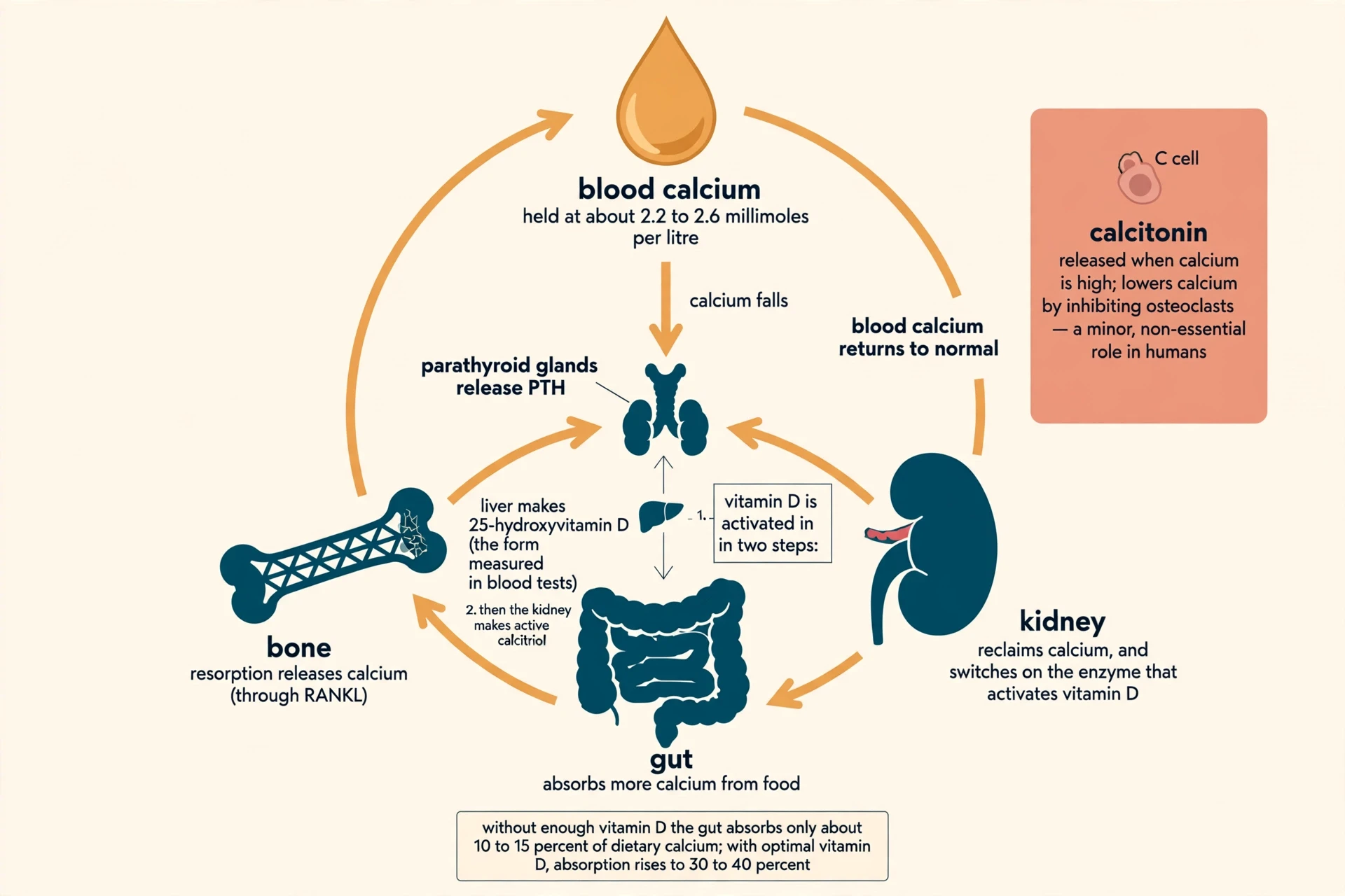
Bone is not only a structure. It is the body's calcium vault, and the vault has a strict manager. About 99 percent of the body's calcium is stored in bone as hydroxyapatite. The small fraction in blood is tightly regulated because calcium is essential for nerve signaling, muscle contraction, and blood clotting. If blood calcium falls too far, nerves misfire and muscles fail. If it rises too far, the heart and kidneys suffer. The skeleton absorbs that risk by giving up calcium when blood levels fall and taking it back when they rise.
9.1 The problem: a narrow range
Blood calcium is held in a narrow range of about 2.2 to 2.6 millimoles per liter (about 10 milligrams per deciliter). Only the ionized fraction, roughly half of total blood calcium, is biologically active and regulated. Three hormones acting on three organs control calcium: parathyroid hormone (PTH), active vitamin D (calcitriol), and calcitonin, acting on bone, gut, and kidney. Three hormones, three organs, one narrow range — and the system rarely misses.
9.2 PTH: the master hormone
When ionized calcium falls, calcium-sensing receptors on the parathyroid glands trigger PTH release. PTH raises blood calcium by resorbing bone (via RANKL), increasing calcium reabsorption in the kidney, and switching on the kidney enzyme that activates vitamin D. It is the fastest responder in the system and the one that calls directly on the skeleton: when blood calcium is low, PTH tells bone to give some up. PTH is phosphaturic: while it raises calcium, it increases phosphate excretion in the urine, so it lowers blood phosphate. The two minerals are managed together because they travel together in hydroxyapatite.
9.3 Vitamin D: the two-step activation
Vitamin D is activated in two steps: it is hydroxylated to 25-hydroxyvitamin D in the liver (the storage form measured in blood tests) and then to active calcitriol, 1,25-dihydroxyvitamin D, in the kidney. Calcitriol's main job is to increase absorption of calcium and phosphate from the gut. Without adequate vitamin D the body absorbs only about 10 to 15 percent of dietary calcium; with optimal vitamin D, absorption rises to 30 to 40 percent. That gap is the reason vitamin D matters so much to bone: calcium in food is useless if the gut cannot absorb it, and vitamin D is what opens the door. For the symptoms, testing, and treatment of deficiency, see our article on Vitamin D Deficiency: Symptoms and Treatment.
9.4 Calcitonin: the minor player
Calcitonin, secreted by C cells of the thyroid gland when calcium is high, inhibits osteoclasts and lowers blood calcium — but it plays only a minor, non-essential role in humans; people without a thyroid maintain calcium balance through PTH and vitamin D alone. It is a backup that the body barely needs, which is worth knowing because calcitonin once had a larger reputation in textbooks than its real role supports.
9.5 The daily numbers
The body cannot manufacture calcium: the only way it enters the body is through the diet, which is why dietary intake matters for the skeleton throughout life. Recommended dietary allowances from the US National Institutes of Health are 1,000 milligrams of calcium per day for adults aged 19 to 50, rising to 1,200 milligrams for women over 50 and men over 70; and 600 IU (15 micrograms) of vitamin D per day up to age 70, rising to 800 IU (20 micrograms) after 70. Those are allowances, not prescriptions — but they are the numbers clinicians use, and they are the floor the diet should meet.
9.6 The wider committee
Estrogen protects bone by restraining osteoclasts, largely by suppressing RANKL and raising OPG. When estrogen falls at menopause, that brake loosens and resorption outpaces formation. Testosterone also supports bone, partly by being converted to estrogen in bone tissue, and partly through its effect on muscle mass, which increases the mechanical load on bone. Other hormones affecting bone include growth hormone and IGF-1 (bone growth), thyroid hormone (too much accelerates turnover and bone loss), cortisol (chronic excess, including from steroid medicines, suppresses bone formation), and leptin from fat tissue, which regulates bone mass through a hypothalamic relay. Bone is the body's phosphate store as well as its calcium store, and phosphate balance is managed alongside calcium by PTH, vitamin D, and the kidney. No single hormone runs the skeleton. A committee does, and the committee argues — which is why bone loss has so many different causes and one common pathway.
10. Bone as an endocrine organ: the osteocalcin surprise
For most of the twentieth century, bone was treated as a structural tissue with a storage sideline. Then a mouse experiment changed the field's mind. Bone is an endocrine organ: osteoblasts secrete osteocalcin, a hormone that acts far beyond the skeleton, influencing insulin secretion, energy metabolism, male fertility, and brain function. This was established by Gerard Karsenty's group at Columbia University, published in Cell in 2007.
The key experiment was disorienting. Mice lacking osteocalcin showed only moderately increased bone mass but had significantly elevated blood glucose, more fat cells, and higher fat mass — the opposite of what a purely structural bone protein would produce. Remove a bone protein and the animal's blood sugar rises: that is not a structural result, it is a metabolic one. Uncarboxylated osteocalcin, the active hormonal form, stimulates insulin production in pancreatic beta cells and adiponectin expression in fat cells, forming a bone-pancreas feedback loop: insulin signaling in osteoblasts raises osteocalcin, which raises insulin secretion (DOI: 10.1016/j.cell.2007.05.047).
Osteocalcin is a 49-amino-acid protein, one of the few proteins made only by osteoblasts. Its carboxylated form binds tightly to hydroxyapatite in bone; the undercarboxylated form circulates in blood as the hormone. The same molecule is a structural glue in one form and a circulating signal in another — a neat trick, and one that makes bone part of the body's energy regulation rather than a bystander to it.
Here is the honest limit, and it matters. Whether the osteocalcin-insulin feedback loop works the same way in humans is still unresolved; most of the evidence comes from mice and laboratory cell studies, and human data are more mixed. The loop is proven in mice. In people, the picture is suggestive but not settled, and any article that presents it as established human physiology is overstating the case. Leptin, a hormone from fat tissue, also regulates bone mass through a relay in the hypothalamus, showing that bone, fat, and the brain form a single regulatory circuit rather than separate systems. The skeleton talks to the rest of the body. How loudly it talks in humans is still being measured.
"Remodelling is the process; almost everything else in metabolic bone disease is either a component of it or a failure of it." — OrthoVellum, Bone Remodeling
That sentence is the pivot of this article. Everything before it was mechanism. Everything after it is what happens when the mechanism drifts, breaks, or is pushed by age, hormones, and disease.
11. Across a lifetime: growth, peak, and slow decline
A skeleton has a biography, and it runs in four acts. Childhood and teens: formation far outpaces resorption, and 90 percent of peak mass is acquired by 18 in girls and 20 in boys. The peak around 30, then a plateau of about 10 years. Then a slow decline from around 40 in both sexes, at first very gradually. Then, for women, a sharp acceleration at menopause.
The menopause numbers deserve to be stated plainly. In women, bone loss accelerates sharply around menopause, when estrogen falls. The Merck Manual puts the post-menopause rate at about 2 percent a year for roughly 10 years before it slows to a steadier age-related pace. Women can lose up to 20 percent of their bone density in the five to seven years following menopause, according to the Bone Health and Osteoporosis Foundation. A fifth of the skeleton's density, gone in less than a decade, driven by the loosening of one hormonal brake. The mechanism is the one from section 6: each remodeling cycle still refills its pit, but more cycles are started, and formation — which takes months per pit — cannot keep up with resorption, which takes weeks. Multiply a small deficit per cycle by thousands of cycles and the result is a skeleton a fifth lighter.
Men lose bone too, later and more slowly. Their decline is steadier, without the sharp menopausal step, which is one reason the disease is under-recognized in men — a point section 12 returns to. The pattern across a lifetime is not a cliff for everyone. It is a long slow slope with one steep stretch, and where you start on the slope — your peak bone mass — decides how far down you can go before trouble begins.
I have watched that slope from the waiting room side. An older relative went for a bone-density scan, and I sat with the result: a T-score, a number I had only ever read about, now attached to a person I knew. The scan itself was uneventful — a quiet machine, a few minutes lying still — but the number it produced changed how the family talked about falls, about stairs, about a rug that had been in the hallway for thirty years. A T-score is an abstraction until it belongs to someone. Then it becomes a reason to move the rug.
12. Osteoporosis: the silent thinning
Osteoporosis is defined by the World Health Organization using bone density measured by DXA (dual-energy X-ray absorptiometry): a T-score of minus 1.0 or above is normal; between minus 1.0 and minus 2.5 is osteopenia (low bone mass); minus 2.5 or below is osteoporosis; and minus 2.5 or below with a fragility fracture is severe osteoporosis. A T-score compares a person's bone mineral density with the average peak bone density of a healthy young adult of the same sex. Each one-standard-deviation fall in bone density roughly doubles fracture risk, and each 1 SD fall in hip density raises hip fracture risk about 2.6-fold. A single standard deviation is the difference between a skeleton that absorbs a fall and one that breaks under it.
The scale is global and large. Worldwide, one in three women and one in five men over the age of 50 will experience an osteoporotic fracture, according to the International Osteoporosis Foundation. The IOF estimates that about 500 million people worldwide are affected by osteoporosis, and up to 37 million fragility fractures occur annually in people aged over 55 — the equivalent of about 70 fractures every minute. Hip fracture numbers are projected to nearly double between 2018 and 2050 as populations age. Those figures are from the IOF's latest releases, including its 2025 World Osteoporosis Day statement, and they are the most current available.
The cascade after a first fracture is the part that rarely reaches the public. After a hip fracture, about one in three patients has a second hip fracture, on average within about 1.5 years, and a prior fracture raises the risk of another fracture by about 86 percent. A fracture is not only an injury. It is a warning that the next one is already more likely, and the window is short. Yet up to 80 percent of patients who suffer a fragility fracture remain undiagnosed and untreated for the underlying osteoporosis, according to the IOF's 2025 World Osteoporosis Day statement. Four out of five people break a bone from fragile bone and never get the disease behind it investigated. That gap — between a treatable cause and an untreated patient — is the largest missed opportunity in bone medicine. It is also what happens when people do not know how bones work: a disease with no symptoms is easy to ignore until the first fracture makes it impossible to ignore.
Osteoporosis is often called a silent disease because bone loss has no symptoms until a bone breaks, often from a minor fall from standing height, or even from everyday stress such as bending or coughing. The most common osteoporotic fracture sites are the hip, spine, and wrist. Spinal (vertebral) fractures can occur without a fall and can cause height loss and a stooped posture — the shrinking height that families notice before any scan is ordered. Risk factors beyond age and sex include a parent who had a hip fracture, low body weight (BMI below 18.5), smoking, heavy alcohol use, an overactive thyroid, coeliac disease, long-term steroid medicine use, and prolonged immobility. Several of those are modifiable. None of them announces itself as a bone problem until the bone breaks.
13. When bones break: fracture healing
A fracture is the remodeling cycle turned up to full volume. Fracture healing is secondary bone repair in action, and it follows four overlapping stages: hematoma formation, soft callus, hard callus, and remodeling. Some clinical sources split it into five by separating inflammation and granulation tissue formation. The four-stage version is used here, with the five-stage split noted where it matters.
13.1 Hematoma and inflammation, days 1 to 5
Blood vessels torn by the fracture form a clot around the broken ends. The clot is not just a plug — it is the biological scaffold of the repair, releasing signaling molecules that summon stem cells and growth factors. Inflammation here is not a side effect to be suppressed. It is the first step of the repair signal, which is why heavy use of anti-inflammatory medicines in the first week can slow healing: the drugs quiet the very signal the bone needs to start rebuilding.
13.2 Soft callus, weeks 1 to 3
Fibrous tissue and cartilage bridge the fracture gap, forming a rubbery bridge that stabilizes the break but is still fragile and invisible on plain X-rays. The bone is held together by a material that does not yet show up on an X-ray, which is why an early X-ray can look unchanged while healing is already under way. The bridge is soft by design: cartilage first, bone later, the same order as endochondral ossification in the embryo.
13.3 Hard callus, weeks 3 to 12
The cartilage scaffold is progressively mineralized into woven bone through endochondral ossification, producing the bulge visible on X-rays around a healing fracture. That bulge is the callus, and it is the reason a healing fracture looks thicker than the bone around it. The body overbuilds first and trims later — a sensible strategy when the priority is stability.
13.4 Remodeling, months to years
Coupled osteoclast resorption and osteoblast formation slowly replace woven bone with strong, organized lamellar bone along the lines of mechanical stress; the callus bump shrinks and the medullary canal reopens. Remodeling after a fracture begins far earlier than most people expect — from roughly day 18 — and continues for months to years, which is why a healed fracture keeps changing on X-rays long after the cast comes off. In children, remodeling can be so complete that even a healed angulation straightens out over a year or two; in adults the correction is more limited.
| Stage | Timing | What happens | What shows on X-ray |
|---|---|---|---|
| Hematoma and inflammation | Days 1 to 5 | Blood clot forms the repair scaffold; signaling molecules summon stem cells and growth factors | The fracture line; no healing yet visible |
| Soft callus | Weeks 1 to 3 (to week 6 in the five-stage model) | Fibrous tissue and cartilage bridge the gap; a rubbery stabilizing bridge | Usually invisible — cartilage does not show on plain X-ray |
| Hard callus | Weeks 3 to 12 | Cartilage mineralizes into woven bone through endochondral ossification | A bulge of new bone around the fracture |
| Remodeling | Months to years (begins around day 18) | Woven bone replaced by organized lamellar bone along stress lines; callus shrinks; canal reopens | Callus gradually shrinking; bone returning to normal shape |
Table 3: The fracture healing timeline. Typical healing times for uncomplicated adult fractures: fingers 3 to 4 weeks, upper limb fractures generally 6 to 8 weeks, and large load-bearing bones such as the femur or tibia 12 to 24 weeks or longer. Children heal roughly twice as fast as adults.
Things that slow healing include smoking, diabetes, poor blood supply, infection, and excessive use of anti-inflammatory medicines in the first week, when inflammation is part of the repair signal. Primary fracture healing — direct bone-to-bone healing without a callus — happens only when the broken ends are rigidly fixed together, for example with surgical plates; most fractures heal by the secondary, callus route. And one common belief deserves a plain correction: a broken bone does not heal back stronger than before. Healing restores the bone to roughly its original strength and shape through remodeling. The callus is temporarily bulkier, which can look stronger on an X-ray, but the finished repair is not stronger than uninjured bone.
I know the cast routine from the family side of it. A relative broke an arm — a clean break, a cast, and then weeks of the small miseries nobody writes about: the itch under the plaster, the arm held at an awkward angle, the careful showering. What I remember most is the series of X-rays: first the sharp dark line of the break, then the cloudy bulge of the callus appearing, then, months later, the bulge slowly shrinking as the bone reshaped itself. The cast came off in week six. The bone kept remodeling for a year. The cast is the visible part of healing. The remodeling is the real part, and it goes on long after anyone is watching.
14. Bones in space: microgravity proves the loading rule
If you want the cleanest experiment on Wolff's law ever run, with humans as the subjects, look at astronauts. In microgravity, weight-bearing bones such as the spine and hips lose between 1 and 1.5 percent of their density each month during four-to-six-month missions, according to NASA, making astronauts susceptible to fractures and premature osteoporosis. A single six-month stay on the International Space Station can therefore strip away as much bone density as roughly two decades of normal aging on Earth. Six months in orbit, twenty years of aging — that is the loading rule running in reverse, at full speed.
The recovery problem is worse than the loss. In a 2022 study of 17 astronauts, nine had still not fully recovered their shin bone density a full year after returning to Earth — a deficit equivalent to a decade or more of age-related bone loss. More than half the crew came home with a skeleton that had not caught up, a year later. Astronauts counter bone loss with about two hours of exercise a day on specialized equipment, including the Advanced Resistive Exercise Device (ARED), installed on the ISS in 2008, which can simulate up to 600 pounds of resistance, plus adequate energy intake, calcium, and vitamin D. Bisphosphonates, the same class of medicines used to treat osteoporosis on Earth, have been tested on ISS crew members and helped mitigate spaceflight-induced bone loss. In space the spine also decompresses without gravity, so astronauts can gain up to about 3 percent in height during a mission — and the calcium freed from thinning bone raises the risk of kidney stones.
Space earns its place in this article because it removes every confounding variable. Same person, same genes, same diet, only the loading changed — and the skeleton responded within weeks. Wolff's law, tested 400 kilometers above the Earth, held.
15. The newest science: 2025 and 2026
Bone medicine is moving from slowing bone loss to rebuilding bone, and the newest work points in that direction — with caveats that deserve the same prominence as the headlines.
In September 2026, a first-in-human trial published in Cell reported a new stem-cell therapy for advanced osteoporosis: a patient's own bone-marrow mesenchymal stem cells were given a temporary sugar "address label" (glycocalyx editing) so they would home to bone marrow blood vessels after a single intravenous infusion. In that trial, 10 women aged 51 to 72 with advanced osteoporosis received one infusion. In the two years before treatment the group had 8 fractures per year; in the two years after, the rate fell to 0.5 fractures per year — a 94 percent reduction — and bone biopsies at about four months showed mean bone tissue area had nearly doubled (DOI: 10.1016/j.cell.2026.08.017).
Those numbers are striking, and they should be read with the trial's limits attached. The same trial reported no serious adverse events related to treatment, but it had only 10 participants, all women, at a single center, with no control group and no blinding — so the fracture reduction cannot yet be attributed to the treatment alone, and larger randomized trials are needed. Ten people, no control group, no blinding: that is a signal, not proof. A signal this strong deserves a large trial. It does not yet deserve to be called a treatment.
A 2026 study in Frontiers in Bioengineering and Biotechnology found that depleting the nuclear protein Lamin B1 in bone-marrow stem cells enhanced their bone-forming ability and improved bone regeneration in osteoporotic mice, a possible route to gene-enhanced cell therapy (DOI: 10.3389/fbioe.2026.1803514). A 2025 review on sclerostin examined its emerging role in bone-muscle crosstalk, suggesting the bone-muscle connection runs in both directions and that sclerostin may coordinate the two tissues. The through-line is consistent: medicine is moving from slowing bone loss to rebuilding bone — the same direction the RANKL and sclerostin drugs already pointed.
16. Keeping your bones strong: what actually works
If you want to know how bones work in practice — what to actually do with this knowledge — the answer starts with load. Bone responds best to physical activity that is high-load and unusual in pattern — the opposite of the cardiovascular response, which improves with long-duration, low-intensity activity. Short bursts of high strain matter more for bone than long gentle sessions. That contrast surprises people who assume all exercise works the same way on every tissue. For the heart, duration matters. For bone, the peak load and its novelty matter.
Tennis players provide the cleanest natural experiment. The bones of a player's racquet arm are far stronger than those of the other arm, even though genetics, nutrition, and hormones are identical for both arms. In a study of 12 former Finnish national-level male tennis players, the playing arm had 14 to 27 percent more bone mineral content than the other arm, with bone strength index 23 to 37 percent higher — and the gain came mostly from the bone growing larger, not denser. In prepubertal tennis players under 12, the playing arm already had 16.3 percent more bone mineral content than the other arm, showing that the bone-building effect of loading starts in childhood. Weight-bearing activities that involve high impacts, such as volleyball and hurdling, are associated with higher bone density than low-impact activities such as orienteering and cross-country skiing. Simple aerobic activities such as walking, jogging, and running also help maintain or increase bone density in women, and resistance training increases bone density in both young and older adults. For a fuller picture of what exercise does across the whole body, see our article on How Exercise Changes the Body: Muscles, Heart, Brain, and Mood.
The supporting cast matters too: calcium and vitamin D at the recommended intakes, enough protein, not smoking, limiting alcohol, and fall prevention at home — good lighting, handrails, no loose rugs. None of these is dramatic. All of them compound over decades, which is the timescale bone works on. For the wider picture of aging well beyond bone, see our science-based guide to Longevity and Healthy Aging.
How bone health is checked: DXA (dual-energy X-ray absorptiometry) is the World Health Organization's gold-standard test for measuring bone mineral density. It uses two low-dose X-ray beams, usually at the hip and lower spine, and reports areal density in grams per square centimeter. DXA measures areal density, not true volumetric density, so it overestimates density in large bones and underestimates it in small ones. Z-scores, which compare a person with age-matched controls, are used instead of T-scores for premenopausal women, men under 50, and children. Bone turnover markers in blood add information DXA cannot give: CTX reflects bone breakdown and P1NP reflects bone formation, so they show whether bone is currently being lost or built.
What the drugs do, briefly, and as information rather than personal advice — any treatment decision belongs with your clinician. Bisphosphonates are the first-line osteoporosis treatment in the American College of Physicians 2023 living guideline; they slow osteoclasts by blocking an enzyme in the cholesterol pathway. Denosumab is second-line for people who cannot take bisphosphonates. Denosumab is a human monoclonal antibody against RANKL — pharmacologically an extra supply of OPG — given as an injection under the skin every six months. It must never be stopped or delayed without a follow-up plan, because bone loss rebounds rapidly and can cause multiple spine fractures. In the FREEDOM trial (Cummings and colleagues, New England Journal of Medicine, 2009), denosumab reduced new vertebral fractures by 68 percent, hip fractures by 40 percent, and non-vertebral fractures by 20 percent over three years in postmenopausal women (DOI: 10.1056/NEJMoa0809493). Romosozumab is a monoclonal antibody against sclerostin, given monthly for 12 months. It is the first drug with a dual action: it increases bone formation and decreases resorption at the same time. In the ARCH trial (Saag and colleagues, New England Journal of Medicine, 2017), romosozumab led to 48 percent fewer new spine fractures and 38 percent fewer hip fractures than alendronate over 24 months, but it carries a boxed warning for heart attack, stroke, and cardiovascular death and is not used after a heart attack or stroke in the past year (DOI: 10.1056/nejmoa1708322). Teriparatide and abaloparatide are bone-building (anabolic) drugs based on parathyroid hormone; they are used for a limited period in people at very high fracture risk, followed by an antiresorptive drug to keep the gains.
17. Myths and mistakes
Myth: Bones are dead, dry structures like a museum skeleton. Living bone is pinkish, warm, full of blood vessels and cells, and replaces about a tenth of itself every year. A museum skeleton is bone with everything living removed. The dry white object in a display case is the leftover architecture, not the tissue.
Myth: Bones stop changing once you finish growing. Remodeling runs for life; the same machinery that builds bone in youth repairs microdamage and rebuilds fractures in old age. Growth ends. Change does not.
Myth: Drinking milk or taking calcium alone will prevent osteoporosis. Calcium is only one input. Without vitamin D the gut absorbs only 10 to 15 percent of dietary calcium, and without mechanical loading and hormones the calcium has nowhere useful to go. A supplement cannot do the job of a skeleton that is never loaded.
Myth: Osteoporosis is only a women's disease. One in five men over 50 will experience an osteoporotic fracture, and men account for a substantial share of hip fractures. Men lose bone later and more slowly, which is why their disease is found later — not why it does not exist.
Myth: A broken bone heals back stronger than before. A healed bone ends up about as strong as it was before the break — no stronger. The callus looks bulky on an X-ray while it is still there, which is where the story comes from, but once remodeling has trimmed it away the repair matches the original bone; it does not beat it. Stronger is a story people tell about the X-ray, not what the bone becomes.
Myth: You cannot build bone after age 30, so exercise is pointless for older bones. Peak mass is set around 30, but loading still stimulates formation at any age — resistance training raises bone density in older adults, and astronauts show the same mechanism in reverse. The bank account stops growing at 30. The withdrawals can still be slowed.
Myth: Bones are solid all the way through. Only the outer shell is dense compact bone. The interior is spongy trabecular bone, a lattice that is mostly empty space filled with marrow — and that lightness is what makes the skeleton strong for its weight. Solid would be heavier, not stronger.
Myth: If your bones feel fine, they must be fine. Osteoporosis has no symptoms until a fracture happens, which is why it is called a silent disease; a DXA scan is the only way to know bone density before a break. Feeling fine is the normal state of a skeleton that is quietly thinning. That is the danger, and the reason screening exists.
18. Frequently asked questions
How do bones work?
Bone is living tissue rebuilt by a five-phase cycle run by osteoblasts, osteoclasts, and osteocytes: activation, resorption, reversal, formation, and termination. About 10 percent of the skeleton is replaced each year — the standard average within a range of 5 to 15 percent — so most of your skeleton is renewed roughly every decade. Osteoblasts build new bone, osteoclasts dissolve old bone, and osteocytes sense mechanical strain and direct the work. The balance between building and removal, controlled by the RANKL/OPG system and by hormones and loading, decides whether bone mass stays stable, grows, or thins.
What are osteoblasts and osteoclasts?
Osteoblasts are the builders: they lay down osteoid, a soft matrix made mostly of type I collagen, and then harden it by adding calcium phosphate mineral. They cannot divide; each one develops from an osteoprogenitor stem cell switched on by the transcription factor Runx2. Osteoclasts are the demolition crew: giant cells with many nuclei, built when several monocyte/macrophage-lineage precursors fuse into one cell, which then dissolves bone with acid and enzymes in a sealed pocket underneath itself. Osteocytes, the most abundant bone cells, supervise both: they sense strain and call the shots through signals such as sclerostin. In healthy bone, the two crews are coupled — what one removes, the other rebuilds.
How does bone remodeling work?
Remodeling runs in small patches, each one handled by a basic multicellular unit — a BMU, a short-lived team of osteoclasts, osteoblasts, blood vessels, and nerves working together in one spot. At any moment, roughly 1 to 2 million BMUs are active in the adult skeleton. Each site follows five phases: activation, when osteocytes detect microdamage and open the site; resorption, when osteoclasts carve a pit of about 0.05 cubic millimeters over 2 to 4 weeks; reversal, when the pit is cleaned and osteoblasts are recruited; formation, when osteoblasts refill and harden the pit over 4 to 5 months; and termination, when the crew disperses and the surface rests. A single site needs 4 to 6 months to complete the cycle in cortical bone; trabecular sites finish sooner. Because formation takes months and resorption takes weeks, any process that starts extra cycles without matching formation causes net bone loss.
Why do bones get weaker with age?
After peak bone mass around age 30, formation slowly falls behind resorption, and bone density begins a slow decline from around age 40 in both sexes. Menopause accelerates the loss sharply as estrogen falls: women can lose up to 20 percent of bone density in the five to seven years after menopause, at about 2 percent a year for roughly a decade. Trabecular bone goes first, because it remodels fastest — about 25 percent a year against 2 to 3 percent for cortical bone. The result is thinner struts, wider gaps in the lattice, and a skeleton that breaks under loads it once absorbed easily.
How long does a broken bone take to heal?
It depends on the bone. As a rough guide for uncomplicated adult fractures, fingers take 3 to 4 weeks, most upper-limb fractures take 6 to 8 weeks, and a femur or tibia needs 12 to 24 weeks or longer. Children heal roughly twice as fast as adults. Healing follows four stages — hematoma, soft callus, hard callus, and remodeling — and the remodeling phase goes on for months to years after the cast comes off, so X-rays of a healed fracture keep changing long after it feels fine. Smoking, diabetes, poor blood supply, infection, and heavy anti-inflammatory use in the first week can all slow healing.
Can you rebuild bone density after 50?
Yes, partially. Resistance and impact exercise stimulate formation at any age, adequate calcium and vitamin D keep the raw materials available, and, where a clinician advises it, bone-building medicines such as teriparatide or romosozumab can add density, while antiresorptive drugs such as bisphosphonates or denosumab slow further loss. Losses can be slowed and some density regained, but the earlier the peak bone mass the better the reserve — which is why youth is the critical window and why treatment after 50 is about defending what remains as much as rebuilding what was lost. Talk to your clinician before starting any treatment or supplement plan.
How much calcium and vitamin D do adults need each day?
Adults aged 19 to 50 should aim for 1,000 milligrams of calcium a day; the target is 1,200 milligrams a day for women past 50 and men past 70. For vitamin D, the target is 600 IU (15 micrograms) a day until age 70, then 800 IU (20 micrograms) a day. Those are the US National Institutes of Health allowances. The body cannot make calcium, so food is the only way in — and vitamin D decides how much of it gets through: with too little vitamin D, the gut takes up only 10 to 15 percent of the calcium eaten, but with enough, uptake rises to 30 to 40 percent.
How is bone density measured?
The standard test is a DXA scan — dual-energy X-ray absorptiometry — which the World Health Organization rates as the gold standard for measuring bone mineral density. It scans the hip and lower spine with two low-dose X-ray beams and gives a T-score: a result of minus 1.0 or better counts as normal, from minus 1.0 down to minus 2.5 counts as low bone mass (osteopenia), and minus 2.5 or lower counts as osteoporosis. For premenopausal women, men under 50, and children, doctors use Z-scores instead — those compare a person with others of the same age rather than with young adults. Blood markers add a further layer: CTX shows breakdown and P1NP shows formation, so together they reveal whether the skeleton is losing bone or building it right now.
19. Sources
- OrthoVellum — Bone Remodeling. https://www.orthovellum.com/topics/bone-remodeling
- Varsity Tutors — Bone Tissue Structure and Remodeling. https://www.varsitytutors.com/learner/subjects/anatomy-and-physiology/lessons/bone-tissue-structure-and-remodeling
- Wikipedia — Bone remodeling (bone metabolism). https://en.wikipedia.org/wiki/Bone_metabolism
- OrthoPaedia — Describe Bone Remodeling. https://www.orthopaedia.com/Describe-bone-remodeling/
- Wikipedia — Wolff's law. https://en.wikipedia.org/wiki/Wolff%27s_law
- Cambridge University Press — Activity patterns: 2. Structural adaptation (Bioarchaeology). https://www.cambridge.org/core/books/abs/bioarchaeology/activity-patterns-2-structural-adaptation/0A91853D7AE44C4A4F3865893B7B225F
- Boyce BF, Xing L. Biology of RANK, RANKL, and osteoprotegerin. Arthritis Research and Therapy. 2007;9 Suppl 1:S1. DOI: 10.1186/ar2165
- Boyce BF, Xing L. Functions of RANKL/RANK/OPG in bone modeling and remodeling. Archives of Biochemistry and Biophysics. 2008;473(2):139-146. DOI: 10.1016/j.abb.2008.03.018
- Boyce BF, Xing L. The RANKL/RANK/OPG pathway. Current Osteoporosis Reports. 2007;5(3):98-104. DOI: 10.1007/s11914-007-0024-y
- International Osteoporosis Foundation — Epidemiology of osteoporosis and fragility fractures. https://www.osteoporosis.foundation/facts-statistics/epidemiology-of-osteoporosis-and-fragility-fractures
- International Osteoporosis Foundation — Osteoporosis: a global health crisis we can no longer ignore (World Osteoporosis Day statement, 17 October 2025). https://www.osteoporosis.foundation/news/osteoporosis-global-health-crisis-we-can-no-longer-ignore-20251020-1320
- University of Baltimore / Pressbooks — Introduction to Human Biology, 6.3 Bone Structure. https://ubalt.pressbooks.pub/introductiontohumanbiology/?p=595
- Osmosis — Skeletal system anatomy and physiology. https://try.osmosis.org/learn/Skeletal_system_anatomy_and_physiology
- OrthoChronicles — How Broken Bones Heal: Stages, Timelines and What Slows Them Down (updated October 2026). https://www.orthochronicles.info/fracture-healing.html
- BoneFractures.org — Fracture Healing Stages: 5 Stages of Bone Healing with Real X-Rays (reviewed August 2026). https://www.bonefractures.org/2021/02/fracture-healing-stages.html
- OrthoVellum — Calcium Homeostasis and Metabolic Bone Disease. https://www.orthovellum.com/topics/calcium-homeostasis-metabolic-bone
- LibreTexts Medicine — 6.7 Calcium Homeostasis. https://med.libretexts.org/Courses/Roosevelt_University/Advanced_Anatomy_and_Physiology/02%3A_Support_and_Movement/2.02%3A_Bone_Tissue_and_the_Skeletal_System/2.2.08%3A_Calcium_Homeostasis_-_Interactions_of_the_Skeletal_System_and_Other_Organ_Systems
- Colorado State University — Endocrine Control of Calcium and Phosphate Homeostasis. https://vivo.colostate.edu/hbooks/pathphys/endocrine/thyroid/calcium.html
- Dexa.report — What Is Bone Mass? Meaning, Normal Ranges and How It Changes. https://dexa.report/blog/what-is-bone-mass
- OrthoVellum — Bone Signaling Pathways (Wnt, RANK-RANKL-OPG, BMP/TGF-beta, Hedgehog, Notch). https://www.orthovellum.com/topics/bone-signaling-pathways
- Sclerostin's role in bone-muscle crosstalk and osteoporosis pathogenesis. Journal of Cachexia, Sarcopenia and Muscle / PMC. 2025. https://pmc.ncbi.nlm.nih.gov/articles/PMC12537467/
- UC Davis — Bone Basics: How Many Bones Are in the Human Body? https://www.ucdavis.edu/research/labs-to-lives/blog/childrens-community-health/bone-basics
- Moraleda JM and colleagues. Glycocalyx-edited mesenchymal stem/stromal cell therapy in advanced osteoporosis. Cell. 2026. DOI: 10.1016/j.cell.2026.08.017
- ScienceAlert — "Living Medication" May Have Restored Bone Strength in 10 Patients With Advanced Osteoporosis (15 September 2026). https://www.sciencealert.com/living-medication-may-have-restored-bone-strength-in-10-patients-with-advanced-osteoporosis
- Frost HM. Bone "mass" and the "mechanostat": a proposal. The Anatomical Record. 1987;219(1):1-9. DOI: 10.1002/ar.1092190104
- Frost HM. Bone's mechanostat: a 2003 update. The Anatomical Record Part A. 2003;275A:1081-1101. DOI: 10.1002/ar.a.10119
- Wikipedia — Mechanostat. https://en.wikipedia.org/wiki/Mechanostat
- MedlinePlus — Calcium, vitamin D, and your bones (US National Library of Medicine). https://medlineplus.gov/ency/patientinstructions/000490.htm
- Wei J, Karsenty G. An overview of the metabolic functions of osteocalcin. Reviews in Endocrine and Metabolic Disorders. 2015;16(2):93-98. DOI: 10.1007/s11154-014-9307-7
- Lee NK and colleagues. Endocrine Regulation of Energy Metabolism by the Skeleton. Cell. 2007;130(3):456-469. DOI: 10.1016/j.cell.2007.05.047
- The Scientist — Endocrine role for skeleton (7 August 2007). https://www.the-scientist.com/endocrine-role-for-skeleton-46225
- NASA — Risk of Spaceflight-Induced Bone Changes. https://www.nasa.gov/reference/risk-of-spaceflight-induced-bone-changes/
- Frontiers in Endocrinology — Osteocalcin: A Versatile Bone-Derived Hormone. 2018. https://www.frontiersin.org/journals/endocrinology/articles/10.3389/fendo.2018.00794/full
- OrthoVellum — Ossification: Intramembranous and Endochondral. https://www.orthovellum.com/topics/ossification-intramembranous-endochondral
- Haapasalo H and colleagues. Exercise-induced bone gain is due to enlargement in bone size without a change in volumetric bone density. Bone. 2000;27(3):351-357. DOI: 10.1016/s8756-3282(00)00331-8
- Bone mass in prepubertal tennis players. PubMed. https://pubmed.ncbi.nlm.nih.gov/20301044/
- Screen My Bones — DEXA Scan Results Explained: What Your T-Score Means. https://www.screenmybones.com/blog/dexa-scan-results-t-score-explained
- OrthoVellum — DEXA Bone Densitometry. https://www.orthovellum.com/topics/dexa-bone-densitometry
- AI DiagMe — Osteoporosis: Symptoms, Causes, Tests, Treatment (summarizing ACP 2023 living guideline, FREEDOM and ARCH trial results). https://aidiagme.com/pathologies-and-diseases/osteoporosis/
- Frontiers in Endocrinology — The RANK/RANKL/OPG system and tumor bone metastasis. https://www.frontiersin.org/journals/endocrinology/articles/10.3389/fendo.2022.1063815/full
- Fu X and colleagues. Genetically reprogrammed BMSCs with Lamin B1 depletion enhance bone regeneration for osteoporosis therapy. Frontiers in Bioengineering and Biotechnology. 2026;14:1803514. DOI: 10.3389/fbioe.2026.1803514
- MedHeads — The Silent Threat of Space Travel: Bone Loss in Astronauts (2 April 2025). https://medheads.media/articles/the-silent-threat-of-space-travel-bone-loss-in-astronauts/
- Knowridge Science Report — New Therapy Can Help Prevent Bone Loss (September 2026). https://www.knowridge.com/2026/09/new-therapy-can-help-prevent-bone-loss/
- Health Council Canada — How Calcium-Regulating Hormones Keep Your Body in Perfect Balance. https://www.healthcouncilcanada.ca/how-calcium-regulating-hormones-keep-your-body-in-perfect-balance/
- ResearchGate — Physiological bases of bone regeneration I. Histology and physiology of bone tissue. https://www.researchgate.net/publication/7383666_Physiological_bases_of_bone_regeneration_I_Histology_and_physiology_of_bone_tissue
- Biology Insights — Osteocytes, Osteoblasts, and Osteoclasts: Bone Remodeling. https://biologyinsights.com/osteocytes-osteoblasts-and-osteoclasts-bone-remodeling/
- Wikipedia — Human skeleton. https://en.m.wikipedia.org/wiki/The_skeletal_system
- LibreTexts Medicine — 30.2B Exercise and Bone Tissue. https://med.libretexts.org/Bookshelves/Anatomy_and_Physiology/Anatomy_and_Physiology_(Boundless)/30%3A_APPENDIX_B%3A_Development_and_Aging_of_the_Organ_Systems/30.2%3A_Bone_Development/30.2B%3A_Exercise_and_Bone_Tissue
- University of Pittsburgh — Physical activity and bone (lecture notes on unilateral loading studies in tennis, baseball and squash players). https://sites.pitt.edu/~super1/lecture/lec6261/016.htm
- Austin M and colleagues. Relationship between bone mineral density changes with denosumab treatment and risk reduction for vertebral and nonvertebral fractures (FREEDOM trial analysis). Journal of Bone and Mineral Research. 2012;27(3):687-693. DOI: 10.1002/jbmr.1472
- The Deep Rabbit Hole — How Much Bone Mass Do Astronauts Lose in Space? (29 October 2026). https://www.thedeeprabbithole.com/videos/astronaut-bone-loss-in-space/
- Bone geometry in response to long-term tennis playing and its relationship with muscle volume (quantitative MRI study in tennis players). Bone. https://www.sciencedirect.com/science/article/pii/S8756328205002504