
Step out of a bright summer street into a dark cinema, and this is what happens. For the first minute you can barely see the seats. Your hand finds the armrest by touch, not by sight. Then, slowly, shapes emerge from the black: the aisle, the rows, the faces of the people already sitting down. By the time the trailers end, you can see clearly. Nothing about the room changed. Your eyes rebuilt their sensitivity from scratch, molecule by molecule, while you sat there in the dark. That quiet rebuilding is the promise of this article. To understand how vision works is to follow a single particle of light from the cinema door all the way to the picture in your brain, through a lens, a chemical cascade, a bundle of nerve fibres, and a cortex that constructs what you see rather than simply receiving it.
I still remember the first time I sat through that change on purpose, watching the dark come apart into shapes instead of reaching for my phone. It felt less like my eyes adjusting and more like a darkroom worker coaxing an image out of nothing, because the chemistry of vision really is a kind of darkroom work: light strikes a pigment, the pigment changes shape, and a chain reaction turns that tiny event into a signal the brain can read.
Vision is not a camera taking a picture. A camera records what falls on its sensor and stops there. Your eye does something stranger. The retina already edits the image before anything leaves the eye. The brain then receives patterns of nerve signals rather than a finished picture, and it builds what you see from those signals plus memory and experience. The camera analogy is useful only for the focusing optics at the front of the eye. Everything after that is closer to a relay race run by chemistry and electricity, and this article follows every leg of it.
1. How Vision Works: The Whole Journey in Sixty Seconds
Here is the whole journey in one paragraph, so you always know where you are. Light enters through the cornea, the clear dome at the front of the eye, and passes through the pupil, the opening in the coloured iris. The lens fine-tunes the focus, bending the light so it lands sharply on the retina, the light-sensitive tissue lining the back of the eyeball. In the retina, photoreceptor cells called rods and cones catch the light and turn it into electrical signals through a chemical cascade called phototransduction. Those signals travel through the optic nerve to the brain, crossing at the optic chiasm, relaying through a structure called the lateral geniculate nucleus, and arriving at the primary visual cortex at the back of the head, where the brain starts pulling lines, edges, colour, and movement out of the raw signal. From there the information splits into streams that answer two different questions: where is it, and what is it.
That is the map. The rest of this article walks the route slowly, because each stop on it is strange enough to deserve its own chapter. The front of the eye is an optical instrument, and a remarkably good one. The retina is a chemical sensor so sensitive it can register a single photon. The brain is an interpreter that receives an upside-down, two-dimensional, hole-punched image and hands you a stable, three-dimensional world in full colour. None of those steps is optional, and a failure at any one of them changes what a person sees. We will come back to the cinema in Section 9, when the chemistry of dark adaptation explains what was happening in your eyes during those first minutes in the dark.
2. The Eye as a Lens System: Cornea, Pupil, and Lens
2.1 The cornea does most of the work
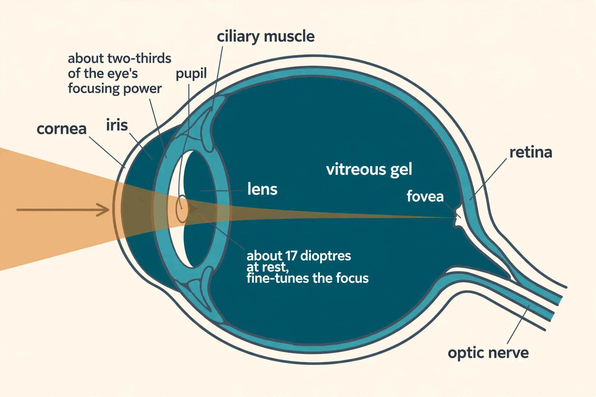
Light enters the eye through the cornea, a clear, dome-shaped layer at the very front. The cornea is the eye's main focusing element, and most people are surprised to learn that. We tend to think of the lens as the part that focuses, because cameras have lenses and the eye has a part called a lens. But the cornea provides about two-thirds of the eye's total focusing power, roughly 43 dioptres out of a total of about 60 dioptres. A dioptre is the unit of refractive power, equal to the reciprocal of the focal length in metres. The reason the cornea does so much of the work is simple physics: light slows down sharply when it passes from air into the dense, curved surface of the cornea, and that sharp change in speed bends the light strongly. The lens, which provides about 17 dioptres at rest, supplies the remaining power and does the fine-tuning.
The cornea is the fixed lens in the system, ground to one shape and left alone, while the lens inside the eye is the adjustable element. If the cornea is damaged or its curve is irregular, no amount of lens adjustment can fully fix the image, because the biggest bend in the whole system happens at the very first surface the light meets.
2.2 The iris and pupil: the aperture that controls how much light gets in
Behind the cornea sits the iris, the coloured part of the eye, and in the middle of the iris is the pupil, the opening through which light passes on its way to the lens. The iris controls how much light enters by widening or narrowing the pupil. In bright light it narrows the opening; in dim light it widens it. This is the fastest adjustment the eye makes, and it works like the aperture of a camera, trading light against sharpness. A wide pupil lets in more light but also lets in rays from the edges of the lens, which focus less precisely. A narrow pupil sharpens the image but admits less light.
The pupil's adjustment is only a small part of how the eye copes with changing light, though. It can change the amount of light entering by a modest factor, while the world itself varies in brightness by more than a billionfold between starlight and sunlight. The heavy lifting of adaptation happens in the retina, in the chemistry of the photoreceptors, and we will come to that in Section 9. The pupil is the doorkeeper. The retina does the real work.
2.3 The lens and the fluids
The lens works together with the cornea to focus light correctly on the retina. It sits behind the iris, held in place by fine fibres called zonules, and its shape is controlled by the ciliary muscle, a ring of muscle around it. When you look at something close, the lens becomes rounder and more powerful. When you look far away, it flattens. Section 3 explains that mechanism in full.
The inside of the eyeball is filled with fluids and gels that keep its shape and keep the optics clear. The vitreous gel is a transparent, colourless mass that fills the rear two-thirds of the eyeball, between the lens and the retina. The front of the eye holds a different fluid, the aqueous humour, a clear watery fluid that fills the chambers between the cornea and the lens. It nourishes the cornea and the lens and helps maintain the eye's shape and internal pressure. Both humours must stay perfectly transparent, because any clouding in them sits directly in the path of the light. A cataract, which we will meet in Section 11, is what happens when the lens itself clouds over.
3. How the Eye Focuses: The Lens That Changes Shape
3.1 The mechanism: accommodation
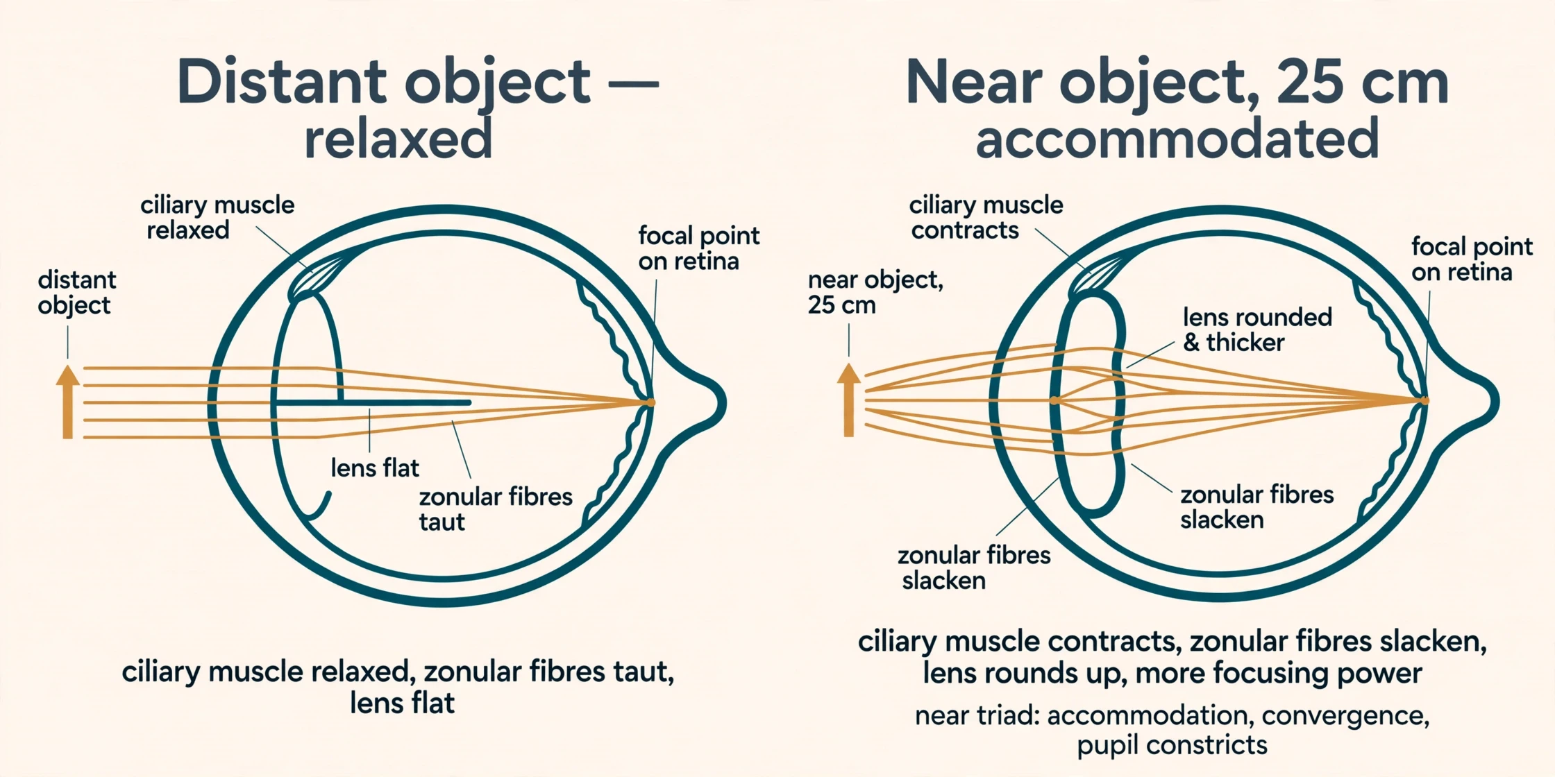
The lens changes shape to focus on near or distant objects, and that process is called accommodation. The mechanism is a small piece of mechanical elegance. When the ciliary muscle contracts, the zonular fibres that hold the lens go slack, and the elastic lens, freed from their pull, becomes rounder and more convex. A rounder lens bends light more strongly, which increases its refractive power and shifts the focal point forward onto the retina, exactly where it needs to be for a near object. For distance vision the ciliary muscle relaxes, the zonules pull taut, and the lens flattens, reducing its power so that parallel rays from a distant object land on the retina.
Near vision is not accommodation alone. It uses a linked trio of responses called the near triad: accommodation as the lens rounds up, convergence as the two eyes turn inward to point at the same near object, and miosis as the pupil constricts. All three happen together, every time you look at something close. Hold a finger in front of your nose and bring it closer: your eyes turn inward, your focus tightens, and your pupils narrow, all without any conscious instruction from you.
A person with normal, ideal vision can see objects clearly at distances ranging from 25 cm to essentially infinity. That range is the whole working life of the eye, from a book held at reading distance to the stars. The total refractive power of the eye is about 60 dioptres, with the cornea contributing about 43 and the lens about 17 at rest. A simplified textbook model that treats the eye as a single thin lens sitting 2.00 cm from the retina gives a lower range, 50.0 to 54.0 dioptres, because the model simplifies the real geometry of the eye. The clinical figure of about 60 dioptres is the one to trust for the real eye.
3.2 When focus fails: myopia, hyperopia, and astigmatism
When the focusing system works as it should, parallel rays from a distant object focus exactly on the retina. That normal state is called emmetropia. The common focusing errors are all failures of that geometry, and each one blurs the world in a different way.
In myopia, or nearsightedness, light focuses in front of the retina, usually because the eyeball is too long or the cornea is too curved. Distant objects blur while near vision stays clear, because light from a near object naturally focuses further back, closer to where the retina actually sits. In hyperopia, or farsightedness, light focuses behind the retina, usually because the eyeball is too short or the cornea is too flat. Near objects blur, and young people can partly compensate by accommodating, pushing the focus forward with extra lens power. That compensation is why farsightedness can stay hidden in a young person until the lens stiffens with age and the trick stops working. In astigmatism, the cornea or the lens is irregularly curved, so light focuses at two different points instead of one, blurring vision at all distances. Imagine a lens shaped more like the back of a spoon than a ball: rays passing through one axis focus at one place and rays through the other axis focus somewhere else, and no single adjustment can bring both into focus at once.
Refractive errors are the most frequent eye problems in the United States. The National Eye Institute states that proper refractive correction could improve vision among 150 million Americans. It means the most common visual problem in one of the world's richest countries is a focusing error, and a large share of it is correctable.
3.3 Presbyopia: the lens stiffens with age
Presbyopia is the age-related loss of near focusing, and it is not a disease. It is a normal part of ageing that occurs in almost all adults around age 40 to 45. The lens stiffens and loses elasticity, so even maximum accommodation cannot bring the focus onto the retina for close objects. The numbers tell the story plainly. The eye's amplitude of accommodation falls from about 14 dioptres at age 10 to about 8 dioptres at age 40 and only 1 to 2 dioptres at age 60. A child can focus on something a few centimetres from the nose. A sixty-year-old, with one or two dioptres of accommodation left, cannot bring a book into focus at normal reading distance without help.
This is why reading glasses appear in so many lives in the mid-forties. Nothing went wrong: the lens simply spent four decades doing a job that depends on elasticity, and elasticity is one of the first things age takes.
"The eye is perhaps the most interesting of all optical instruments. The eye is remarkable in how it forms images and in the richness of detail and color it can detect." — OpenStax, College Physics, section 26.1 "Physics of the Eye"
4. The Retina: 126 Million Light Sensors and One Tiny Sharp Spot
4.1 About 120 million rods and about 6 million cones
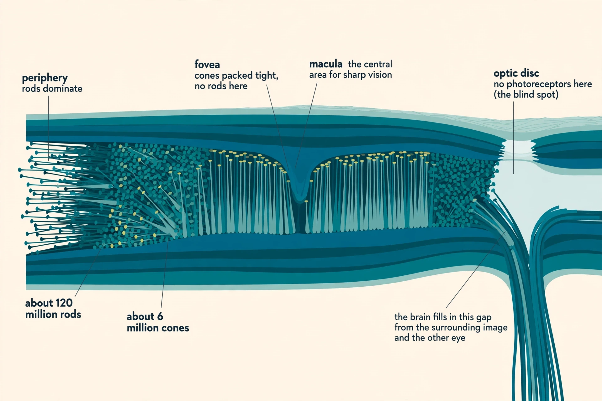
The retina is a thin layer of tissue lining the back of the eyeball, and it is the light-sensitive tissue that converts light into electrical impulses sent to the brain through the optic nerve. Inside it sit the photoreceptors, and their numbers are startling. Each retina holds about 120 million rods and about 6 million cones, roughly twenty times more rods than cones. The rods are distributed throughout the retina except for the fovea, while the 6 to 7 million cones are concentrated in the fovea and the central retina.
The fovea is a small central pit in the retina, and cones are densely packed into it. It is the part of the retina we use to look directly at objects and perceive fine detail, and it has the greatest density of light receptors and the greatest sharpness in the visual field. The proportion of rods increases toward the periphery, so the edges of your visual field are rod country: sensitive to dim light and movement, but poor at detail and blind to colour. There are no rods in the fovea at all. Look directly at a faint star and it vanishes, because you are pointing your rod-free fovea at it. Look slightly to one side and it reappears, because the rods in your peripheral retina can see what your sharp central spot cannot.
4.2 What each does: rods for dim light, cones for bright light and colour
Rods and cones divide the work of vision between them. Rods are for dim-light vision, the kind of seeing you do by starlight or in a dark room. They are far more sensitive to light than cones, but they detect no colour, only shades of grey. Cones are for bright-light vision, fine detail, and colour. The difference comes down to their pigments. Rods contain a single photopigment, rhodopsin, which registers brightness only and cannot distinguish wavelengths. Cones contain one of three photopigments, each tuned to a different part of the spectrum, and colour vision comes from comparing their outputs.
The macula is the small, sensitive area of the retina needed for central vision, and it contains the fovea. When the macula deteriorates, as it does in age-related macular degeneration, central vision goes first: reading, recognising faces, seeing fine detail. The periphery keeps working, because the periphery is rod country and the rods are elsewhere. That pattern, a hole in the middle of the visual world with the edges intact, is the direct result of the retina's geography.
| Feature | Rods | Cones |
|---|---|---|
| Number per retina | About 120 million | About 6 million |
| Location | Throughout the retina except the fovea; dominant in the periphery | Concentrated in the fovea and central retina |
| Pigment | One photopigment, rhodopsin | One of three photopigments (S, M, or L) |
| Light level | Dim light (scotopic vision) | Bright light (photopic vision) |
| Colour | None — shades of grey only | Yes — colour comes from comparing cone outputs |
| Detail | Poor; many rods share one output | Fine; dense packing in the fovea gives sharp vision |
| Speed of dark adaptation | Slow — full sensitivity takes 30 to 45 minutes or longer | Fast — maximum sensitivity in about 9 to 10 minutes |
4.3 The third photoreceptor: cells that do not form images
The human retina contains a third, much rarer class of photoreceptor alongside the rods and cones: the intrinsically photosensitive retinal ganglion cells, known as ipRGCs. They make up under 5 percent of ganglion cells, and they do not form images at all. Instead, they signal overall light levels for subconscious reflexes such as pupil constriction and for entrainment of the body's circadian rhythms. Some of them project directly to the suprachiasmatic nucleus, the brain's master circadian clock, through the retinohypothalamic tract, telling the clock whether it is day or night.
The ipRGCs use a different photopigment, melanopsin, and a different signalling route from the rods and cones. Where rods and cones hyperpolarise in response to light, ipRGCs depolarise. They respond slowly and are relatively insensitive, which makes sense for their job: they are measuring the general brightness of the world over minutes and hours, not catching the flicker of a moving edge. Their discovery, in 2002, rewrote the textbook account of the retina, and Section 12 tells the story of how it happened.
4.4 The blind spot: where the optic nerve leaves
Where the optic nerve leaves the eye there are no photoreceptors at all. That patch is the optic disc, or blind spot, and it is a genuine hole in the sensor: more than a million nerve fibres have to leave the eye somehow, and they leave through a gap in the photoreceptor layer. Yet you never notice a hole in your vision, because the brain fills in the missing patch from the surrounding image and from the other eye.
You can prove the blind spot exists with a simple two-dot test, closing one eye and moving a page until one dot vanishes, and the same test proves your brain papers over the gap so smoothly you would never know it was there. The blind spot is the clearest everyday evidence that what you see is not a raw recording of the world but a construction, edited in real time.
5. Phototransduction: The Moment Light Becomes a Signal
5.1 Rhodopsin and the photon
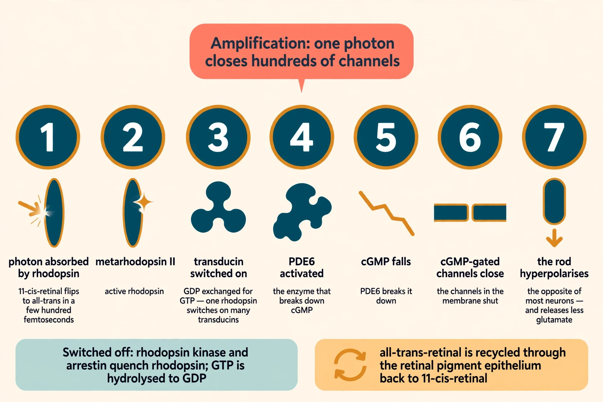
Inside every rod sits a pigment called rhodopsin, and rhodopsin is where light stops being light and starts being information. Rhodopsin is an opsin protein with a chromophore, a light-absorbing molecule called 11-cis-retinal, bound to it. Absorbing a photon isomerises the 11-cis-retinal to all-trans-retinal in a few hundred femtoseconds, with a quantum efficiency of about 0.7, and the opsin changes shape into its active form, metarhodopsin II.
The chromophore is the aldehyde of vitamin A, which is why vitamin A is the raw material of the visual pigment and why vitamin A deficiency causes night blindness. Without enough vitamin A, the eye cannot make enough rhodopsin, and the rods cannot do their job in dim light. But the relationship runs in one direction only. Once you have enough vitamin A, extra does not sharpen normal vision, because the limiting factor is no longer the raw material. Section 13 returns to that point, because it is one of the most persistent myths about eyesight.
5.2 The cascade, step by step
What follows the photon's absorption is a cascade, and the cascade is the reason a single photon can be detected at all. Active metarhodopsin II activates the G protein transducin by catalysing the exchange of GDP for GTP. Transducin then activates the enzyme phosphodiesterase 6, known as PDE6. This is the first amplification step: one activated rhodopsin activates many transducin molecules, and each of those activates PDE6.
Activated PDE6 hydrolyses cyclic GMP, or cGMP, to GMP, so the intracellular cGMP concentration falls. That fall is the signal. In darkness, high cGMP keeps cGMP-gated cation channels open, and the photoreceptor sits in a steady depolarised state, releasing the neurotransmitter glutamate continuously. When cGMP falls, the channels close and the photoreceptor hyperpolarises. That is the opposite of most neurons, which depolarise when they fire. A rod signals light by quieting down. In darkness, photoreceptors steadily release glutamate; light, by hyperpolarising the cell, reduces that release. The signal is therefore a graded change in transmitter release, not an all-or-nothing spike.
Inside the retina, photoreceptors pass their signals to bipolar cells, which pass them to ganglion cells. The ganglion cells' axons bundle together to form the optic nerve. Horizontal and amacrine cells modulate the signal sideways at the two synaptic layers, so the retina already does substantial processing before anything leaves the eye. For more on how the rest of the nervous system carries signals like these, see How the Nervous System Works: Brain, Spinal Cord, and Nerves.
5.3 Amplification and switch-off
The cascade amplifies enormously. One photon can close hundreds of channels by activating many transducin and PDE molecules, and each closed channel changes the voltage of the cell. That amplification is what makes the rod a near-perfect photon counter.
A signal that cannot be switched off is useless, so the response is switched off in two steps. Activated rhodopsin is phosphorylated by rhodopsin kinase and then quenched by arrestin, while transducin and PDE6 are deactivated when their bound GTP is hydrolysed to GDP. Bleached rhodopsin must then be regenerated before full sensitivity returns: all-trans-retinal is recycled through the retinal pigment epithelium back to 11-cis-retinal, in a loop called the visual cycle. That recycling is slow, and its slowness is the reason dark adaptation takes minutes rather than seconds. We will come back to it in Section 9.
5.4 How sensitive is it, really?
The numbers are difficult to believe until you see how they were measured. A dark-adapted human can reliably detect a flash when only about 5 to 7 photons are absorbed across a population of about 500 rods, so each rod must be responding to a single photon. That result was shown behaviourally by Hecht, Schlaer and Pirenne in 1942, and confirmed directly more than thirty years later by recording from single rods. In 2016, researchers using a quantum light source showed directly that humans can detect a single photon incident on the cornea with a probability significantly above chance.
Rods are near-perfect photon counters. They combine high quantum efficiency, enough amplification to produce a measurable response, low dark noise, and low trial-to-trial variability, matching or beating man-made detectors in several ways. A just-detectable flash delivers about 50 photons at the cornea, of which only a small fraction are absorbed. In starlight, fewer than 10 out of every 10,000 rods absorb a photon during the roughly 200-millisecond integration time of rod signals. Your visual system is counting individual particles of light, in the dark, with a detector built from meat and salt water.
When I first read Wald's notebooks on this chemistry, what stayed with me was not the elegance of the cascade but the stubbornness behind it. In Otto Warburg's Berlin laboratory in the early 1930s, Wald extracted retinas with chloroform, reacted the extract with antimony trichloride, and the solution turned bright blue, with an absorption curve typical of vitamin A. He had found vitamin A inside the visual pigment. He then confirmed it in cow, sheep, and pig retinas with Paul Karrer in Zurich. Wald went on to identify the decisive chemical step: the 11-cis isomer of retinal is the chromophore of rhodopsin, and light isomerises it to the all-trans form, the first known case of cis-trans isomerisation playing a role in biology. He also named the cone pigment iodopsin. For that work, and for discoveries concerning the primary physiological and chemical visual processes in the eye, Wald won a share of the 1967 Nobel Prize in Physiology or Medicine, with Ragnar Granit and Haldan Keffer Hartline. He was explaining chemistry that Boll and Kuhne had first glimpsed sixty years earlier, when Boll discovered the rod visual pigment in 1876 and Kuhne showed in 1878 that light bleaches it from reddish-purple through yellow to colourless. Wald's chemistry explained what they had seen.
6. Colour Vision: Three Kinds of Cone and the Colours They Build
6.1 Trichromacy: three cones sampling the spectrum
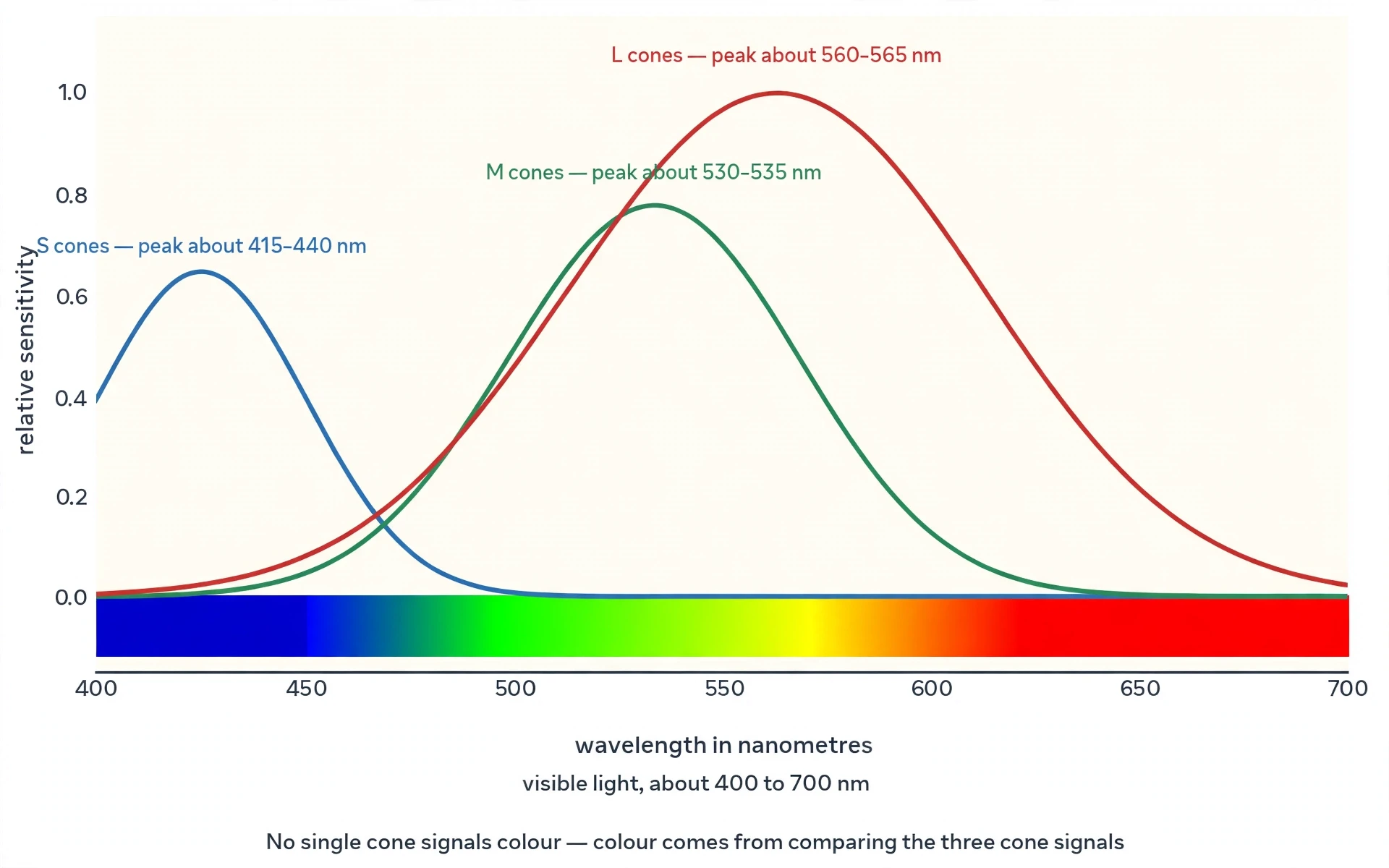
Human colour vision is trichromatic. Three cone classes sample the visible spectrum, roughly 400 to 700 nanometres, with broadly overlapping sensitivities. The S cones peak in the short wavelengths, the M cones in the middle, and the L cones in the long wavelengths. The peaks are best given as ranges, because different measurement methods give slightly different numbers: S cones peak at about 415 to 440 nanometres, M cones at about 530 to 535, and L cones at about 560 to 565. The spread comes from the difference between measuring single cones directly and measuring colour matching by human observers, and no single number is the correct one.
The trichromatic theory is usually credited to Thomas Young, who proposed it in 1802, and Hermann von Helmholtz in the nineteenth century. Young inferred from colour-matching experiments that the eye must contain a small number of receptor types, each tuned to a different part of the spectrum, because any visible colour could be reproduced by mixing just three primary lights. He proposed this on purely behavioural grounds, more than 150 years before single-cone recordings confirmed that exactly three cone classes exist. It is one of the great deductions in the history of science: the number three, derived from what people could match with coloured lights, turned out to be the number of cone types in the retina.
6.2 Why one cone cannot see colour
No single cone can signal colour, because each cone is univariant: it reports only how many photons it caught, not their wavelength. A cone that catches a hundred photons of green light gives the same signal as a cone that catches a hundred photons of red light, if the two wavelengths happen to stimulate it equally. Colour comes from comparing the relative activity of the three cone types. If the L cones fire much harder than the M cones, the light is probably long-wavelength; if all three fire together, it is probably white. The brain reads the ratios, not the absolute counts.
After the cones, the signals are recoded into opponent channels: red-versus-green, blue-versus-yellow, and light-versus-dark. That recoding explains why some colour pairs seem opposed to each other, and why afterimages take the complementary hue. Stare at a red square for thirty seconds, then look at a white wall, and you see a green square, because the red-green channel has adapted and its opponent shows through.
6.3 The cone mosaic: mostly L, some M, few S
The three cone types differ sharply in number. L cones are about 60 to 64 percent of all cones, M cones about 30 to 33 percent, and S cones only about 5 to 10 percent, yet the proportions vary greatly from person to person, especially between L and M. Two people with normal colour vision can have quite different cone mosaics, because the brain calibrates itself to whatever ratios it is given.
S cones are essentially absent from the central 1 degree of the retina, the foveola, and reach their peak density at about 1 to 2 degrees of eccentricity, falling away with distance from the centre. In the long-wavelength region, where S cones barely respond, colour vision is effectively dichromatic. The three cone classes were mapped directly in the living human eye by Roorda and Williams in 1999, using adaptive optics to image individual cones, and the map they produced showed a mosaic far more irregular than anyone had drawn in textbooks: clumps of L cones here, patches of M there, S cones scattered thinly between them.
6.4 Colour blindness: losing or shifting one cone type
Colour blindness usually results from losing one cone type or from a shift in one cone type's spectral sensitivity. Most forms reflect inherited defects in the L and M photopigment genes, which sit on the X chromosome, which is why red-green deficiency affects about 8 percent of men of Northern European descent but only about 0.5 percent of women. Colour vision deficiency affects about 5 percent of people overall, about 1 in 20, and about 8 percent of men.
The common forms have precise names. Protanopia is the absence of working L cones. Deuteranopia is the absence of working M cones. Tritanopia is the absence of working S cones. The milder anomalous forms are protanomaly, deuteranomaly, and tritanomaly, and deuteranomaly is the most common of all. Total absence of colour vision, achromatopsia, is rare. Red-green deficiency accounts for roughly 95 percent of all congenital cases. And the most persistent myth about it is simply wrong: almost all colour-blind people see colours. They confuse specific pairs, most often reds and greens. The world does not drain to black and white for them. It loses some of its distinctions, and keeps the rest.
7. How Vision Works Beyond the Eye: From Retina to Brain
7.1 Retinal processing first
Nothing leaves the eye unprocessed. The retina is not a passive sensor but a small piece of brain that happens to sit at the back of the eyeball, and the wiring of Section 5.2 already does substantial computation before it sends anything onward. By the time the signal reaches the optic nerve, a bundle of more than 1 million nerve fibres carrying visual messages from the retina to the brain, it is no longer a pixel-by-pixel copy of the light. It is already a processed summary: edges emphasised, steady backgrounds suppressed, changes flagged.
A camera sensor reports light levels. The retina reports a first draft of meaning.
7.2 The route: optic nerve, chiasm, LGN, visual cortex
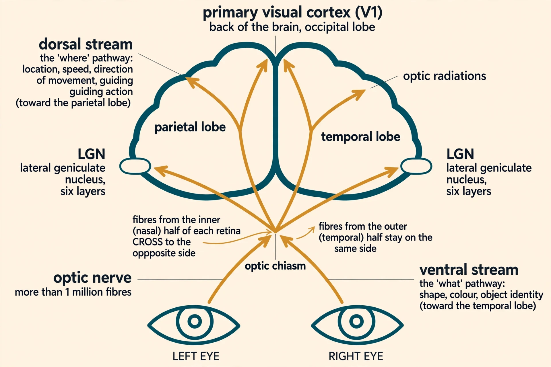
The route from the eye to the cortex runs through a series of relay stations, and each one matters. The path is: retina, optic nerve, optic chiasm, optic tract, lateral geniculate nucleus of the thalamus, optic radiations, and finally the primary visual cortex, known as V1 or Brodmann area 17, in the occipital lobe at the back of the head.
At the optic chiasm, fibres from the nasal, inner half of each retina cross to the opposite side, while fibres from the temporal, outer half stay on the same side. The result is that each hemisphere of the brain receives the opposite visual field: the left visual field is processed by the right visual cortex and vice versa. The lateral geniculate nucleus, the LGN, has six layers, which keep inputs from the two eyes and from different cell types separate. It is the thalamic relay where visual information is first processed beyond the retina before reaching V1.
7.3 V1 and the feature detectors
The primary visual cortex, V1, also called the striate cortex, is the first cortical region to integrate visual information. It detects basic features such as lines, edges, colours, and motion, and from V1 information flows onward to visual association areas. What Hubel and Wiesel showed, in work that earned them a share of the 1981 Nobel Prize in Physiology or Medicine, is that cortical neurons are feature detectors. Individual V1 neurons respond best to lines, bars, and edges at specific orientations, a property called orientation selectivity, and neurons with similar orientation preferences are grouped into columns. Each hypercolumn covers about 2 by 2 millimetres of cortex.
The visual system analyses the image step by step. Contrasts, line patterns, and movement are extracted in successive stages, from retina to LGN to V1 and beyond, rather than the brain receiving a finished picture. Hubel and Wiesel also showed that the visual cortex is not ready-made: patterned visual input in early life is needed for normal development, and light stimulation alone is not enough. The retinal image must have pattern and contours. A cortex that never sees edges never learns to see them properly.
"The message about the image falling on the retina undergoes a step-wise analysis in a system of nerve cells stored in columns. In this system each cell has its specific function and is responsible for a specific detail in the pattern of the retinal image." — Nobel Assembly at Karolinska Institutet, press release for the 1981 Nobel Prize in Physiology or Medicine
When I first heard the story of how Hubel and Wiesel found orientation selectivity, it changed the way I think about accidents in science. They were recording from cells in the visual cortex of an anaesthetised cat, and the cells failed to respond to spots of light. Then a slide edge moved across the screen, and a cell fired vigorously. The cells did not care about dots of light. They cared about oriented edges and lines. A slide edge moving across the screen, at the wrong moment, revealed the organising principle of the visual cortex. Their 1962 paper reporting receptive fields, binocular interaction, and functional architecture in the cat's visual cortex is one of the founding papers of visual neuroscience, and it began with a cell ignoring everything the experimenters expected it to respond to.
For a fuller picture of the brain region where all of this happens, see Human Brain Anatomy and Functions: A Complete Guide.
7.4 Two streams: where and what
Beyond V1 there are two main streams, and they answer different questions. The dorsal stream, the "where" pathway, runs from V1 toward the posterior parietal lobe and handles location, speed, and direction of movement, guiding action. It tells you where something is and how to reach for it. The ventral stream, the "what" pathway, runs toward the inferior temporal lobe and handles shape, colour, and object identity. It tells you what you are looking at. A patient with damage to one stream can sometimes recognise an object but not reach for it accurately, or reach accurately for something they cannot name. The two questions really are answered separately.
The optic nerve itself carries two main fibre types. Parvo fibres carry fine detail and colour. Magno fibres carry motion and coarse, fast-changing information. And there is a second, older route besides the main pathway from the retina through the LGN to the cortex: the tectal pathway, which runs from the retina to the superior colliculus in the midbrain and on via the pulvinar to visual association areas, supporting orienting and attention to sudden movements. It is the route that makes you turn your head toward a flicker at the edge of your vision before you know what flickered.
8. Why the Image Is Upside Down on Your Retina — and Why You Never Notice
8.1 The optics: a convex lens inverts the image
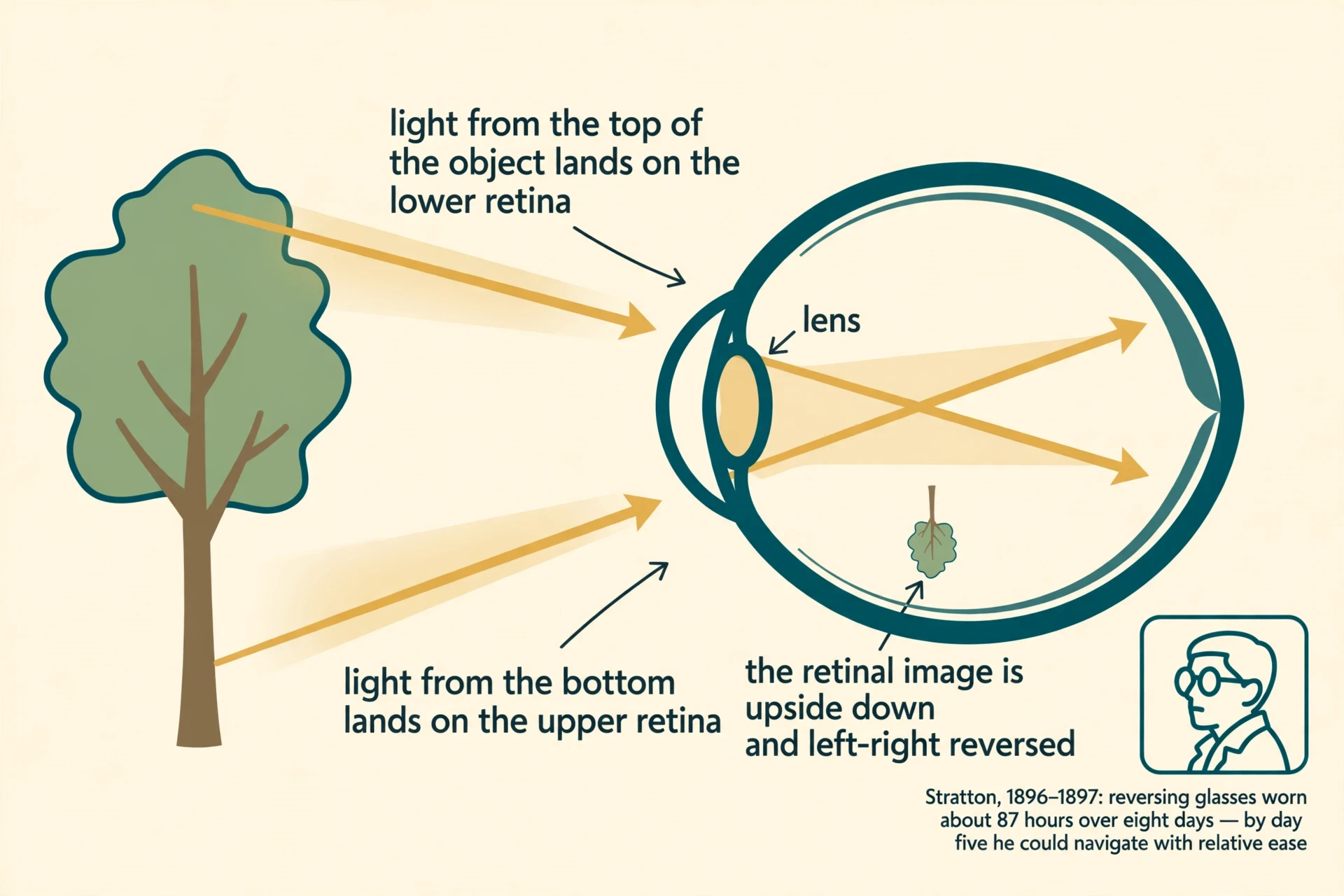
The cornea and lens act like a convex lens, so the image projected onto the retina is inverted, upside down, and reversed left-to-right. Light from the top of an object crosses over with light from the bottom as it passes through the lens, landing on the lower retina, while light from the bottom lands on the upper retina. Every image your retina has ever received has been upside down. Descartes proved the inversion in the seventeenth century by replacing the retina of an excised bull's eye with a screen: the image that appeared was a smaller, inverted copy of the scene.
The camera eye itself, with its convex lens and inverted image, is a product of evolution, and its history is a story in its own right. For how this design arose from light-sensitive cells over hundreds of millions of years, see Evolution of the Eye: From Light-Sensitive Cells to Eyes.
8.2 The correction is not a flip
Here is the point most explanations get wrong. The brain does not literally flip the image like rotating a photograph. There is no mechanism in the brain that takes a picture and turns it over. Instead, the brain interprets patterns of nerve signals using spatial relationships, memory, and prior experience, and it learns to associate activity in the lower retina with "up" in the world. Modern neuroscience describes this as interpretation and learned mapping, not a flip. The image on each retina is also a flat, two-dimensional projection, and the brain overlays the two images to build a single three-dimensional percept. Uprightness is something the brain constructs, not something it receives.
8.3 Stratton's experiment: eight days upside down
George Stratton tested this directly in 1896 and 1897 by wearing reversing lenses that made his retinal image upright, so that the world itself appeared upside down. At first everything looked upside down and movement was clumsy. Over days he adapted, and by the fifth day of his eight-day experiment he could navigate with relative ease, with moments of upright vision. After removing the glasses, the world briefly looked upside down again. His total wearing time in the 1897 experiment was about 87 hours over eight days.
Reading Stratton's own account, what struck me was not the adaptation itself but how ordinary it became. By the fifth day he could navigate with relative ease, and at moments the world he saw through the glasses had simply become upright. The mapping had rewritten itself. Then he removed the glasses, and the familiar world tilted upside down for a while, until the old mapping reasserted itself. The brain does not contain a fixed picture of up and down. It contains a learned agreement between the retina and the world, and that agreement can be renegotiated in days. That is the strongest evidence there is that seeing is something the brain does, not something the eye delivers.
9. Dark Adaptation: Why the Cinema Takes Twenty Minutes
9.1 The range: more than a billionfold
The visual system works across more than nine orders of magnitude of light intensity, from bright sunlight to dim starlight. Much of this adaptation happens in the retina itself, in the photoreceptors. Four mechanisms together give the eye its huge operating range: the duplex retina, with rods for dim light and cones for bright; changes in pupil size; photochemical adaptation, the bleaching and regeneration of photopigment; and neural adaptation, changes in retinal responsiveness, receptive-field size, and temporal summation.
The pupil is the fastest of the four and the least powerful; the chemistry is the slowest and the most powerful, and it explains the cinema.
9.2 The biphasic curve: cones first, rods second
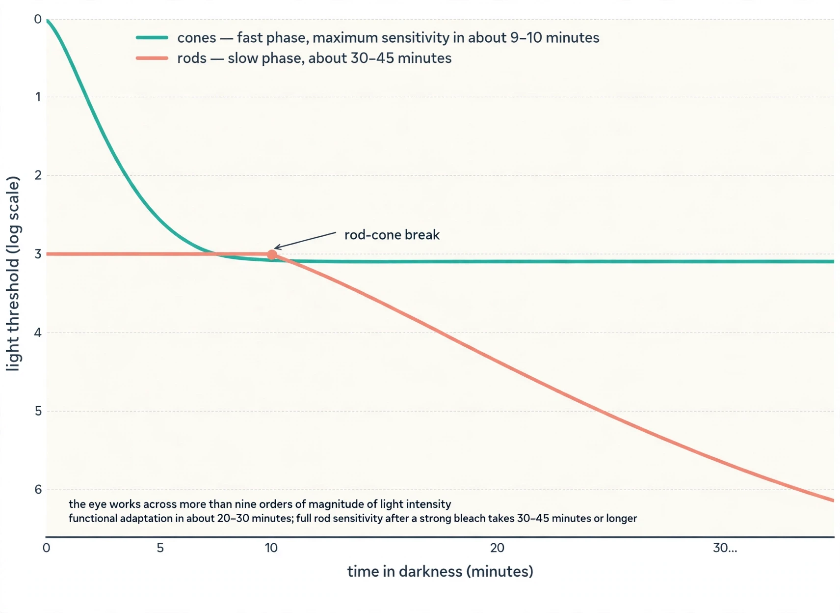
Dark adaptation is the slow recovery of sensitivity after bright light bleaches photopigment, and the recovery curve is biphasic: a fast cone phase first, then a slower rod phase. Cones regain maximum sensitivity in about 9 to 10 minutes of darkness. Rods take about 30 to 45 minutes, and after a near-total bleach, full rod recovery can take over 30 minutes. Put together, functional adaptation takes about 20 to 30 minutes, while full rod sensitivity after a strong bleach takes 30 to 45 minutes or longer. There is no single number that is "the" time for dark adaptation.
The slow step is the regeneration of photopigment. Bleached opsin without its chromophore, known as apo-opsin or free opsin, keeps activating phototransduction weakly, acting like a background light that desensitises the cell. Sensitivity returns only as 11-cis-retinal is delivered back to the photoreceptors at a limited rate. Light adaptation, by contrast, is far faster than dark adaptation, a matter of seconds, because it mainly involves shutting down sensitivity that is already present rather than rebuilding pigment. That asymmetry is why stepping out of a dark cinema into sunlight is blinding for a moment, while stepping into the cinema leaves you effectively blind for minutes.
9.3 Each eye adapts on its own
Dark adaptation can proceed independently in each eye. One eye can be light-adapted while the other is dark-adapted, which proves the process happens in the retina, before the optic chiasm where the two eyes' signals mix. Dark adaptation is also faster and deeper in young people than in the elderly, one more way the ageing visual system loses ground quietly.
So here is the cinema again, with the chemistry filled in. When you stepped out of the summer street, your photopigment was bleached by the sunlight, and your rods were desensitised by free opsin acting as a false background light. Your pupils widened within seconds, and that helped a little. But real vision in the dark had to wait for the visual cycle to deliver fresh 11-cis-retinal to your rods, molecule by molecule, and that takes tens of minutes. By the time the trailers ended, the cones had finished their fast phase and the rods were well into theirs, and the room that had been black was now full of shapes. The room itself had not changed at all. Your eyes did.
10. Two Eyes, Depth, and Eyes That Never Sit Still
10.1 Stereopsis: two views, one world
Binocular vision works because the two eyes see the world from slightly different angles. The brain fuses the two slightly different retinal images into a single percept and uses the differences between them, binocular disparity, to infer depth. That depth from disparity is called stereopsis. It is most useful for nearby objects and becomes virtually useless for depth beyond about 50 to 200 feet, where monocular cues take over.
Depth perception also uses monocular cues that work with one eye alone: the known size of objects, occlusion, where one object hides another, perspective, and motion parallax, where near objects sweep across the retina faster than far ones when you move. Close one eye and you can still judge distance from the monocular cues, but you lose the fine sense of depth at arm's length: threading a needle, catching a ball, pouring water into a glass. That is stereopsis, and it is a near-range sense.
10.2 Eye movements: fixations, saccades, and the tremor underneath
Your eyes never sit still, even when you think they do. Humans make two to three fixations per second while exploring a scene, and each fixation requires the two eyes to converge accurately on the new target, or double images result. Between fixations, the eyes make saccades, rapid jumps from one fixation point to another. Saccades last about 10 to 80 milliseconds, and vision is suppressed during a saccade, a phenomenon called saccadic suppression, so we do not perceive the blur of the jump. You are, in effect, briefly blind several times a second, and you never notice.
Even when we try to hold the eyes still, tiny fixational movements continue: microsaccades once or twice per second, slow drift, and tremor. Microsaccades counteract visual fading, help scan small informative regions, and improve acuity by precisely relocating the fovea. Without them, a perfectly stabilised image would fade from view within seconds, because the retina is built to report change. Smooth pursuit is the slow tracking movement that keeps a moving object on the fovea, and it is distinct from saccades: it works only when something is actually moving. Abnormalities of binocular vision include strabismus, misaligned eyes, and amblyopia, the "lazy eye" in which one eye has poor vision, often because the brain suppresses its input in childhood.
11. When Vision Goes Wrong: What Breaks at Each Step of the Journey
11.1 Focus failures and the myopia surge
The focusing errors from Section 3 are the commonest problems in vision, and one of them is rising fast. Myopia is rising worldwide: an estimated 22.9 percent of the world's population was myopic in 2000, and modelling projects 49.8 percent, about 4.758 billion people, by 2050, with high myopia at 9.8 percent, about 938 million people. Myopia is an optical mismatch, usually an eyeball that is too long for its focusing power. Near work and screen time are associated with higher risk, and outdoor time is protective, but the mechanism is not "reading damages the eye". A 2025 meta-analysis found a dose-response relationship between screen time and myopia risk in children and adolescents, with risk rising from one to four hours of screen time per day, and a separate 2025 meta-analysis of 15 studies with over 16,000 participants confirmed that more outdoor time protects against myopia. A 2026 study proposes the mechanism may be how much light reaches the retina rather than what is being looked at. Treat that mechanism as proposed, not proven, and do not present screens as the proven cause of myopia.
High myopia carries risks beyond blurred distance vision. High myopia, about minus 6 dioptres or worse, carries a heightened risk of retinal detachment, glaucoma, cataracts, and permanent blindness. A longer eyeball is thought to stretch the retina, leaving it more vulnerable. The focusing errors of Section 3 belong here too, not as diseases but as the commonest ways the optics fail: an eyeball too short, a cornea curved irregularly, or a lens that has lost its elasticity with age.
| Condition | What goes wrong | What blurs | Why |
|---|---|---|---|
| Myopia (nearsightedness) | Light focuses in front of the retina | Distant objects; near vision stays clear | Eyeball too long or cornea too curved |
| Hyperopia (farsightedness) | Light focuses behind the retina | Near objects; young people can partly compensate by accommodating | Eyeball too short or cornea too flat |
| Astigmatism | Light focuses at two different points instead of one | Vision at all distances | Irregularly curved cornea or lens |
| Presbyopia | Lens stiffens and loses elasticity | Near objects, from around age 40 to 45 | Normal ageing; accommodation falls to 1–2 dioptres by age 60 |
11.2 The lens clouds: cataract
A cataract is the clouding of the eye's natural lens. Around age 40, the proteins in the lens start to break down and clump together, forming cloudy areas that scatter and block light. It is the leading cause of blindness worldwide and the leading cause of vision loss in the United States. The main risk factors, in descending order of importance, are increasing age, exposure to UV-B light, and topical or internal corticosteroids. Diabetes also contributes, through glycation and sorbitol accumulation in the lens. The lens must stay transparent for the whole optical system to work, and the proteins that keep it clear spend a lifetime slowly losing that battle.
11.3 The retina fails: macular degeneration and diabetic retinopathy
Age-related macular degeneration, or AMD, is deterioration of the macula, the central retina needed for sharp central vision. Dry AMD, 85 to 90 percent of cases, involves fatty deposits called drusen and slow atrophy of the retinal pigment epithelium. Wet AMD, 10 to 15 percent, involves abnormal blood vessels growing under the retina, leaking blood or fluid, and it accounts for about 90 percent of AMD-related blindness. AMD is the most common cause of irreversible vision loss in people over 50 in developed countries. In AMD, damage begins in the retinal pigment epithelium and Bruch's membrane near the edge of the macula, reducing the passage of nutrients from the choroid to the sensory retina and leading to degeneration of the cones.
Diabetic retinopathy damages the retina's blood vessels in diabetes and ranks among the main drivers of sight loss in the United States, alongside AMD, cataract, and glaucoma.
11.4 The nerve is damaged: glaucoma and retinitis pigmentosa
Glaucoma is a group of diseases that damage the optic nerve, usually but not always in association with raised pressure inside the eye. Fluid builds up in the front of the eye, pressure rises, and the optic nerve is progressively damaged, often with no early symptoms, which is why it is called the "silent thief of sight". Lost vision from glaucoma cannot be recovered. It is a leading cause of irreversible blindness worldwide.
Retinitis pigmentosa is a group of inherited disorders in which the light-sensing photoreceptor cells gradually degenerate, typically beginning with night blindness and peripheral vision loss in childhood or adolescence and progressing toward severe visual impairment or total blindness by adulthood. It affects more than 1.5 million people worldwide. The rods die first, which is why night blindness comes first, and the cones follow.
11.5 The big picture
The leading causes of blindness and low vision in the United States are primarily age-related eye diseases: age-related macular degeneration, cataract, diabetic retinopathy, and glaucoma. Refractive errors remain the most frequent eye problem overall, and proper correction could improve vision among 150 million Americans. Most vision loss is not a single dramatic event. It is a slow failure at one stage of the journey: the optics, the retina, the nerve, or the brain.
| Stage of the journey | What happens there | What breaks it |
|---|---|---|
| Optics: cornea and lens | Light is bent and focused onto the retina | Myopia, hyperopia, astigmatism, presbyopia, cataract |
| Retina: rods, cones, and pigment epithelium | Light becomes an electrical signal | Macular degeneration, diabetic retinopathy, retinitis pigmentosa, vitamin A deficiency |
| Pathway: optic nerve and relay stations | Signals travel to the visual cortex | Glaucoma, optic nerve damage |
| Brain: visual cortex and the two streams | Signals become objects, places, and movement | Lack of patterned visual input in early life, which the cortex needs to develop normally |
Eye symptoms deserve a professional eye examination. This article explains mechanisms only. It does not diagnose, and it offers no treatment advice.
12. The Newest Science: Restoring Sight in 2025 and 2026
12.1 PIONEER: optogenetic therapy for late-stage retinitis pigmentosa
In October 2026, the PIONEER trial was published in the New England Journal of Medicine. Ten legally blind patients with late-stage retinitis pigmentosa received an injection of an adeno-associated virus carrying the gene for ChrimsonR, a light-sensitive protein, into the worse eye, plus light-stimulating goggles. Light sensitivity improved in 7 of 10 patients, by a factor of 2.0 to 62.3, and 6 of 10 reached a clinically meaningful improvement. The trial was open-label, not randomised, and designed primarily for safety, and the authors stress the results are exploratory. Seven of ten improving in a small, open-label trial is a signal, not a verdict, but for people who were legally blind, a signal is a great deal.
12.2 The Nobel connection: optogenetics wins the 2026 prize
The PIONEER publication came days after the 2026 Nobel Prize in Physiology or Medicine was awarded to Karl Deisseroth, Peter Hegemann, and Georg Nagel for foundational work on light-gated ion channels and optogenetics, the science the therapy is built on. The Nobel Assembly announced the prize on 5 October 2026, "for their discoveries concerning light-gated ion channels and optogenetics". Hegemann and Nagel discovered channelrhodopsin, a light-gated channel in a single-celled alga, and Deisseroth turned it into a switch that can turn nerve cells on with light. In a little over two decades, a basic discovery about how an alga swims toward light became a therapy being tested in blind patients. That arc, from algae to a human retina, is the same arc the PIONEER trial is walking.
12.3 MCO-010: three-year results presented at ARVO 2026
A second optogenetic approach, MCO-010, reported three-year results at the ARVO 2026 annual meeting. In the REMAIN extension of the phase 2b RESTORE trial in retinitis pigmentosa, visual acuity gains were sustained to three years, with patients able to make out objects on a table or walk down a hallway. It is gene-agnostic, working regardless of the underlying mutation, and is delivered by injection into the vitreous rather than under the retina. This was a conference presentation, not yet a peer-reviewed paper, so these results should be read as presented, not as established.
12.4 PRIMA: a chip under the retina
The PRIMA retinal prosthesis takes a different route entirely: a tiny wireless chip implanted under the retina, paired with camera-equipped glasses. In a multicentre trial it achieved clinically meaningful gains in visual acuity in about 80 percent of 38 patients with geographic atrophy from advanced AMD, and the developer has applied for a CE mark in Europe.
12.5 Stem cells: early work in animal models
Stem-cell approaches are advancing, but they are further from the clinic. A 2026 study in the Journal of Translational Medicine isolated a subgroup of human induced pluripotent stem-cell-derived retinal progenitor cells, known as hRPC1, that migrated into the retina and produced structural preservation and measurable visual recovery in animal models of retinal degeneration over 4 to 8 weeks. That is animal-model work, not a human treatment.
12.6 Dark adaptation's open questions
Even the oldest questions in vision science are still open. A 2025 review of dark-adaptation science summarised the newest findings: free opsin after bleaching acts as an equivalent background that desensitises rods, and cone dark adaptation involves a second, faster pathway using the RGR opsin. Open questions remain about the exact delivery route of 11-cis-retinal to cones. The chemistry you use to see in the dark still holds details nobody has nailed down.
What stopped me when I first read Berson's 2002 paper was the sheer stubbornness of the experiment. David Berson and colleagues wanted to know whether some retinal ganglion cells were themselves light-sensitive, so they traced cells projecting to the suprachiasmatic nucleus, recorded from them while blocking all rod and cone signalling with drugs, and the cells still responded to light. Then they went further: they physically isolated the cells from the rest of the retina, and the cells still answered to light. A cell that should have been blind, by every textbook account of the time, was seeing. That experiment established the third photoreceptor class, the ipRGCs, and it did it by refusing to accept the textbook's word for what a ganglion cell could do.
13. Eight Things People Get Wrong About Vision
Myth 1: Your eyes work like a camera, and your brain just looks at the photo. A camera records a picture; the eye does not. The retina already processes the image before anything leaves the eye, the brain receives patterns of nerve signals rather than a picture, and it constructs what you see from those signals plus memory and experience. The camera comparison holds only for the focusing optics at the front of the eye.
Myth 2: Your brain flips the upside-down retinal image the right way up, like rotating a photo. There is no literal flip. The brain learns a consistent mapping between retinal positions and the world through experience, which is why Stratton could adapt to reversing glasses over days. The modern description is interpretation, not flipping.
Myth 3: Colour-blind people see the world in black and white. Almost all colour-blind people see colours. What they lose is the ability to tell certain pairs apart, most often reds from greens. Total colour blindness, achromatopsia, is rare, and red-green deficiency affects about 8 percent of men.
Myth 4: Eating lots of carrots, or taking extra vitamin A, will sharpen your eyesight. Vitamin A is the raw material of rhodopsin, and deficiency causes night blindness. But once your body has enough, extra vitamin A does not make normal vision sharper, because the raw material is no longer the limiting factor.
Myth 5: You can see colours in dim light if you look hard enough. In dim light only rods work, and rods feed a single brightness channel. They cannot distinguish wavelengths at all. That is why the world drains to grey at dusk, and why reds are the first colours to vanish.
Myth 6: You have a hole in your vision where the blind spot is. The optic disc has no photoreceptors, but you never notice a gap, because the brain fills in the missing patch from surrounding detail and from the other eye. A simple two-dot test proves the blind spot is real, and the same test proves you never normally notice it.
Myth 7: Myopia is caused by reading too much or by screens. Myopia is a mismatch between the eye's focusing power and the length of the eyeball, usually an eyeball that is too long. Near work and screen time track with higher risk, and time outdoors is protective, yet the mechanism is not "reading damages the eye". The leading current hypothesis involves how much light reaches the retina, and it is proposed, not proven.
Myth 8: Dark adaptation is just your pupils widening. Pupil widening is fast and contributes only a small part. Real dark adaptation is the slow chemical rebuilding of bleached photopigment, with functional adaptation taking about 20 to 30 minutes and full rod sensitivity after a strong bleach taking 30 to 45 minutes or longer. That is why stepping from sunlight into a dark cinema leaves you effectively blind for minutes, long after your pupils have opened.
14. Seeing Is Something Your Brain Does
Back in the cinema, the trailers are ending and the room is fully visible now. The seats, the aisle, the faces. Nothing about the room changed while you sat there. What changed was chemistry: pigment bleached by sunlight, rebuilt molecule by molecule in the dark, until rods that had been desensitised by free opsin could count photons again. The same dark room, seen by the same eyes, became a different room, because seeing is not a property of the room or of the eyes alone. It is something the whole system does together: cornea and lens bending light, rods and cones turning photons into signals, a cortex assembling those signals into objects, places, and movement.
Follow the journey once more, in three sentences. Light enters through the cornea and lens and lands on the retina, where a photon flips a single molecule and a chemical cascade amplifies that event into an electrical signal. The signal travels through the optic nerve, across the chiasm, through the thalamus, and into the visual cortex, which pulls lines, edges, colour, and movement out of it and splits the work into streams that ask where something is and what it is. And the system that builds your whole visual world out of that relay can register a single particle of light. A dark-adapted person can spot a flash when only about 5 to 7 photons are absorbed, and in 2016 an experiment with a quantum light source proved that a single photon landing on the cornea can be detected. Your entire visual world, every colour and face and horizon you have ever seen, is built from events that small.
15. Frequently Asked Questions About How Vision Works
How does vision work?
Vision works as a relay from light to brain. Light enters through the cornea, passes through the pupil and the lens, and lands on the retina, where rods and cones turn it into electrical signals through phototransduction. Those signals travel through the optic nerve, cross at the optic chiasm, relay through the lateral geniculate nucleus, and arrive at the primary visual cortex, where the brain extracts lines, edges, colour, and movement and builds the picture you see. The short version: optics focus the light, chemistry converts it, and the brain constructs the image.
What do rods and cones do?
Rods and cones are the retina's two main photoreceptors, and they divide the work. Rods, about 120 million per retina, work in dim light and detect only shades of grey, using a single pigment, rhodopsin. Cones, about 6 million per retina, work in bright light and give us fine detail and colour, using one of three pigments each. Cones are packed into the fovea, the retina's sharp central spot, while rods dominate the periphery, which is why you see faint stars better out of the corner of your eye.
How does the eye focus?
The eye focuses by changing the shape of its lens, a process called accommodation. When the ciliary muscle contracts, the zonular fibres slacken and the elastic lens becomes rounder, increasing its power for near objects. For distance vision the muscle relaxes, the zonules pull taut, and the lens flattens. The cornea does most of the focusing, about two-thirds of the eye's total power of about 60 dioptres, while the lens fine-tunes it. A person with normal vision can see clearly from 25 cm to essentially infinity.
Why is the image upside down on the retina?
The cornea and lens work together as a convex lens, bending the rays so the top of the scene ends up on the lower retina and the image arrives inverted and left-right reversed. You never notice because the brain does not flip the image; it interprets the pattern of nerve signals through learned mappings between retinal positions and the world, built from experience. Stratton's reversing-glasses experiment in 1896–1897 showed the mapping can even be relearned in days.
Why can't I see colours in the dark?
In dim light only your rods are working, and each rod carries just one pigment, rhodopsin, which records brightness and nothing else: it cannot tell one wavelength from another. Colour vision needs cones, and cones need bright light. When the light falls, the cones drop out and the rods take over, feeding the brain a single brightness channel, which is why the world drains to grey at dusk and reds vanish first.
Why does it take so long to see in the dark?
Dark adaptation is slow because it is chemical, not mechanical. Bright light bleaches photopigment, and the leftover free opsin keeps activating the signalling cascade weakly, acting like a background light that desensitises the rods. Full sensitivity returns only as fresh 11-cis-retinal is delivered back to the photoreceptors, and that rebuilding takes time. Cones bounce back in about 9 to 10 minutes. Usable vision in the dark returns over roughly 20 to 30 minutes, but the rods need 30 to 45 minutes or more after a strong bleach before they reach full sensitivity.
Can carrots improve your eyesight?
Carrots contain vitamin A, and vitamin A is the raw material of rhodopsin, the visual pigment, so a deficiency causes night blindness and correcting it restores night vision. But once intake is adequate, eating more does not make normal vision sharper, because a shortage of the raw material is no longer what holds vision back. A balanced diet protects your sight by preventing deficiency; it does not upgrade eyes that already have what they need.
What is the difference between the dorsal and ventral streams?
The two streams carry visual information from V1 to different parts of the brain for different jobs. The dorsal stream, the "where" pathway, heads for the posterior parietal lobe and deals with where things are, how fast they move, and which way they are going, so you can reach for them. The ventral stream, the "what" pathway, heads for the inferior temporal lobe and deals with shape, colour, and identity, so you know what you are looking at. When one stream is damaged the other can keep working, so someone may reach accurately for an object they cannot name, or recognise something they cannot reach for.
16. Sources
- National Eye Institute — How the Eyes Work
- National Eye Institute — About the Eye
- National Eye Institute — Eye Handout (See Well for a Lifetime)
- OpenStax College Physics — 26.1 Physics of the Eye
- Opterio — How the Eye Focuses Light: A Paraoptometric Guide
- Basic Physiology — Vision: The Eye
- Centers for Disease Control and Prevention — About Common Eye Disorders and Diseases
- Holden et al. 2016 — Global prevalence of myopia and high myopia (Ophthalmology)
- Khan Academy — Photoreceptors (rods vs cones)
- ScienceDirect — Retina Cone
- Scientific American — Why does it take so long for our vision to adjust to a darkened theater?
- Reactome — Activation of the phototransduction cascade
- Reactome — Visual phototransduction
- University College London — Phototransduction cascade (Stockman)
- Anatixa — Phototransduction: How Light Becomes a Neural Signal
- Wikipedia — Visual phototransduction
- Hecht, Schlaer and Pirenne 1942 — Energy, quanta, and vision (Journal of General Physiology)
- Tinsley et al. 2016 — Direct detection of a single photon by humans (Nature Communications)
- Rieke and Baylor 1998 — Single-photon detection by rod cells of the retina (Reviews of Modern Physics)
- Kiani and Ala-Laurila 2020 — Seeing With a Few Photons
- NobelPrize.org — George Wald, Facts (1967 Nobel Prize)
- Journal of Biological Chemistry — Retrospective on George Wald
- National Academy of Sciences — Biographical Memoir of George Wald
- Scholarpedia — Color Vision
- EffiScientifig — The biology behind color vision
- Cognitive Psychology — Color Vision: Trichromacy, Opponent Channels, and the Cone Mosaic
- Psychology Town — The Science of Colour Vision
- Khubani et al. 2025 — Prevalence of Colour Vision Defect (Zenodo)
- Roorda and Williams 1999 — The arrangement of the three cone classes in the living human eye (Nature)
- Webvision — Melanopsin-expressing, Intrinsically Photosensitive Retinal Ganglion Cells
- Do and Yau 2012 — Melanopsin and Mechanisms of Non-visual Ocular Photoreception (Journal of Biological Chemistry)
- Berson, Dunn and Takao 2002 — Phototransduction by retinal ganglion cells that set the circadian clock (Science)
- Hattar et al. 2002 — Melanopsin-containing retinal ganglion cells (Science)
- Knowt — The Central Visual System
- LEA-Test — Visual Pathways
- ShareTechnote — Neuro Science, Visual Path
- NobelPrize.org — 1981 Nobel Prize in Physiology or Medicine, press release (Hubel and Wiesel)
- Hubel and Wiesel 2012 — David Hubel and Torsten Wiesel (Neuron)
- Hubel and Wiesel 1962 — Receptive fields, binocular interaction and functional architecture in the cat's visual cortex (Journal of Physiology)
- Harvard Crimson — Why They Won Nobel Prizes (1981)
- Mental Floss — How Our Eyes See Everything Upside Down
- Optometry Australia — Ask An Optom: do our eyes really see the world upside down?
- AccessScience — Visual System I: The Eye
- Stratton 1896 — Some preliminary experiments on vision without inversion of the retinal image (Psychological Review)
- Stratton 1897 — Vision without inversion of the retinal image (Psychological Review)
- Fain et al. 2025 — The physiology of dark adaptation (Progress in Retinal and Eye Research)
- Lamb et al. 2022 — Human retinal dark adaptation tracked in vivo with the electroretinogram (Journal of Physiology)
- Wikipedia — Adaptation (eye)
- Brainscape — Dark Adaptation flashcards
- Martinez-Conde et al. 2014 — Fixational eye movements and binocular vision (Frontiers in Integrative Neuroscience)
- Gibaldi et al. 2019 — Binocular Eye Movements Are Adapted to the Natural Environment (Journal of Neuroscience)
- UC Berkeley Psych 129 — Depth
- Guyton and Hall — Eye Physiology Part 4 (SlideShare)
- Color Atlas of Physiology — Eye Movements and Stereoscopic Vision
- LibreTexts — Diseases of the Eyes
- National Eye Institute via HealthDiscovery — Understanding Vision through Three Eye Diseases
- The Free Library — Common Ocular Conditions in Clinical Practice: Macular Degeneration
- Eye Surgery Guide — Understanding the Six Common Eye Conditions
- Ophthalmology Times — Myopia's global impact, by the numbers
- TrialMedPath — Optogenetic Therapy Restores Partial Light Perception in 7 of 10 Patients With Advanced Retinitis Pigmentosa
- Sahel et al. 2026 — Optogenetic Therapy for Restoring Aspects of Visual Function (New England Journal of Medicine, PIONEER)
- GenSight Biologics — NEJM publication on GS030 phase 1/2 trial results in late-stage retinitis pigmentosa
- Earth.com — Optogenetics: blind patients regain vision
- ZME Science — Restoring blindness: cell retina
- NobelPrize.org — The 2026 Nobel Prize in Physiology or Medicine, press release
- Medscape — Gene-Agnostic Therapy Sees 3-Year Gain in Severe Vision Loss (ARVO 2026)
- Nordic Life Science — Will gene therapy, implants and stem cells bring back sight?
- Liu et al. 2026 — SULT1A4 and human iPSC-derived retinal progenitor cells (Journal of Translational Medicine)
- TechFixated — Half the World's Population Will Be Nearsighted by 2050
- NLM Medical Subject Headings — Aqueous Humor
- Field — The Ophthalmic Study Guide for Nurses and Health Professionals (accommodation amplitude)
- SlideShare — Comprehensive Visual Pathways Notes
- Philosophical Transactions of the Royal Society B — Behavioural and physiological limits to vision in mammals