

1. The Breath You Never Notice
Hold your breath for ten seconds. Go on — I will wait.
One. Two. Three. By four, your chest feels tight. By seven, something deep in your brain starts to protest. By ten, you are glad to let the air rush back in. That urgency is not weakness. It is your respiratory system doing its job with a ruthlessness that keeps you alive around the clock, and it does it about 20,000 times a day without ever asking your permission.
I first felt that urgency as a teenager, sprinting the last hundred metres of a school race with my lungs burning and my legs begging me to stop. I crossed the line, bent double, hands on my knees, and gasped — great, ragged pulls of air that sounded like a broken bellows. My coach laughed and said my lungs were "catching up." He was right, in a way I only understood years later in a physiology lab: my muscles had run up an oxygen debt, and my breathing was paying it back, breath by breath.
The respiratory system is the body's air-handling department. Its headline job is simple to state and astonishing to watch in action: bring oxygen in, hand it to the blood, and carry carbon dioxide out. But that simple sentence hides a branching tree of airways, a sheet of muscle that works like a piston, six hundred million tiny air sacs, and a control centre in the brain that never sleeps. This article follows one breath through that whole machine — and shows what happens when the machine breaks down, and how to keep it running well for a lifetime.
2. What the Respiratory System Actually Is
2.1 A definition that does the work
The respiratory system is the network of organs and tissues that moves air into and out of the body and swaps gases between the air and the blood. Oxygen comes in. Carbon dioxide goes out. That swap — called gas exchange — is the whole point of the system, and everything else in it exists to make the swap fast, safe, and constant.
People often use "breathing" and "respiration" as if they meant the same thing. They do not, and the difference matters. Breathing, or ventilation, is the mechanical part: air moving in and out of the lungs. Respiration is the wider business of gas exchange — first between air and blood in the lungs, then between blood and the body's tissues. And cellular respiration is something else again: the chemical process inside each cell that burns fuel with oxygen to release energy. Your lungs do not burn anything. They deliver the oxygen that lets your cells do the burning, and they haul away the carbon dioxide the burning produces. For the cell-level story, see our guide to how cells work, from organelles to energy.
2.2 Two jobs, one system
The respiratory system has two headline jobs, and it never gets a day off from either:
Gas exchange: move oxygen from inhaled air into the blood, and carbon dioxide from the blood into exhaled air.
Air conditioning and defence: warm, moisten, and filter the air on its way in, and trap or expel dust, germs, and other irritants before they reach the delicate lung tissue.
It also does quiet side work that rarely gets mentioned: it helps you speak, laugh, and sing by pushing air across your vocal cords; it helps regulate the acidity of your blood by controlling how much carbon dioxide you hold on to; and it even helps you smell the world, because scent molecules ride in on the same air you breathe.
2.3 Upper and lower tract: a useful map
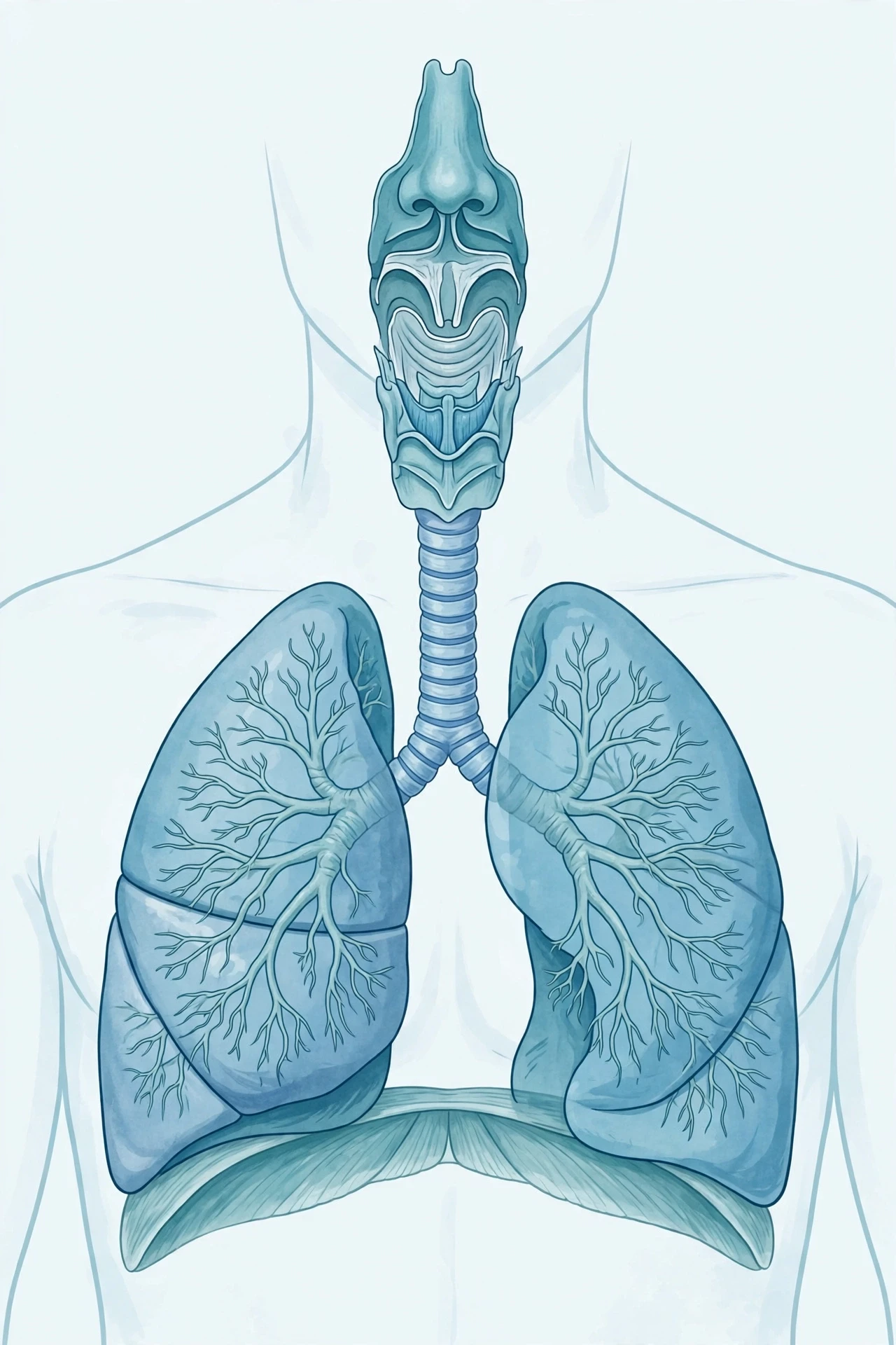
Anatomists split the respiratory system into two parts. The upper respiratory tract — nose, mouth, nasal cavity, sinuses, pharynx (throat), and larynx (voice box) — sits outside the chest and handles intake and filtering. The lower respiratory tract — trachea (windpipe), bronchi, bronchioles, and lungs — sits inside the chest and handles the actual exchange. The Cleveland Clinic's overview of the respiratory system lays out these parts clearly, and it is a good companion to this article if you want a second, clinical voice on the anatomy.
3. The Journey of a Breath: Nose to Alveoli
3.1 The nose: more than a doorway
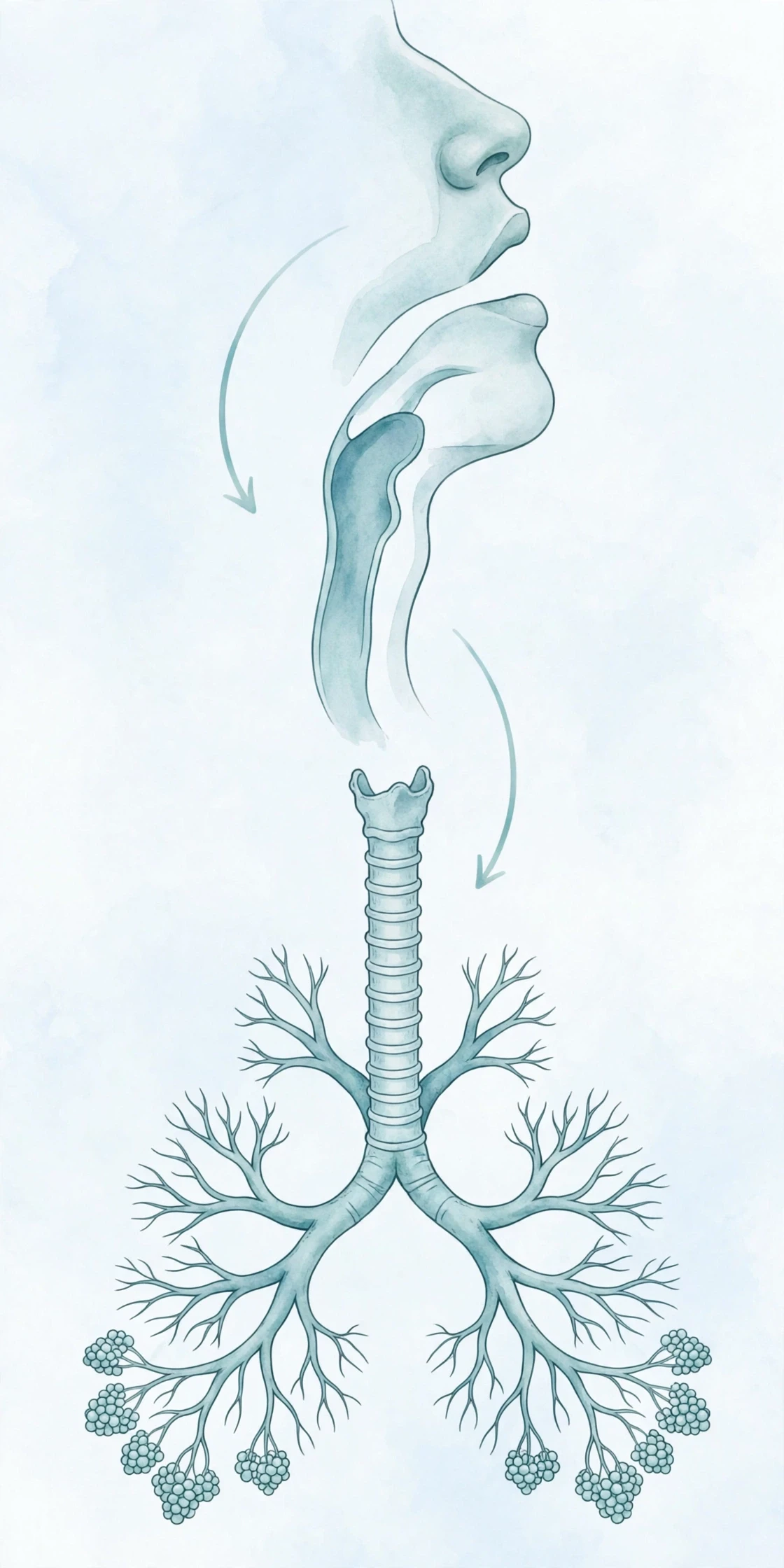
A breath usually starts at the nose, and the nose earns its keep. Inside each nostril, tiny hairs catch large dust particles, while a sticky layer of mucus traps smaller ones. The nasal passages are lined with blood vessels that warm the air to body temperature, and glands add moisture to it. Cold, dry winter air arrives at your lungs warm and damp — a small mercy your airways repay by staying healthy.
Mouth breathing skips most of this treatment. That is why breathing through your mouth on a freezing morning can leave your throat raw: the air arrives cold and dry, and the lower airways have to cope with it unprepared. Your nose is not just a doorway. It is the system's first filter and first heater.
3.2 Down the throat, past the voice box
From the nose or mouth, air enters the pharynx, the shared hallway where the paths for air and food cross. This is the spot where the body has to make a choice, many times a day, between breathing and swallowing — and it almost always gets it right. A flap of cartilage called the epiglottis folds down over the windpipe each time you swallow, steering food toward the oesophagus instead of the lungs. When it misfires, you cough — violently, and for good reason.
Below the pharynx sits the larynx, the voice box. Two folds of tissue inside it — the vocal cords — vibrate as air passes over them, and the muscles around them stretch and relax to change pitch. Every word you have ever spoken rode out of your body on exhaled air. Speech is, quite literally, breathing put to work.
3.3 The trachea and the branching tree
The trachea, or windpipe, is a tube about as wide as your thumb, held open by rings of cartilage so it cannot collapse when you breathe in hard. I remember the first time I held a preserved trachea in an anatomy lab: those cartilage rings felt like the ridges of a vacuum-cleaner hose, springy and stubborn. It was a small, strange thrill to touch the engineering directly.
At the bottom of the trachea, the airway splits into two bronchi, one for each lung. Each bronchus then branches again and again — into smaller bronchi, then bronchioles — in a pattern anatomists call the bronchial tree. It looks like an upside-down tree, and it works like one too: a single trunk dividing into millions of twigs, so that air can reach every corner of the lung. The National Heart, Lung, and Blood Institute describes this branching system in its guide to how the lungs work, and the OpenStax anatomy text gives a full tour of the organs and structures of the respiratory system.
3.4 The alveoli: where the magic happens
At the end of each bronchiole sits a cluster of alveoli — tiny, grape-like air sacs with walls so thin that gases can pass straight through them. Each lung holds hundreds of millions of alveoli, and if you flattened them all out, their combined surface would cover roughly half a tennis court. That enormous surface is the point: gas exchange needs area, and the alveoli provide it in a package small enough to fit inside your chest.
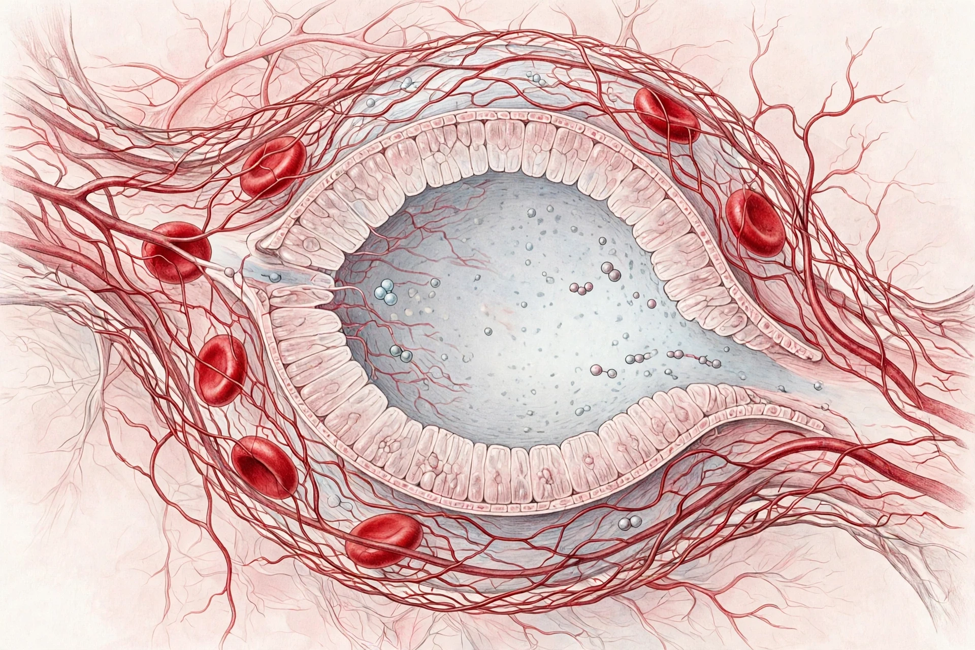
Each alveolus is wrapped in a net of capillaries, the smallest blood vessels in the body. The wall between the air inside the alveolus and the blood inside the capillary is about one cell thick on each side. Oxygen crosses it in one direction. Carbon dioxide crosses it in the other. There is no pump, no valve, no moving part at this stage — just physics, working at a scale you could not see without a microscope.
4. How Breathing Works: The Diaphragm and the Physics of Air
4.1 Your lungs cannot move themselves
Here is a fact that surprises most people the first time they hear it: your lungs have almost no muscle of their own. They cannot inflate themselves. They are soft, spongy, elastic bags that sit inside a sealed chest cavity, and they only expand because the chest cavity around them expands first — and drags them open with it.
Think of a balloon inside a sealed jar. If you pull the bottom of the jar downward, the space inside the jar grows, the pressure inside it falls, and the balloon swells as air rushes in to fill it. That is, almost exactly, how your chest works. The jar is your rib cage. The moving bottom is your diaphragm.
4.2 Inhalation: the diaphragm pulls down
The diaphragm, the respiratory system's main muscle, is a dome-shaped sheet of muscle that separates your chest from your abdomen. When you breathe in, it contracts and flattens, moving downward like a piston. At the same time, the muscles between your ribs pull the rib cage upward and outward. The chest cavity gets bigger. Pressure inside it falls below the pressure of the outside air. And air — obedient to physics — flows down the pressure gradient, through your nose and airways, and into your lungs.
Quiet breathing uses the diaphragm for most of the work. It is a remarkably efficient arrangement: one muscle, contracting a few centimetres, moves half a litre of air with every breath.
4.3 Exhalation: mostly letting go
Breathing out, at rest, is the opposite of effort. The diaphragm relaxes and domes back upward. The rib muscles relax. The elastic lungs, stretched by the breath in, recoil like a released rubber band. The chest cavity shrinks, pressure inside rises above the outside air, and air flows out the way it came in. No muscle needs to push it.
When you need to breathe out hard — blowing up a balloon, coughing, singing a long note — extra muscles join in: the abdominal muscles squeeze the belly upward, and the inner rib muscles pull the ribs down. But at rest, exhalation is a letting-go, not a push.
4.4 Inhalation versus exhalation at a glance
Feature | Inhalation (breathing in) | Exhalation (breathing out) |
|---|---|---|
Diaphragm | Contracts and flattens, moves down | Relaxes and domes back up |
Rib cage | Rib muscles lift ribs up and out | Ribs fall back down as muscles relax |
Chest volume | Increases | Decreases |
Pressure in lungs | Falls below outside air pressure | Rises above outside air pressure |
Airflow | Air flows into the lungs | Air flows out of the lungs |
Effort at rest | Active — muscles work | Passive — elastic recoil does the work |
Main muscles | Diaphragm, external intercostals | None at rest; abdominals when forced |
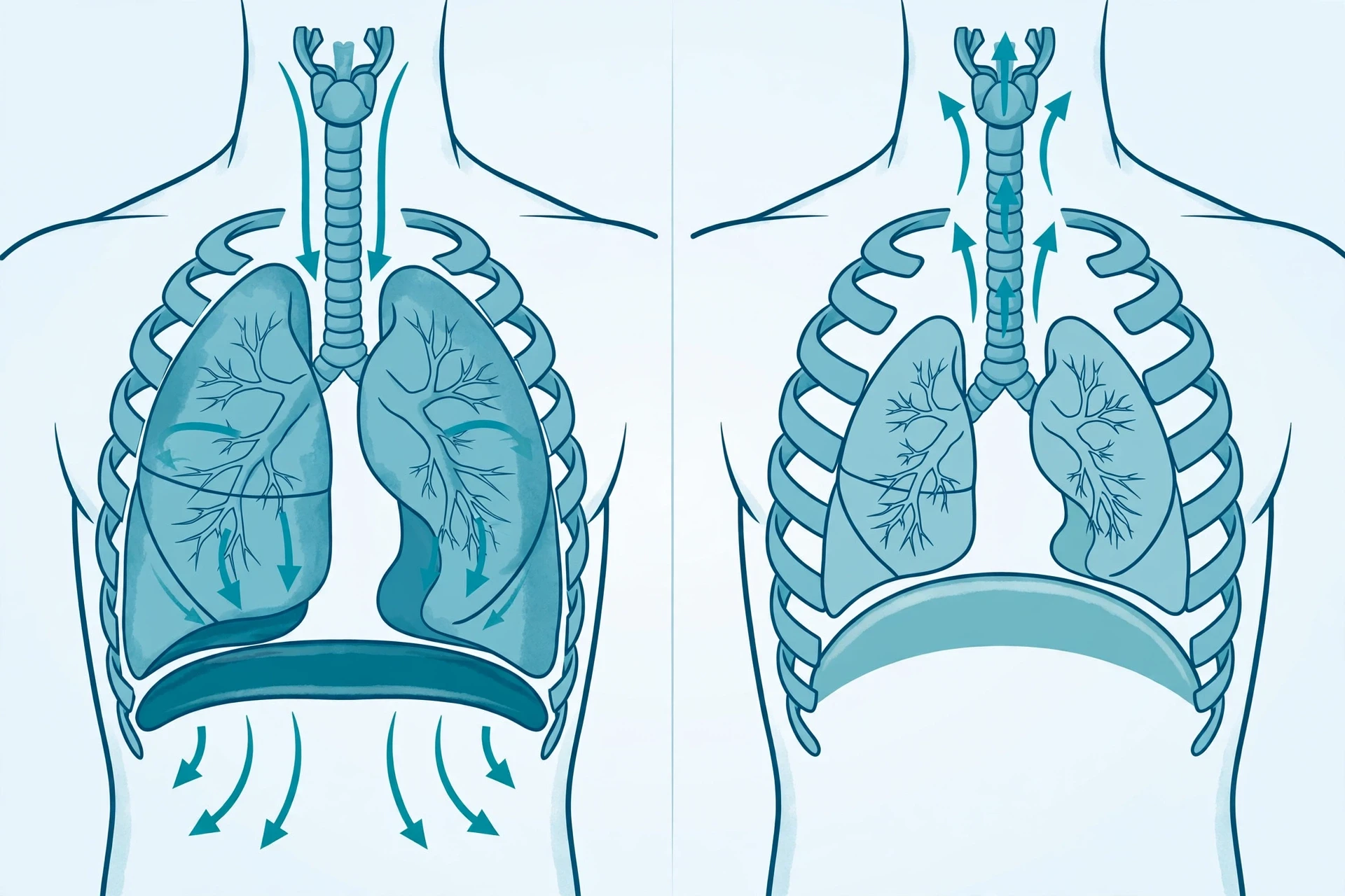
Breathing looks like something the lungs do. It is really something the chest does to the lungs — and the diaphragm is the muscle that makes it happen.
4.5 Why you sigh, yawn, and hiccup
Every so often, your breathing pattern breaks its own rhythm, and each break has a reason. A sigh — a slow, deep breath taken every few minutes — re-inflates alveoli that have partly collapsed during shallow breathing. A yawn is a long, deep inhalation that may help cool the brain and stretch the jaw and chest muscles; researchers still argue about its exact purpose, and that honest uncertainty is part of what makes it interesting. A hiccup is a sudden, involuntary spasm of the diaphragm, followed by the vocal cords snapping shut — the familiar "hic" — and it usually fades on its own when the diaphragm settles.
None of these are faults. They are the respiratory system tuning itself.
5. Gas Exchange: Oxygen In, Carbon Dioxide Out
5.1 Diffusion: the quiet engine
Gas exchange in the respiratory system runs on diffusion — the tendency of molecules to move from where they are crowded to where they are scarce. In the alveoli, oxygen is plentiful in the inhaled air and scarce in the blood arriving from the body. So oxygen drifts across the thin alveolar wall into the capillaries. Carbon dioxide is plentiful in that same blood and scarce in the fresh air, so it drifts the other way, into the alveolus, ready to be breathed out.
No energy is spent on this crossing. The system pays for it in advance, by keeping fresh air flowing in and fresh blood flowing past. As long as both keep moving, diffusion does the rest — silently, continuously, about 20,000 times a day.
5.2 The alveolar-capillary membrane: thinner than paper
The barrier between air and blood is astonishingly thin: the alveolar wall plus the capillary wall together measure roughly a micrometre or two — far thinner than a sheet of paper, and about a fiftieth the width of a human hair. That thinness is what makes gas exchange fast enough to keep up with a sprint, a climb, or a sudden fright. It is also what makes the lungs fragile: fluid, inflammation, or scarring in that wall slows the crossing, and the whole body feels it within seconds.
A soapy substance called surfactant, made by cells in the alveoli, keeps those tiny sacs from collapsing. Without it, the surface tension of the thin fluid lining each alveolus would pull the sac shut every time you breathed out — like a wet plastic bag sealing itself. Premature babies, whose lungs have not yet made enough surfactant, struggle to keep their alveoli open, and that single missing substance was once a leading cause of death in newborns. Modern medicine now gives surfactant directly, and it is one of the quiet triumphs of respiratory care.
5.3 External and internal respiration
Physiologists split gas exchange in the respiratory system into two stages. External respiration is the swap in the lungs: oxygen into the blood, carbon dioxide out of it. Internal respiration is the swap at the tissues: oxygen out of the blood and into the cells, carbon dioxide out of the cells and into the blood. The OpenStax chapter on gas exchange walks through both stages in detail, including the partial-pressure gradients that drive them.
The two stages are mirror images, driven by the same rule — gases move from high concentration to low — and the blood is the courier between them.
6. Oxygen's Journey: From Lung to Cell and Back
6.1 Haemoglobin: the oxygen taxi fleet
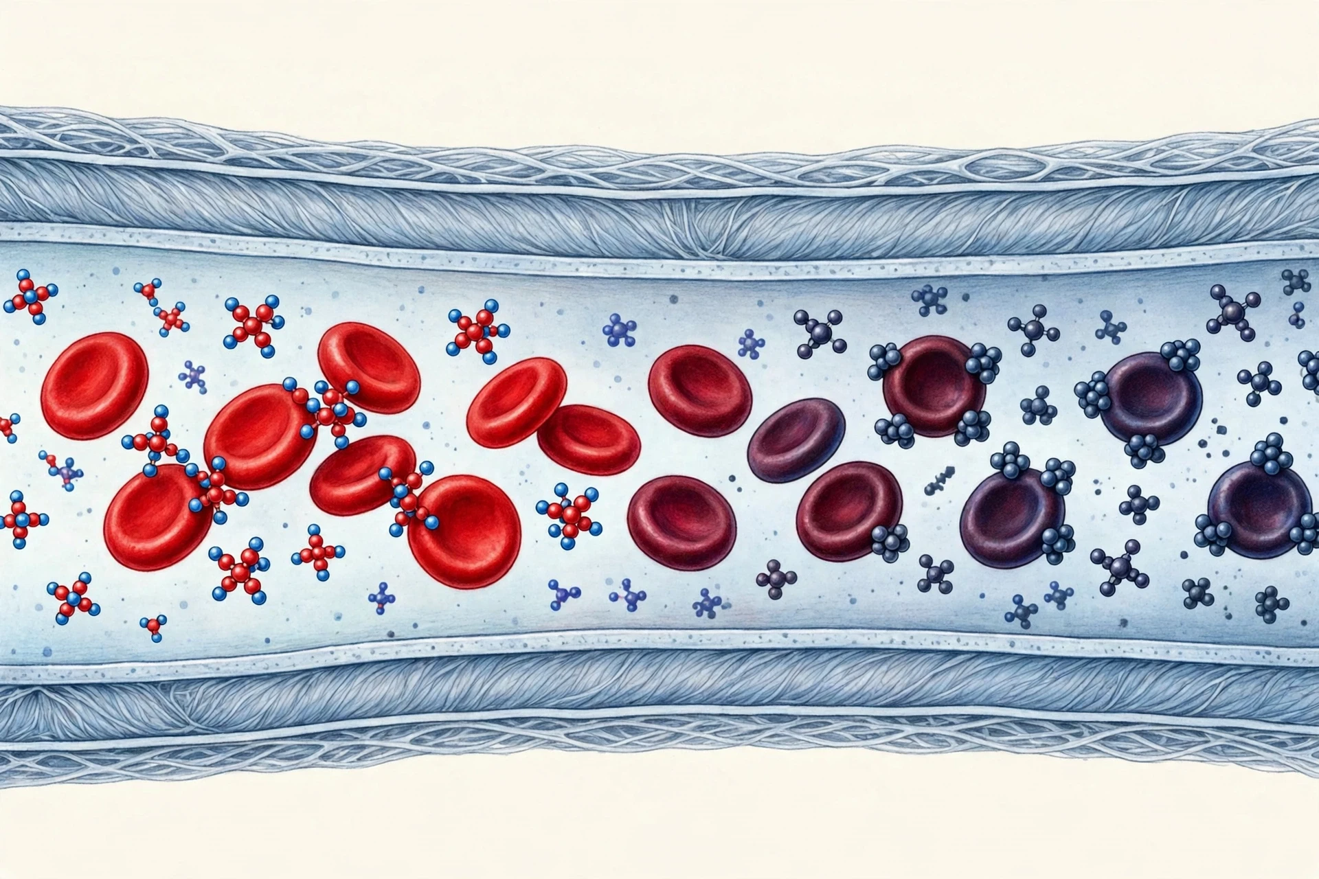
Oxygen does not dissolve well in blood plasma. Left to dissolve on its own, your blood could carry only a tiny fraction of what your body needs. The solution is haemoglobin, the iron-rich protein packed inside red blood cells. Each haemoglobin molecule can bind four oxygen molecules, and each red blood cell carries hundreds of millions of haemoglobin molecules. The result is a fleet of taxis so large that blood can carry roughly seventy times more oxygen than plasma alone could manage.
In the lungs, where oxygen is plentiful, haemoglobin loads up. In the tissues, where oxygen is scarce and carbon dioxide is rising, it lets go. The release is helped along by the tissues themselves: working muscles produce carbon dioxide and acid, and both nudge haemoglobin to unload its cargo exactly where it is needed most. It is a delivery system that responds to demand without any central instruction.
6.2 The round trip through the heart
Oxygen-rich blood leaves the lungs and flows to the left side of the heart, which pumps it out to the whole body through the arteries. At the tissues, capillaries thinner than a hair let oxygen slip out to the cells and carbon dioxide slip in. The now oxygen-poor blood returns through the veins to the right side of the heart, which pumps it back to the lungs for another load. The loop never stops, and the respiratory system sits at its centre like a refuelling station.
I once watched this loop on a monitor during a hospital visit with a relative — the pulse oximeter clip on a fingertip, the number hovering near 98 percent. That number is the share of haemoglobin carrying oxygen, and watching it hold steady while a person simply lay there breathing was oddly moving. The whole system, invisible, doing its sums.
6.3 Carbon dioxide: the return cargo
Carbon dioxide travels home in three forms: a small amount dissolved in the plasma, a share bound to haemoglobin, and the largest share converted into bicarbonate ions in the blood. At the lungs, the bicarbonate converts back into carbon dioxide, which diffuses into the alveoli and leaves with the next breath out. The body treats carbon dioxide as waste, but it is waste with a second job: its level in the blood is the main signal your brain uses to decide how fast to breathe, as the next section explains.
6.4 What cells do with the oxygen
Inside each cell, oxygen feeds cellular respiration — the controlled burning of glucose that releases the energy cells run on. The waste from that burning is carbon dioxide and water. So the breath you just took is, in a real sense, the fuel delivery for every heartbeat, every thought, and every muscle twitch you will make in the next few seconds. The ecosystems article shows the same oxygen–carbon dioxide swap running at planetary scale, in the air, the forests, and the oceans that produced the oxygen you are breathing now.
7. Who Controls Your Breathing? The Brain's Quiet Autopilot
7.1 The brainstem: a control room that never sleeps
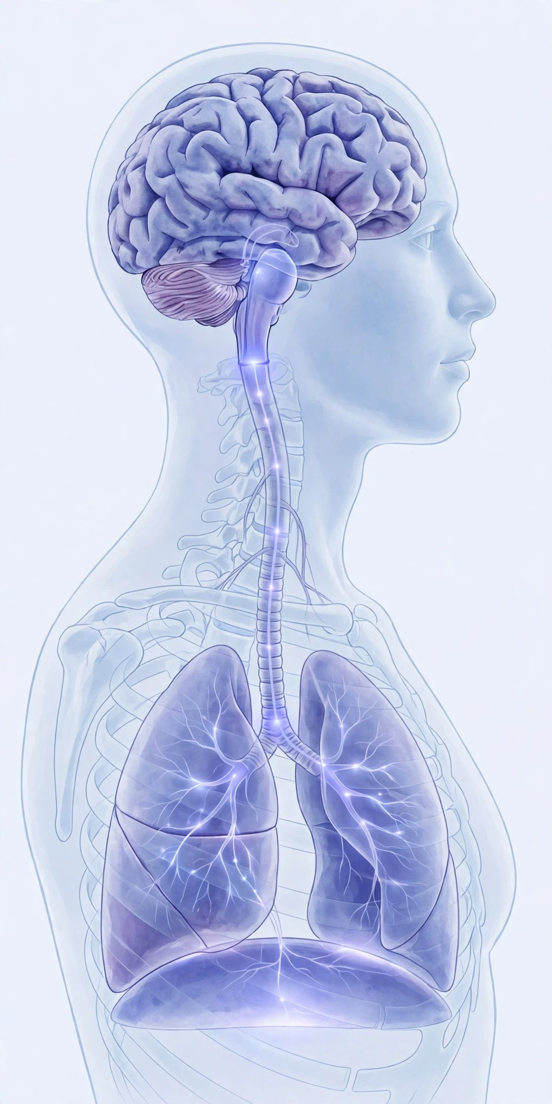
You can hold your breath on purpose, and you can take a deep breath on purpose — but you cannot forget to breathe. The reason sits at the base of your brain, in the brainstem: clusters of nerve cells in the medulla and pons that generate the basic rhythm of breathing, automatically, from birth to death. They send signals down to the diaphragm and rib muscles about twelve to twenty times a minute at rest, and they adjust the rate and depth the moment conditions change.
Our guide to human brain anatomy and functions maps the brainstem and its neighbours in detail; the breathing centres live in the lowest, oldest part of that map, which is fitting — breathing is older than thought.
7.2 Carbon dioxide, not oxygen, is the main alarm
Most people assume the brain watches oxygen levels and speeds up breathing when oxygen runs low. The truth is almost the opposite. The respiratory system's main sensor watches carbon dioxide. Special receptors in the brainstem and in the large arteries detect rising carbon dioxide — which makes the blood slightly more acidic — and they order faster, deeper breathing to blow the excess off.
That is why holding your breath becomes unbearable long before your oxygen runs dangerously low: it is the rising carbon dioxide, not the falling oxygen, that triggers the desperate urge to breathe. And it is why breathing pure oxygen can be dangerous for some people with chronic lung disease — it can blunt the backup oxygen sensors and, in rare cases, slow the drive to breathe. The body has a hierarchy of alarms, and carbon dioxide sits at the top.
7.3 When you take over — and when you cannot
Conscious control sits on top of the autopilot. You can speak, sing, hold your breath underwater, or slow your breathing to calm yourself — all by overriding the brainstem for a while. But the override has limits. Hold your breath long enough and the autopilot wins: carbon dioxide climbs, the urge becomes overwhelming, and breathing resumes whether you want it to or not. Freedivers train for years to stretch that limit, and even they are negotiating with the same alarm system.
Sleep hands control back to the autopilot entirely. That is why breathing disorders that show up at night — like sleep apnoea, where the airway collapses repeatedly during sleep — can go unnoticed for years while they quietly strain the heart. Poor sleep and breathing problems feed each other, which is one reason disrupted breathing shows up so often alongside the sleep troubles described in our article on chronic insomnia and the brain.
8. How Much Air Do Your Lungs Hold? Volumes and Capacities
8.1 The breaths you take, measured
A normal, quiet breath moves about half a litre of air in and out. That is your tidal volume — the everyday unit of breathing. But your lungs hold much more than that, and the extra volumes have names that doctors use to test lung health:
Inspiratory reserve volume: the extra air you can pull in with a maximal deep breath, beyond a normal one.
Expiratory reserve volume: the extra air you can force out after a normal breath out.
Residual volume: the air that stays in your lungs no matter how hard you breathe out — about a litre or more — which keeps the alveoli from collapsing entirely.
Vital capacity: the total you can breathe out after the deepest possible breath in — tidal volume plus both reserves. It is the headline number of a spirometry test, and it shrinks with age and with lung disease.
Add the residual volume to the vital capacity and you get the total lung capacity: everything the lungs can hold, roughly six litres in an average adult man and a little less in an average adult woman. Height, sex, age, fitness, and smoking history all shift these numbers, which is why lung-function tests always compare you with people like you.
8.2 Spirometry: the test that listens to your breath
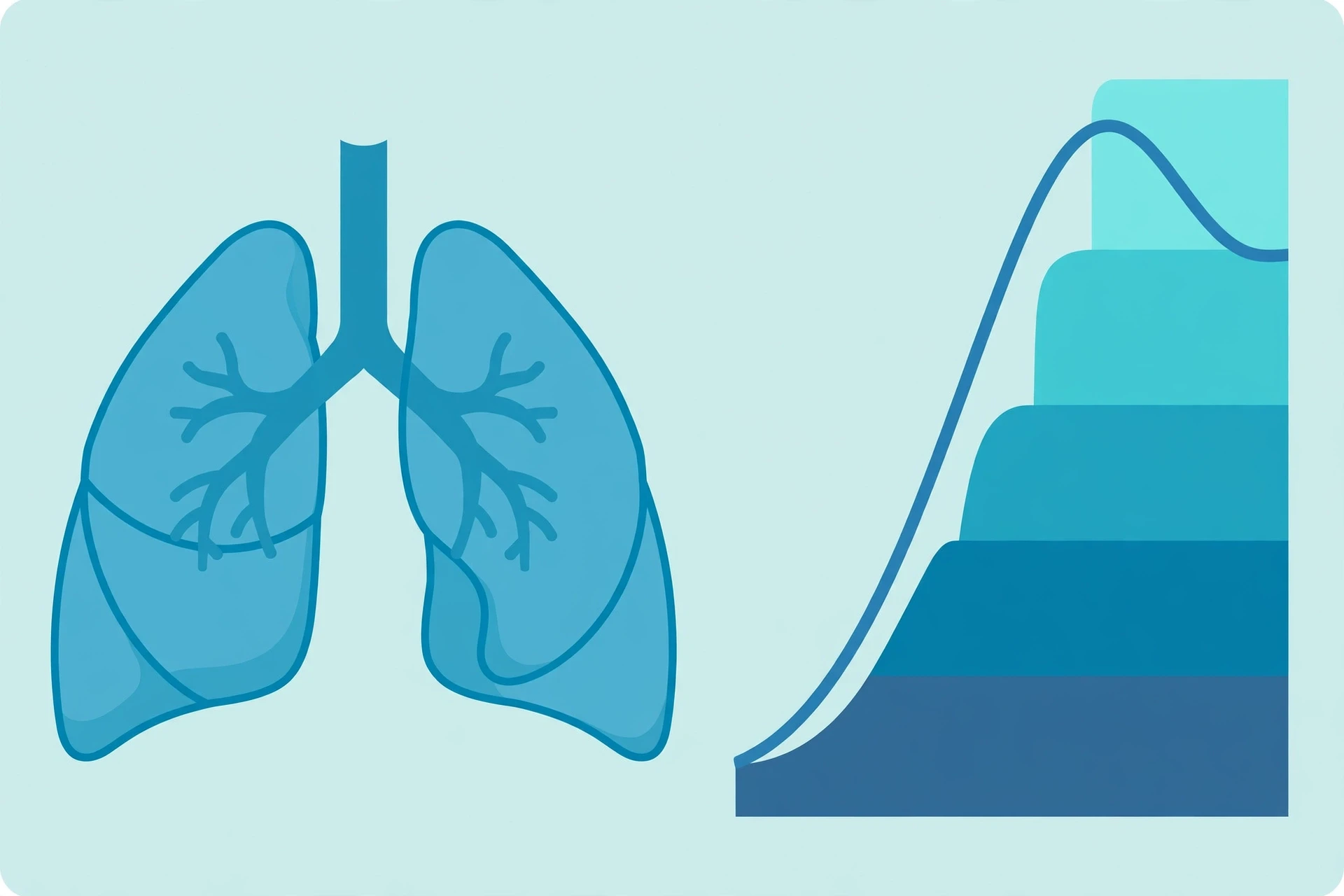
A spirometer is a simple device: you breathe into a tube as hard and as fast as you can, and it records how much air you move and how quickly. The trace it draws — a rising and falling wave — tells a trained reader a great deal. In asthma, the air comes out slowly because narrowed airways resist the flow. In fibrosis, where lung tissue has stiffened and scarred, the total volume shrinks. One test, two very different stories, read from the shape of a breath.
I took a spirometry test once during a university health screening, and I remember the technician's instruction — "blast it out, keep going, keep going" — and the odd feeling of emptying my lungs further than I thought possible. The printout looked like a small mountain range. It was the first time I had seen my own breathing as data.
9. The Lungs' Defence System
9.1 Filters, mucus, and the escalator
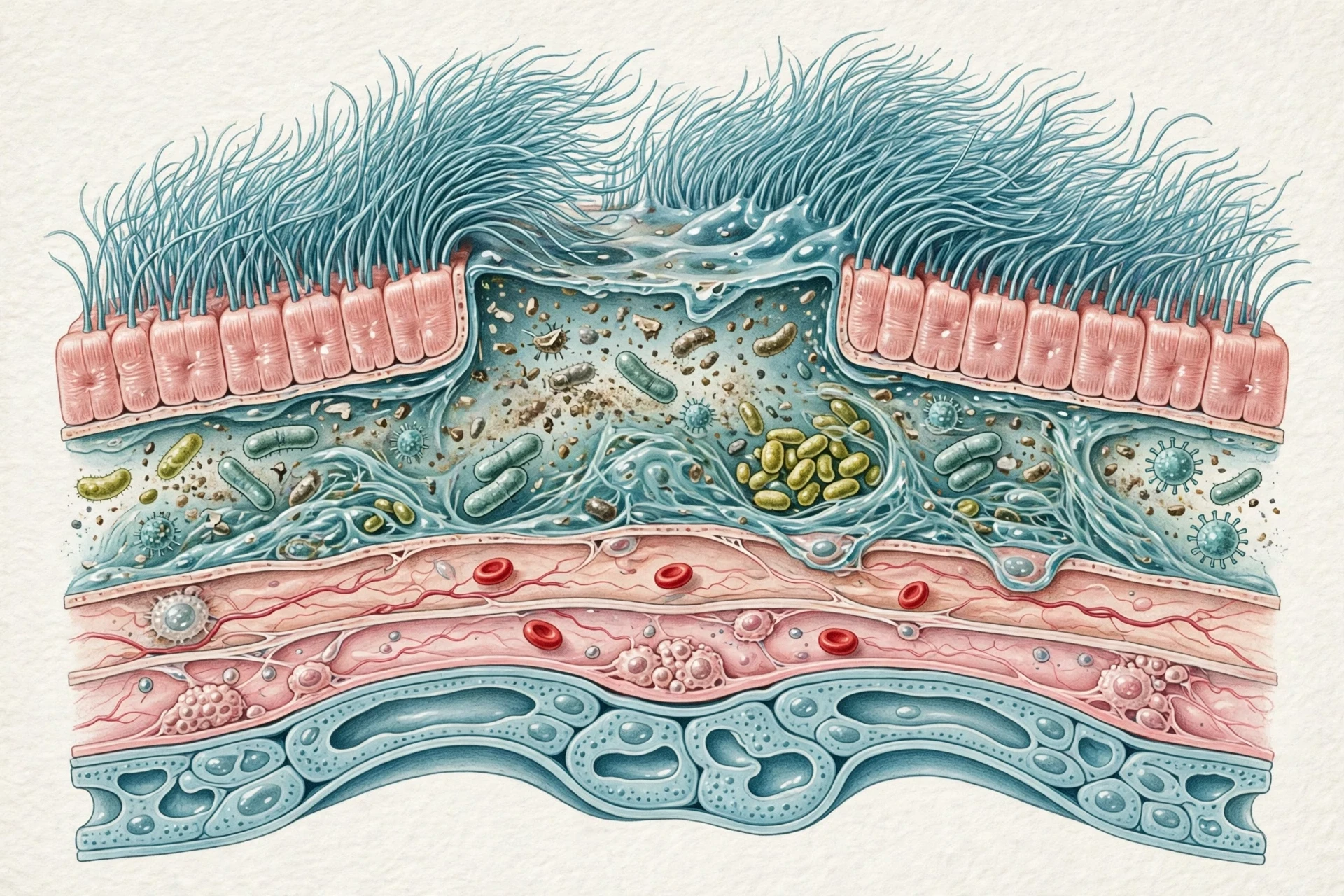
Every breath carries more than air: dust, pollen, smoke, bacteria, and viruses all ride in. The respiratory system meets them with layered defences. The nose filters the largest particles. Sticky mucus lining the airways traps the rest. And cilia — microscopic hair-like structures on the airway cells — beat in coordinated waves, sweeping the mucus and its trapped cargo upward toward the throat, where it is swallowed or coughed out. Physiologists call this the mucociliary escalator, and it runs all day, every day, moving a thin sheet of mucus at roughly a centimetre a minute.
Coughing and sneezing are the emergency version of the same system: a sudden, forceful blast of air that clears an airway faster than the escalator ever could. A sneeze can drive air out at speeds that rival a motorway car — which is why covering your mouth matters.
9.2 The immune system joins in
When germs get past the physical defences, the immune system takes over. Immune cells patrol the alveoli, engulfing bacteria and debris; antibodies and inflammation wall off infections; and the lymph nodes near the lungs act as checkpoints. The respiratory and immune systems are partners, not neighbours, and a respiratory infection is really a story about both of them. Our article on how the human immune system works explains the defences that step in when the lungs' own barriers are breached.
9.3 When defences are overwhelmed
Smoke is the great enemy of the respiratory system's mucociliary escalator: it paralyses the cilia, so mucus pools and infections take hold more easily. That is a large part of why smokers cough in the morning — the cilia recover overnight and spend the morning clearing the backlog. Air pollution does similar damage on a smaller scale, day after day. The weather itself plays a part too: cold, dry air and sudden temperature swings can irritate airways, and the meteorology behind air quality, humidity, and pollution episodes shapes how much your lungs have to defend against on any given day.
10. Common Misconceptions About Breathing
The respiratory system attracts its share of myths, and most of them sound sensible until you look closely.
10.1 "We only use a small part of our lungs"
Most people assume they use only a fraction of their lung capacity, and that the rest sits idle. What the data actually show is subtler: at rest you use a small fraction, because a small fraction is all you need. The reserve is not wasted space — it is the margin that lets you sprint, climb stairs, sing, or recover from illness. Using your full capacity all the time would be like driving everywhere in first gear.
10.2 "Deep breathing is always better"
Slow, deep breathing can calm the nervous system and help with stress — that much is well supported. But deliberately over-breathing, or hyperventilating, blows off too much carbon dioxide, which makes the blood more alkaline and can cause dizziness, tingling, and even fainting. The goal is not maximum air. It is the right amount of air, matched to what the body is doing. Your brainstem is usually better at that maths than your conscious mind.
10.3 "Holding your breath trains your lungs"
Holding your breath trains your tolerance for carbon dioxide, not your lung capacity. Lung size is mostly set by your body size, your genes, and your history; no breathing exercise will grow new alveoli in an adult. What exercise does improve is the efficiency of the whole system — stronger breathing muscles, better oxygen delivery, and a heart that pumps more blood with each beat.
10.4 "Coughing is always a sign of illness"
A cough is a defence, not a disease. Healthy people cough to clear mucus, dust, or a crumb that went down the wrong way. It becomes a warning sign when it lasts for weeks, brings up blood, or comes with fever, weight loss, or breathlessness — then it deserves a doctor's attention, not a cough syrup and a shrug.
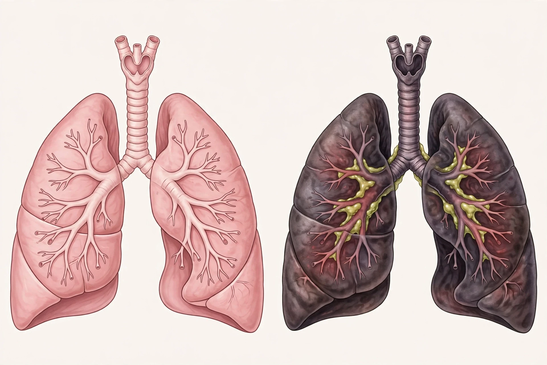
11. When Breathing Goes Wrong
The respiratory system is sturdy, but it is not indestructible. When any part of it fails — the airways, the alveoli, the muscles, or the control centre — the whole body feels it quickly.
11.1 Asthma: airways that overreact
In asthma, the airways are twitchy: cold air, pollen, exercise, or a cold can trigger inflammation and tightening of the muscles around the bronchi. The airways narrow, air gets trapped, and breathing out becomes hard work. The wheeze of asthma is the sound of air squeezing through a narrowed tube. Inhalers work by relaxing those muscles or calming the inflammation — two different tools for two different parts of the problem. The NHLBI's pages on asthma explain the condition, its triggers, and its treatment in plain language.
11.2 COPD: slow damage, heavy cost
Chronic obstructive pulmonary disease (COPD) — mostly chronic bronchitis and emphysema — is usually the result of years of smoking or long exposure to polluted air. In emphysema, the walls between alveoli break down, so the lungs lose surface area and elastic recoil; air gets trapped, and breathing out becomes a struggle. In chronic bronchitis, the airways make too much mucus and stay inflamed. COPD develops slowly and cannot be reversed, which is why stopping smoking, even late, still matters: it slows the decline.
11.3 Pneumonia and infections
Pneumonia is an infection that fills the alveoli with fluid and pus, blocking gas exchange at exactly the place it needs to happen. Breathing becomes fast and shallow as the body tries to compensate, and oxygen levels can fall dangerously. It is the reason a chest infection in an older person is treated with urgency: the margin for error shrinks when the lungs are already working harder. MedlinePlus's overview of pneumonia and lung health is a reliable starting point for readers who want the clinical picture.
11.4 Sleep apnoea and the night shift
In obstructive sleep apnoea, the muscles of the throat relax during sleep and the airway collapses, again and again, through the night. Each collapse briefly stops breathing until the brain wakes the person just enough to reopen the airway — often without them remembering it. The result is fragmented sleep, daytime exhaustion, and long-term strain on the heart and blood pressure. It is common, underdiagnosed, and very treatable.
12. Keeping Your Lungs Healthy for Life
12.1 What actually helps
The advice for a healthy respiratory system is unglamorous, and that is the point — it works:
Do not smoke, and avoid secondhand smoke. It is the single largest preventable cause of lung damage, and quitting at any age slows further decline.
Move your body. Regular aerobic exercise strengthens the diaphragm and rib muscles, improves oxygen delivery, and raises the ceiling on what your lungs can do for you.
Watch the air you breathe. Check local air-quality forecasts on high-pollution days, ventilate indoor spaces, and use a mask when dust or fumes are unavoidable.
Get vaccinated. Flu, pneumonia, and COVID-19 vaccines protect the lungs from the infections that damage them most.
Treat breathing problems early. A cough that lasts more than three weeks, wheezing, or breathlessness on mild exertion all deserve a medical check, not a wait-and-see.
12.2 Exercise: training the bellows
Exercise does not enlarge your lungs, but it transforms what they can do. A trained athlete's heart pumps more blood per beat, their muscles extract oxygen more efficiently, and their breathing muscles fatigue more slowly. That is why a fit person can climb the same hill breathing easily while an unfit person gasps: the lungs are similar, but everything around them works better. I learned this the slow way, returning to running after years away — the first weeks were humiliating, the lungs burning on the smallest hill, and then, almost without noticing, the hill stopped hurting. The system adapts. It just asks for patience.
12.3 Ageing lungs
Lungs age like everything else. The chest wall stiffens, the diaphragm weakens slightly, alveoli lose some elasticity, and the cough reflex dulls. Vital capacity falls gradually from early adulthood onward. None of this is a verdict: people who stay active, avoid smoke, and treat infections promptly keep far more of their lung function into old age than people who do not. The decline is real, but its speed is negotiable.
13. What Researchers Are Learning Now
Research on the respiratory system is moving fast, and three directions stand out. First, researchers are mapping the lung's repair systems — the stem-like cells that rebuild alveoli after injury — and asking whether they can be nudged to repair damage from diseases like emphysema, where repair currently fails. Second, imaging is getting sharper: new scanning techniques can show ventilation in different regions of the lung in real time, letting doctors see exactly which parts of a lung are working and which are not. Third, the long shadow of respiratory infections is under study, as scientists track how severe lung infections can leave the airways more sensitive for months afterward, and what helps them recover.
There is also honest disagreement. Researchers still debate how much of asthma's rise in recent decades comes from air pollution, how much from changes in childhood infections and hygiene, and how much from diet and indoor living. The debate matters because the answer decides where prevention money should go. Good science says so when it does not yet know — and on this question, it does not yet know.
The lungs are not just bags of air. They are a meeting place — where the outside world, the blood, and the brain negotiate, breath by breath, for the whole of a life.
14. The Last Breath of This Article
Go back to that held breath from the start. Ten seconds without air, and your whole body noticed. Now think about what happened in those ten seconds: your brainstem kept its rhythm ready, your diaphragm waited for the signal, your blood kept carrying its cargo, and the moment you let go, the system reset itself without a single conscious instruction. You have done that roughly 20,000 times today, and you will do it again tomorrow, and the day after, for as long as you live.
The respiratory system is easy to take for granted precisely because it works so well. It asks for almost nothing — clean air, a little exercise, no smoke — and in return it carries out the most fundamental exchange in your body: the outside world in, the waste out, twenty thousand times a day. Look after it, and it will look after everything else.
Frequently Asked Questions
How does the respiratory system work?
The respiratory system works by moving air into the lungs, swapping oxygen into the blood and carbon dioxide out of it in the alveoli, and then moving the stale air back out. The diaphragm and rib muscles drive the airflow, diffusion handles the gas swap, and the brainstem controls the rhythm automatically.
What are the main parts of the respiratory system?
The main parts are the nose and mouth, pharynx, larynx, trachea, bronchi, bronchioles, lungs, alveoli, and diaphragm. The upper tract (nose to larynx) filters and warms the air; the lower tract (trachea to lungs) carries it to the alveoli, where gas exchange happens.
How do lungs exchange oxygen and carbon dioxide?
In the alveoli, oxygen diffuses across a wall only a couple of cells thick into the surrounding capillaries, where haemoglobin in red blood cells picks it up. Carbon dioxide diffuses the opposite way, from the blood into the alveolus, and leaves the body on the next breath out. The process runs on concentration gradients, not on muscular effort.
What controls breathing?
Breathing is controlled mainly by the brainstem, which sets the basic rhythm and adjusts it in response to carbon dioxide levels in the blood. Rising carbon dioxide is the strongest signal to breathe faster and deeper. You can override the rhythm consciously — to speak, sing, or hold your breath — but only temporarily.
Why do we breathe faster during exercise?
Muscles working hard consume more oxygen and produce more carbon dioxide, so the brainstem orders faster, deeper breathing to match supply to demand. The heart also beats faster, moving blood through the lungs more quickly. This is why breathlessness during exercise is normal, and why fitness makes the same effort feel easier over time.
Can you improve your lung capacity?
You cannot greatly increase the physical size of your lungs as an adult, but you can improve how well the system works: regular aerobic exercise strengthens breathing muscles, improves oxygen delivery, and raises endurance. Avoiding smoke and air pollution protects the capacity you have, and breathing exercises can improve control and efficiency.
Sources
National Heart, Lung, and Blood Institute — How the Lungs Work: https://www.nhlbi.nih.gov/health/lungs
Cleveland Clinic — Respiratory System: Organs, Facts, Anatomy & Function: https://my.clevelandclinic.org/health/articles/21205-respiratory-system
MedlinePlus (U.S. National Library of Medicine) — Lung health and pneumonia: https://medlineplus.gov/ency/article/000052.htm
OpenStax, Anatomy & Physiology 2e — Organs and Structures of the Respiratory System: https://openstax.org/books/anatomy-and-physiology-2e/pages/22-1-organs-and-structures-of-the-respiratory-system
OpenStax, Anatomy & Physiology 2e — Gas Exchange: https://openstax.org/books/anatomy-and-physiology-2e/pages/22-4-gas-exchange
National Heart, Lung, and Blood Institute — Asthma: https://www.nhlbi.nih.gov/health/asthma