
The bread on your breakfast table is changing before you eat it. Enzymes in your saliva are already cutting its starch into sugars while you chew. By the time you swallow, the chemistry of your body has taken over a job that started in a wheat field, in sunlight, in soil. That quiet handover — from a plant's chemistry to your chemistry — is the territory of biochemistry, and it never pauses. It runs while you sleep, while you run, while you read this sentence.
Biochemistry is the study of the chemical processes inside living things. It asks how molecules are built, how they change, how they store and spend energy, and how they carry instructions from one generation to the next. It sits exactly where chemistry meets biology, and it refuses to treat those two subjects as separate. A cell is not a bag of chemicals that happens to be alive. A cell is chemistry organised so precisely that the result is life itself.
I first felt that precision as a student, watching a clear liquid in a test tube turn cloudy the moment an enzyme was added. Nothing dramatic happened — no flash, no smoke — yet a reaction that would have taken hours on its own finished in seconds. The tube looked the same, but I knew something invisible and exact had just happened. That is the feeling biochemistry keeps giving back: ordinary-looking substances doing extraordinary, tightly controlled work.
This article follows that work from the ground up. It starts with what biochemistry is and how the field grew, then moves through the four great families of molecules that build every living thing — carbohydrates, lipids, proteins, and nucleic acids. It explains how DNA stores information, what enzymes do in the body, how cells earn and spend energy, and why water and pH matter so much. It ends with the places biochemistry touches daily life: medicine, food, farming, and the search for new treatments. Along the way, the story keeps returning to one idea: life is chemistry with a memory, a shape, and a purpose.
1. What Is Biochemistry?
1.1 A definition that actually helps
Biochemistry is the branch of science that studies the chemical reactions and molecular structures that make life possible. Chemists study how atoms bond and react. Biologists study how organisms grow, reproduce, and respond to their surroundings. Biochemists study the overlap — the molecules inside a cell, the reactions between them, and the rules that keep those reactions in balance.
That overlap is enormous. Every breath you take, every thought you form, every wound that heals, and every meal you digest is a set of biochemical reactions running in sequence. When you feel hungry, hormones — themselves biochemical messengers — are signalling your brain. When your muscles ache after a run, lactic acid chemistry and repair processes are already at work. When a cut closes, proteins called clotting factors are assembling in a cascade so precise that a single missing step can cause dangerous bleeding.
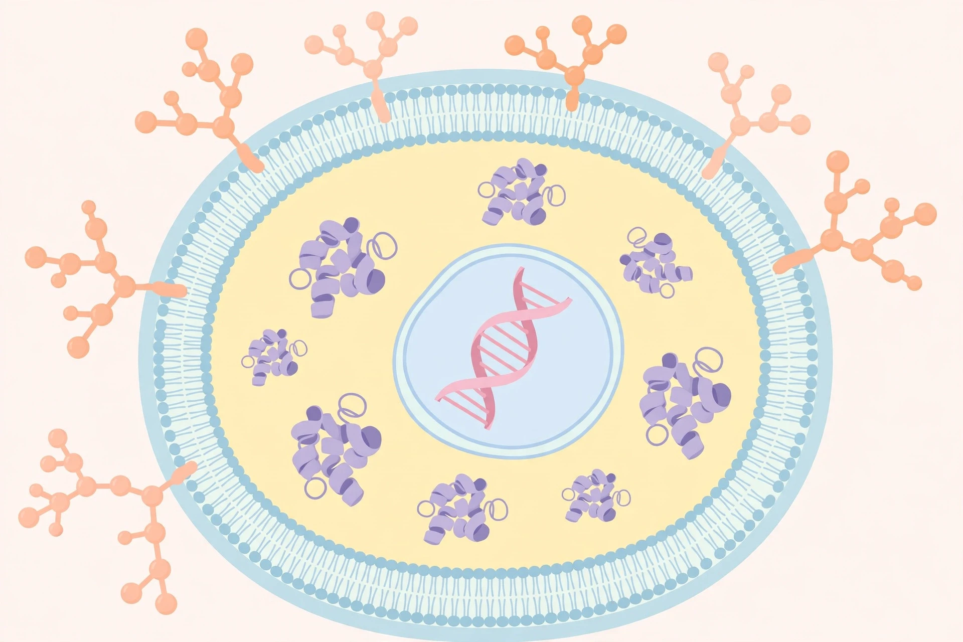
A useful way to picture the field is to think of a cell as a small city. Its streets are membranes, its power stations are mitochondria, its factories are ribosomes, its archives are the nucleus, and its delivery vans are vesicles. Biochemistry is the study of what those buildings are made of, how goods move between them, and who gives the orders. Without the chemistry, the city is only a map. With it, the city works.
1.2 How the field grew
For a long time, many scientists believed living things ran on a special force that chemistry could never explain. That idea had a name — vitalism — and it held that organic substances could only be made by living organisms. It fell apart in 1828, when Friedrich Wöhler made urea, a substance found in urine, from inorganic starting materials in a laboratory. Urea was not a living thing, and no kidney was involved. The wall between living chemistry and laboratory chemistry had a crack in it, and the crack kept widening.
Later in the nineteenth century, Eduard Buchner showed that fermentation — the conversion of sugar to alcohol — could happen in a cell-free extract of yeast. No living yeast cells were needed, only the molecules they contained. That result pointed straight at enzymes, the protein catalysts that drive the reactions of life. In 1926, James Sumner crystallised the enzyme urease and showed it was a protein, proving that a catalyst of life could be isolated, purified, and studied like any other chemical. Each of these steps shrank the mystery and enlarged the map.
The twentieth century then gave biochemistry its most famous structure. In 1953, James Watson and Francis Crick, building on the X-ray work of Rosalind Franklin and Maurice Wilkins, proposed the double-helix structure of DNA. Suddenly the storage of hereditary information had a chemical shape: two strands, paired bases, a code written in four letters. The decades that followed filled in the machinery — how DNA is copied, how its code is read into RNA, and how RNA is translated into protein. That sequence, DNA to RNA to protein, became the central framework of molecular biology, and biochemistry supplied the chemical detail that made it real.
I remember the first time I held a plastic model of the double helix and twisted it in my hands. The model was clumsy and the colours were wrong, but the logic was beautiful. Two strands held together by simple pairing rules could carry the instructions for a whole organism. It felt less like a discovery and more like finding the key to a house I had lived in all my life without ever seeing the lock.
1.3 What biochemists actually study
Modern biochemistry has three broad questions, and every experiment in the field answers one of them in some form. First, what are living things made of — which molecules, in which amounts, arranged in which shapes? Second, how do those molecules change — which reactions transform them, how fast, and in what order? Third, how is all of it controlled — what switches reactions on and off, and what happens when control fails?
The first question leads to structural biochemistry: the shapes of proteins, the packing of DNA, the layout of membranes. The second leads to metabolism: the networks of reactions that build molecules up and break them down. The third leads to regulation and signalling: hormones, second messengers, feedback loops, and gene control. A single disease can touch all three. Diabetes, for example, involves the structure of insulin, the metabolic pathways that handle glucose, and the signalling that tells cells to take glucose in. To understand it, you need all three views at once.
Biochemistry also borrows tools from everywhere. It uses physics to measure molecular shapes with X-rays and magnetic resonance, engineering to build instruments that read DNA letter by letter, and computing to model proteins that are too large to draw by hand. The field is a meeting point, and that is its strength. A question that looks purely medical — why does a drug work in one person and not another — often turns out to be a biochemical question about an enzyme variant, a transport protein, or a metabolic rate.
2. The Four Biomolecules: Life's Building Blocks
2.1 What are the four biomolecules?
Every living thing on Earth — a bacterium, a mushroom, a whale, a wheat plant, a human — is built from the same four families of large molecules, called biomolecules or macromolecules. They are carbohydrates, lipids, proteins, and nucleic acids. Each family has its own building blocks, its own shapes, and its own jobs, yet they work together so closely that none of them makes sense alone. Carbohydrates supply quick energy. Lipids store energy for later and build membranes. Proteins do the work: they catalyse reactions, carry molecules, send signals, and give structure. Nucleic acids store and transmit the instructions for making everything else.
The building blocks are small. Carbohydrates are built from simple sugars. Lipids are built from fatty acids and glycerol. Proteins are built from amino acids. Nucleic acids are built from nucleotides. Life assembles these small units into long chains, then folds, twists, and packs those chains into working shapes. The same principle repeats at every scale: small parts, joined by chemical bonds, folded into a structure whose shape decides its function.
That shape-function link is the single most useful idea in biochemistry. A protein that is folded wrong cannot do its job, even if every amino acid is present in the right order. A DNA strand with a single wrong letter can change a protein and alter a trait. A membrane made of the wrong lipids can leak. Biochemistry is, in large part, the study of how shape is earned and how it is lost.
2.2 The elements of life
Only a handful of elements do almost all the work of life. Carbon, hydrogen, oxygen, and nitrogen make up the great bulk of every organism, with smaller amounts of phosphorus and sulfur, and traces of elements such as calcium, potassium, sodium, magnesium, iron, and zinc. Carbon is the star of the show because it can form four stable bonds, link to itself in chains and rings, and hold the single and double bonds that give molecules their variety. No other element offers that combination, which is why the chemistry of life is, at its core, the chemistry of carbon.
Water is the stage on which all of this happens. Cells are mostly water, and most biochemical reactions take place dissolved in it. Water's polarity — its uneven sharing of electrons — lets it dissolve salts and sugars, form hydrogen bonds, and carry heat. Proteins fold in water the way they do partly because some amino acids hide from water while others face it. Membranes form because lipids refuse to mix with water. Even the double helix of DNA is stabilised by its surroundings. To study biochemistry without water is like studying swimming without a pool.
2.3 A comparison of the four families
The table below gives a quick map of the four biomolecules. The sections that follow take each family in turn, because each one deserves its own story.
Biomolecule | Building block | Main jobs | Everyday examples |
|---|---|---|---|
Carbohydrates | Simple sugars (monosaccharides) | Quick energy, energy storage, structure | Glucose, starch, cellulose, glycogen |
Lipids | Fatty acids and glycerol | Long-term energy storage, membranes, signalling | Fats, oils, cholesterol, phospholipids |
Proteins | Amino acids | Catalysis, transport, structure, signalling, defence | Enzymes, haemoglobin, collagen, antibodies |
Nucleic acids | Nucleotides | Storing and transmitting genetic information | DNA, RNA |
Notice what the table leaves out: the sheer number of different molecules in each family. There are only twenty common amino acids, yet they combine into proteins of astonishing variety. There are only four DNA bases, yet they spell out every genome on Earth. Biochemistry is a science of combinations. A small alphabet, endlessly rearranged, writes a library large enough for all of life.
A cell is not a bag of chemicals that happens to be alive. A cell is chemistry organised so precisely that the result is life itself.
3. Carbohydrates: Fast Fuel and Strong Fibre
3.1 Sugars, starch, and fibre
Carbohydrates are molecules made of carbon, hydrogen, and oxygen, usually in a ratio that gives them their old name — hydrates of carbon. The simplest carbohydrates are monosaccharides: single sugar units such as glucose, fructose, and galactose. Glucose is the most important of these in human biochemistry. It circulates in the blood, enters cells, and is broken down to release energy. Fructose is the sugar in fruit and honey. Galactose is part of the sugar in milk.
Two monosaccharides can join to form a disaccharide. Glucose plus fructose makes sucrose, ordinary table sugar. Glucose plus galactose makes lactose, the sugar in milk. Glucose plus glucose makes maltose, the sugar that appears when starch is broken down. These small sugars taste sweet, dissolve easily in water, and are quick to use — which is why a sweet drink can lift blood sugar within minutes.
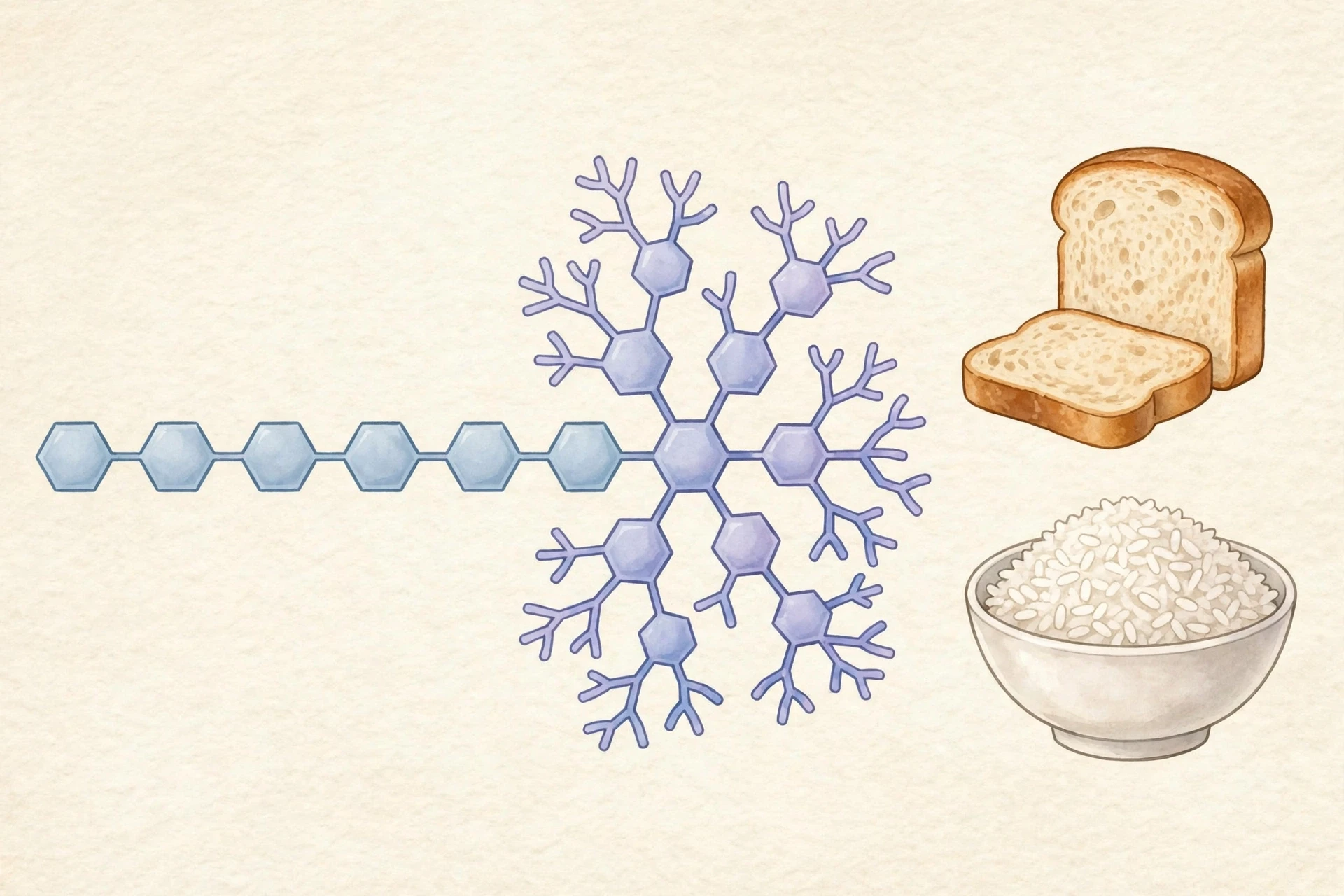
Long chains of monosaccharides are polysaccharides. Starch is the energy store of plants: long chains of glucose packed into grains inside seeds, roots, and tubers. When you eat bread, rice, or potatoes, you are eating plant starch, and your digestive enzymes cut it back into glucose. Glycogen is the animal equivalent, stored mainly in the liver and muscles as a branched, compact reserve that can be mobilised quickly. Cellulose is also a glucose polymer, but its glucose units are linked differently, in straight chains that stack into strong fibres. Plants use cellulose to build cell walls. Humans cannot digest it — we lack the enzyme that breaks its particular bond — so cellulose passes through us as fibre, feeding gut microbes and helping digestion along the way.
3.2 How the body handles glucose
Glucose is so central to human biochemistry that the body guards its blood level within a narrow range. After a meal, blood glucose rises, and the pancreas releases insulin, a protein hormone that tells cells — especially muscle, liver, and fat cells — to take glucose in. Inside the liver, excess glucose is stored as glycogen. When blood glucose falls, between meals or during exercise, the pancreas releases glucagon, which tells the liver to break glycogen down and release glucose back into the blood. This push and pull keeps the brain supplied, because the brain runs almost entirely on glucose and has almost no store of its own.
When that control system fails, the result is diabetes. In type 1 diabetes, the insulin-producing cells of the pancreas are destroyed, so insulin must be supplied from outside. In type 2 diabetes, cells respond poorly to insulin, and the pancreas cannot always keep up. Both forms leave blood glucose too high, and over years that excess damages blood vessels, nerves, kidneys, and eyes. The biochemistry is the same in both cases — glucose handling gone wrong — but the causes differ, which is why the treatments differ too. The story of diabetes is a reminder that biochemistry is not abstract. It is the difference between a body in balance and a body slowly coming apart.
3.3 Carbohydrates beyond energy
It would be a mistake to think of carbohydrates only as fuel. They also serve as identity markers on cell surfaces. Chains of sugars attached to proteins and lipids form a sugary coat on the outside of cells, and other cells read that coat to decide whether a cell belongs, whether it is a friend or a threat. Blood groups are a famous example: the A and B blood types differ in the sugar chains on the surface of red blood cells. A transfusion with the wrong type triggers an immune attack because the recipient's antibodies recognise the foreign sugar pattern.
Carbohydrates also protect and cushion. The slippery mucus that lines your airways and gut is rich in sugar-coated proteins that trap dust and microbes. The tough outer shells of insects and the cell walls of fungi contain chitin, a carbohydrate polymer related to cellulose. Even the joints in your body rely on sugar-rich molecules that hold water and absorb shock. Fuel is the headline, but the supporting roles are just as real.
I once watched a student taste a cracker, chew it slowly, and announce that it had turned sweet. Nothing had been added. The class had simply given the amylase in saliva time to cut starch into maltose, a sugar. It was a small moment, but it made the abstract concrete: chemistry happens in your mouth, in seconds, every time you eat.
4. Lipids: Membranes, Stores, and Signals
4.1 What lipids are
Lipids are a diverse family defined less by a shared structure than by a shared behaviour: they do not dissolve well in water. That single property explains almost everything they do. Fats and oils store energy. Phospholipids build membranes. Steroids act as hormones. Waxes waterproof feathers, leaves, and skin. What unites them is a large share of carbon and hydrogen, with relatively little oxygen, and a tendency to cluster together away from water.
The most familiar lipids are triglycerides: three fatty acids attached to a glycerol backbone. A fatty acid is a long chain of carbon atoms with a carboxyl group at one end. If the chain has no double bonds, it is saturated, and it packs neatly into a solid — think of butter or the fat on a cut of meat. If it has one or more double bonds, it is unsaturated, and the kinks in the chain keep it liquid — think of olive oil. That difference in shape, a bend in a chain, decides whether a fat is solid or liquid at room temperature, and it matters in the body too, because membrane fluidity depends on the mix of saturated and unsaturated fatty acids in the membrane.
4.2 The membrane that makes a cell a cell
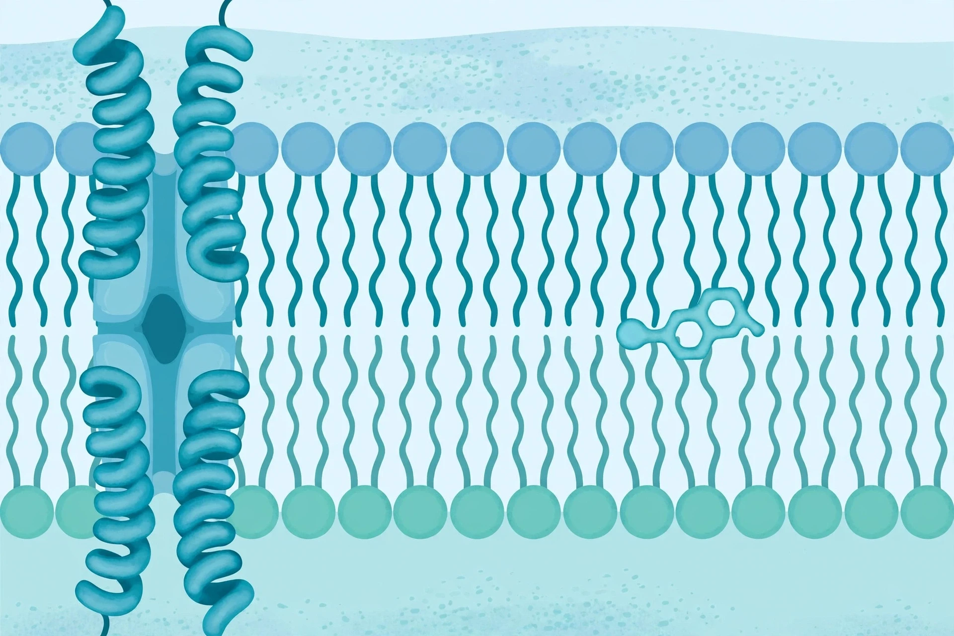
The most important lipid structure in biochemistry is the membrane, and its building block is the phospholipid. A phospholipid has a water-loving head and two water-fearing tails. Put many of them in water and they arrange themselves into a double layer — a bilayer — with the heads facing the water on both sides and the tails hidden in the middle. That bilayer is the boundary of every cell. It keeps the inside in and the outside out, while letting selected substances pass through protein channels and transporters.
Membranes are not rigid walls. They are fluid, like a two-dimensional sea, with proteins drifting in them and lipids moving past one another. Cholesterol, a steroid lipid, sits among the phospholipids and tunes that fluidity, stiffening the membrane when it is warm and keeping it flexible when it is cold. The membrane also carries identity: the sugars and proteins on its outer face tell other cells what this cell is and where it belongs. A cell without a membrane is not a cell at all — it is a spill. The invention of the membrane, early in the history of life, was the invention of the inside.
For a deeper look at how membranes and organelles work together inside the cell, see the Sciory guide to Cell Biology.
4.3 Fats as fuel and as messengers
Gram for gram, fats store more than twice as much energy as carbohydrates. That is why the body keeps its long-term reserves as fat. When food is plentiful, excess energy is converted into triglycerides and stored in fat cells. When food is scarce, those triglycerides are broken down into fatty acids, which travel to tissues and are burned for energy. This system is efficient, but it has a cost: fat stores are almost unlimited, and in a world of constant food, the store keeps growing.
Lipids also carry messages. Steroid hormones — including cortisol, oestrogen, testosterone, and aldosterone — are built from cholesterol and travel through the blood to switch genes on and off in distant cells. Prostaglandins, made from fatty acids, act locally to trigger inflammation, pain, and fever; that is why drugs that block their production can ease pain. Even vitamins can be lipids: vitamins A, D, E, and K are fat-soluble, which is why they are stored in body fat and why a very low-fat diet can leave a person short of them.
Most people assume fat is simply something to avoid, but what the data actually show is more subtle: the type of fat, the amount, and the overall diet matter far more than any single food. Unsaturated fats from plants and fish behave differently in the body from the saturated fats in processed meats and fried foods, and the biochemistry of each — how it is digested, transported, and stored — explains why. A molecule's shape, again, decides its fate.
5. Proteins: The Workers of the Cell
5.1 Amino acids and the peptide bond
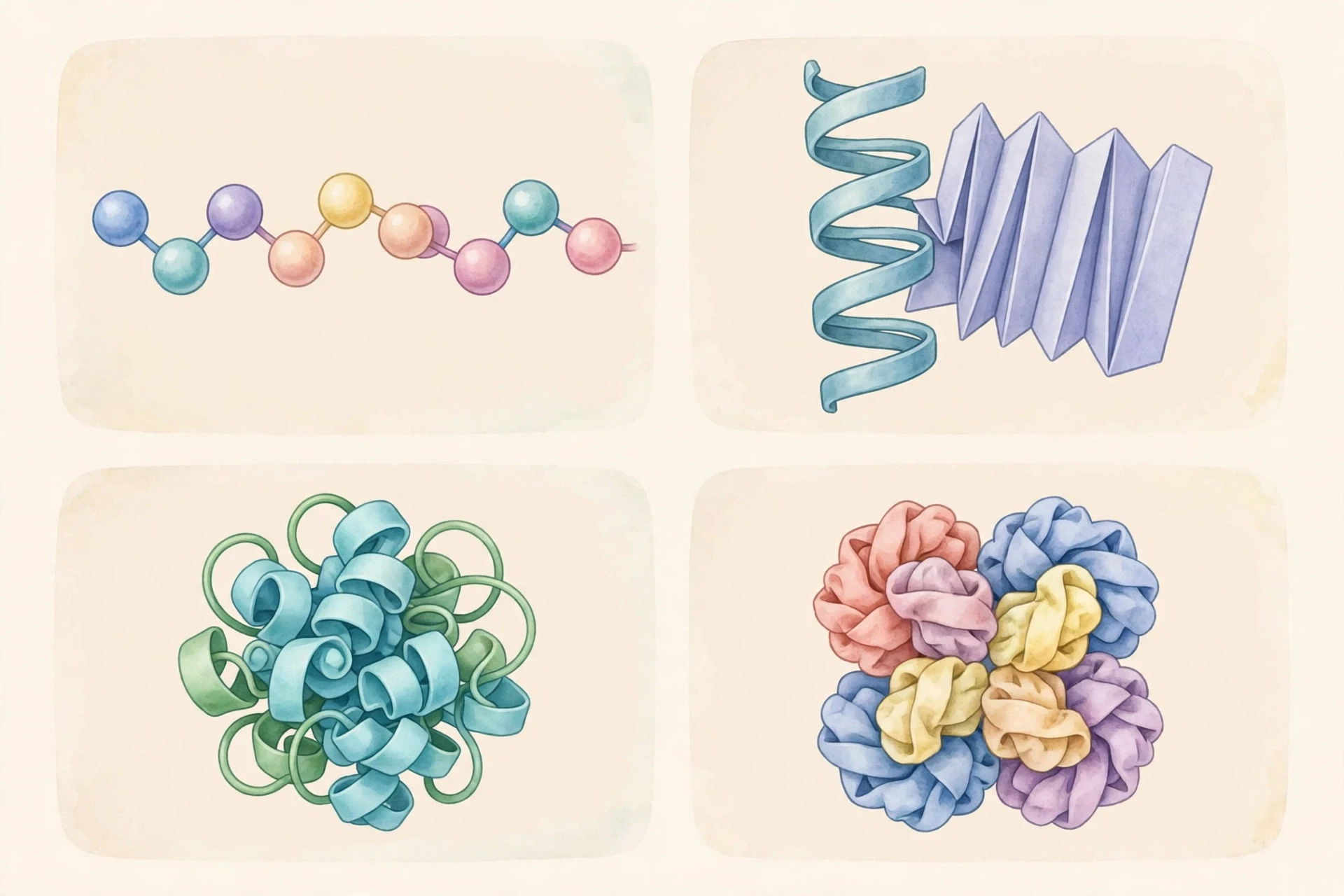
Proteins are the most versatile biomolecules, and they are built from just twenty common amino acids. Each amino acid has the same backbone — a central carbon attached to an amino group, a carboxyl group, a hydrogen atom, and a side chain — and it is the side chain that makes each one different. Some side chains are water-loving, some water-fearing, some positively charged, some negatively charged, some large, some small. The order of amino acids in a chain, called the primary structure, is written in DNA, and it decides everything that follows.
Amino acids join through peptide bonds, formed when the carboxyl group of one meets the amino group of the next, releasing a molecule of water. A short chain is a peptide; a long chain is a polypeptide; a polypeptide folded into a working shape is a protein. The bond itself is simple, but the folding is not. As the chain emerges from the ribosome, it begins to fold, guided by the attractions and repulsions between its side chains and by the water around it. Water-fearing side chains tuck inward, away from the surrounding water. Water-loving side chains face outward. Hydrogen bonds, ionic attractions, and disulfide bridges lock the shape in place.
5.2 Four levels of structure
Biochemists describe protein structure in four levels. The primary structure is the sequence of amino acids. The secondary structure is the local folding — the alpha helix, a tight coil, and the beta sheet, a flat pleated arrangement — held together by hydrogen bonds along the backbone. The tertiary structure is the overall three-dimensional shape of a single polypeptide chain, the result of interactions between side chains that may be far apart in the sequence. The quaternary structure is the assembly of several polypeptide chains into one working unit.
Haemoglobin is the classic example of quaternary structure: four chains, two of each of two types, working together to carry oxygen. When one chain binds oxygen, the shape of the whole molecule shifts slightly, making it easier for the next chain to bind oxygen too. That cooperation, called cooperativity, makes haemoglobin far better at picking up oxygen in the lungs and releasing it in the tissues than a single chain could ever be. It is a beautiful piece of engineering, and it emerges entirely from shape.
5.3 When folding goes wrong
A protein's function depends on its shape, so a protein that folds wrong is a protein that fails. Sometimes the cell catches the error: chaperone proteins help new chains fold correctly, and quality-control systems destroy chains that cannot be fixed. Sometimes the error escapes, and the consequences can be serious. In sickle cell disease, a single amino acid change in haemoglobin makes the protein stick together into fibres that distort red blood cells into a sickle shape, blocking blood flow. In Alzheimer's disease and related conditions, proteins misfold and clump into aggregates that damage brain cells. In cystic fibrosis, a faulty protein fails to reach the cell membrane, so salt and water transport goes wrong.
These examples show why protein folding is one of the most active areas of biochemistry. Predicting how a chain will fold from its sequence alone was a grand challenge for decades, and recent computational tools have made large strides — yet the cell still does it in seconds, at body temperature, without a supercomputer. The gap between what a cell does routinely and what our best tools can match is a useful measure of how much there is still to learn.
5.4 The many jobs of proteins
Proteins do almost every job in the cell. Enzymes catalyse reactions. Transport proteins carry oxygen, iron, and other cargo through the blood. Motor proteins walk along molecular tracks, pulling chromosomes apart during cell division and moving vesicles through the cell. Structural proteins such as collagen give tendons and skin their strength, while keratin builds hair and nails. Antibodies recognise and neutralise invaders. Receptor proteins sit in membranes and receive signals from outside. Hormones such as insulin are themselves proteins that carry messages between organs.
6. Nucleic Acids: How DNA Stores Information
6.1 What are nucleic acids?
Nucleic acids are the information molecules of life. There are two kinds: deoxyribonucleic acid, or DNA, and ribonucleic acid, or RNA. Both are built from nucleotides, and each nucleotide has three parts: a five-carbon sugar, a phosphate group, and a nitrogen-containing base. In DNA the sugar is deoxyribose; in RNA it is ribose. DNA carries four bases — adenine (A), thymine (T), guanine (G), and cytosine (C). RNA also carries four, but uses uracil (U) instead of thymine.
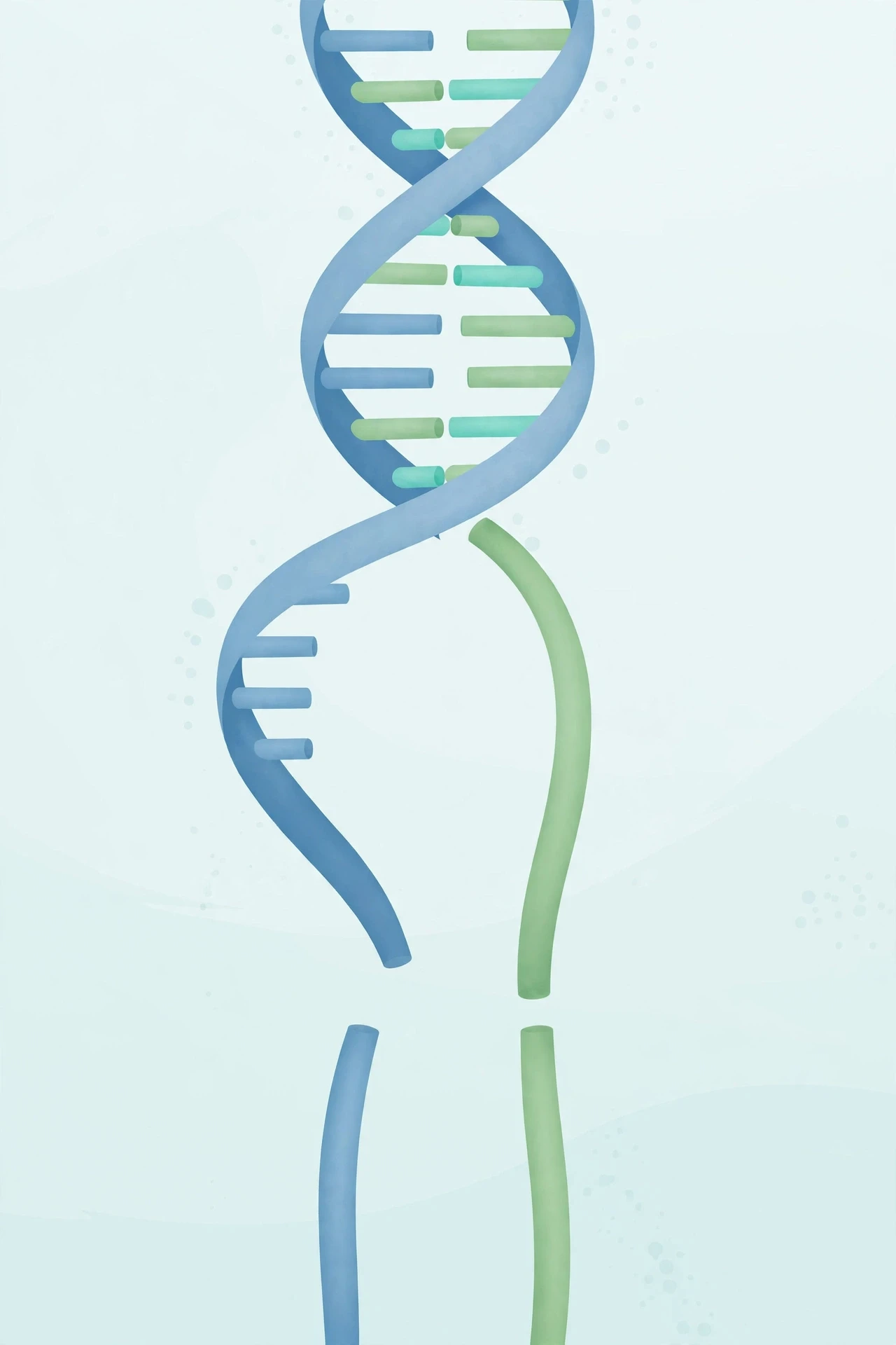
Nucleotides join into long chains through phosphodiester bonds between the sugar of one nucleotide and the phosphate of the next, forming a sugar-phosphate backbone. The bases stick out from that backbone like the teeth of a zip. In DNA, two chains wind around each other in a double helix, and the bases pair up: A always pairs with T, and G always pairs with C, held together by hydrogen bonds. That pairing rule is the key to everything DNA does. It means each strand is a template for the other, and it means the information can be copied accurately.
The structure and copying of DNA are covered in depth in the Sciory article on DNA, and the way genes are read and inherited is explained in the Sciory guide to Genetics.
6.2 How does DNA store information?
DNA stores information the way a book stores a story: in a sequence of symbols. The symbols are the four bases, and the sequence of bases along a gene spells out the sequence of amino acids in a protein. Three bases in a row — a codon — specify one amino acid, or a signal to start or stop. With four bases and three positions, there are sixty-four possible codons, more than enough for twenty amino acids plus start and stop signals, so most amino acids are specified by more than one codon. That redundancy is not waste; it is a buffer against errors.
The amount of information is staggering. The human genome contains around three billion base pairs, spread across forty-six chromosomes in most cells. If the bases were printed as letters, the genome would fill hundreds of thick books. Yet all of it fits inside a nucleus a few millionths of a metre across, because DNA is coiled and packed around proteins called histones into structures called chromosomes. Packing is not just storage — it is control. Genes that are packed tightly are hard to read; genes that are unpacked are available. The cell opens and closes its archive as needed, which is one reason the same genome can produce a skin cell, a liver cell, and a neuron.
6.3 From DNA to RNA to protein
The flow of information in most cells follows a path often called the central dogma: DNA is copied into RNA, and RNA is read to build protein. The copying step is transcription. An enzyme called RNA polymerase reads one strand of DNA and builds a matching strand of messenger RNA, or mRNA, using the same pairing rules — except that RNA uses uracil where DNA uses thymine. The mRNA then travels to a ribosome, a large molecular machine made of RNA and protein, where it is translated: transfer RNA molecules bring amino acids one by one, matching each codon to its amino acid, and the ribosome links them into a growing chain.
Not all RNA is a messenger. Ribosomal RNA forms the core of the ribosome itself. Transfer RNA carries amino acids. Small regulatory RNAs switch genes on and off. The picture that has emerged is richer than the simple one-way flow once imagined: RNA can regulate, catalyse, and carry information in its own right. Some scientists argue that early life ran on RNA alone, before DNA and proteins took over their modern roles — a hypothesis known as the RNA world. It is unproven, but it is a reminder that the molecules we see today are the survivors of a long evolutionary experiment.
6.4 Mutations, repair, and variation
DNA is copied with remarkable accuracy, but it is not perfect. Mistakes happen — a wrong base inserted, a base skipped, a segment duplicated or deleted. Most mistakes are caught and fixed by repair systems that scan the new strand, spot the mismatch, and cut it out. When a mistake survives, it is a mutation. Most mutations are harmless or are repaired in the next round. Some are harmful, causing disease. A few are helpful, and those are the raw material of evolution.
Mutations are also why no two people, except identical twins, have the same genome. The small differences between us — in height, in eye colour, in how we respond to a drug — trace back to variations in DNA sequence. Biochemistry studies those variations at the molecular level: which base changed, which protein changed as a result, and what that change does to a cell. That chain of cause and effect, from a single letter to a whole person, is the heart of medical biochemistry.
7. Enzymes: What Do Enzymes Do in the Body?
7.1 Catalysts that make life fast enough
Enzymes are proteins that speed up chemical reactions without being used up themselves. They are catalysts, and without them life would be too slow to exist. Many of the reactions in a cell would take hours, days, or years on their own at body temperature. With the right enzyme, the same reaction can finish in milliseconds. The difference is not a small tweak; it can be a speed-up of millions of times.
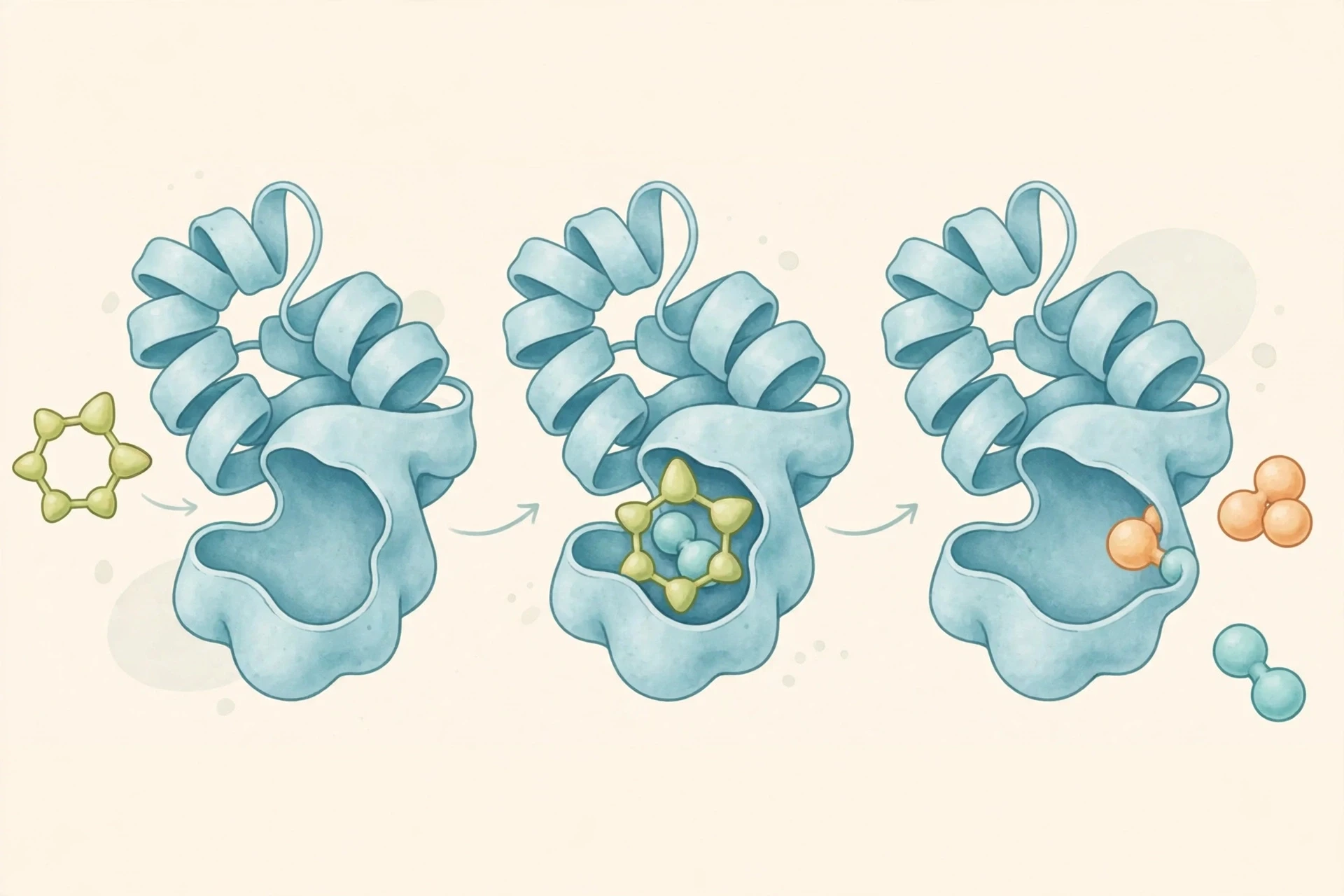
Enzymes work by lowering the activation energy of a reaction — the energy barrier that reactants must cross before they can change into products. They do this by binding the reactants, called substrates, in a pocket called the active site, holding them in exactly the right orientation, and stabilising the in-between state of the reaction. When the reaction is done, the products leave, and the enzyme is free to take the next substrate. A single enzyme molecule can process hundreds or thousands of substrate molecules every second.
The classic picture is the lock and key: the substrate fits the active site the way a key fits a lock. The fuller picture is induced fit: the enzyme changes shape slightly as the substrate binds, closing around it like a hand around a ball. Both pictures capture part of the truth. The active site is specific — an enzyme that cuts starch will not cut protein — but it is also flexible, and that flexibility is part of how it works.
7.2 What do enzymes do in the body?
Enzymes do nearly everything that happens quickly in the body. Amylase in saliva starts breaking starch into sugars. Pepsin in the stomach and trypsin in the small intestine cut proteins into smaller pieces. Lipase breaks fats into fatty acids and glycerol. Lactase breaks lactose, the sugar in milk, into glucose and galactose. DNA polymerase copies DNA. RNA polymerase builds RNA. ATP synthase makes the cell's energy currency. The list runs into the thousands, and every step of metabolism depends on at least one enzyme.
Enzymes also clean up. Catalase breaks hydrogen peroxide — a toxic by-product of metabolism — into water and oxygen, so fast that a drop of blood on a wound can fizz visibly as oxygen bubbles escape. Superoxide dismutase disarms another dangerous by-product. Without these defences, the chemistry that keeps us alive would also poison us.
I learned this the hard way in a teaching lab, when a student left an enzyme solution on a hot bench and came back to find it dead. The enzyme had denatured — its folded shape had unravelled in the heat — and no amount of cooling brought the activity back. The amino acids were all still there, in the right order. Only the shape was gone, and with it, the function. It was a lesson no textbook could have made as clear: in biochemistry, shape is function.
7.3 How enzymes are controlled
Because enzymes are so powerful, the cell controls them carefully. The simplest control is availability: the cell makes more of an enzyme when it is needed and breaks it down when it is not. A faster control is allosteric regulation: a molecule binds to a site away from the active site and changes the enzyme's shape, switching it on or off. Feedback inhibition is a common pattern: the end product of a pathway switches off the first enzyme in that pathway, so the cell stops making something it already has enough of.
Enzymes can also be switched by adding or removing a chemical group, such as a phosphate, to a specific amino acid. This is how hormones often work: a hormone binds a receptor, a chain of signals follows, and an enzyme is phosphorylated and activated within seconds. Covalent modification is fast, reversible, and precise — exactly what a cell needs when conditions change suddenly.
Conditions matter too. Each enzyme works best at a particular temperature and pH, matching the place where it lives. Pepsin works in the acid of the stomach, around pH 2. Trypsin works in the slightly alkaline small intestine, around pH 8. Move an enzyme far from its optimum and its activity falls; move it far enough and it denatures. That is why a high fever is dangerous: it is not only the infection that harms the body, but the heat itself, pushing enzymes away from the conditions they evolved for.
7.4 Enzymes in medicine and industry
Enzymes are targets for many medicines. Drugs that block an enzyme can slow a disease process: some blood-pressure drugs block the enzyme that makes a vessel-tightening hormone; some cholesterol drugs block the enzyme that makes cholesterol in the liver; some antiviral drugs block the enzymes a virus needs to copy itself. The challenge is specificity — blocking the right enzyme without blocking the dozens of similar enzymes the body also needs.
Outside the body, enzymes are industrial workhorses. They are used to make cheese, brew beer, bake bread, soften fabrics, and clean laundry at low temperatures. They are used in diagnostic tests: a blood test for a heart attack measures enzymes released by damaged heart muscle. They are used in DNA testing and in the polymerase chain reaction, the technique that copies tiny amounts of DNA into amounts large enough to study. The same molecules that run a cell can run a factory, a laboratory, and a hospital.
For a clear overview of how enzymes work, the OpenStax Biology textbook explains enzymes and the energy of metabolism, and the LibreTexts biological chemistry collection covers biochemistry in depth.
8. Metabolism: How Cells Earn and Spend Energy
8.1 Anabolism and catabolism
Metabolism is the sum of all the chemical reactions in a cell or organism. It has two directions. Catabolism breaks large molecules down into smaller ones, releasing energy. Anabolism builds large molecules from smaller ones, spending energy. The two directions are linked: the energy released by catabolism pays for the building work of anabolism, and the molecules released by catabolism become the raw materials for anabolism. A cell is always doing both at once, in balance.
Think of a city again. Catabolism is demolition and power generation: tearing down old buildings, burning fuel, feeding the grid. Anabolism is construction: raising new buildings from bricks, steel, and glass. A city that only demolished would be a ruin; a city that only built, with no power, would be a shell. Life needs both, and biochemistry is the study of how the two are coordinated, hour by hour, in every cell.
8.2 ATP: the energy currency
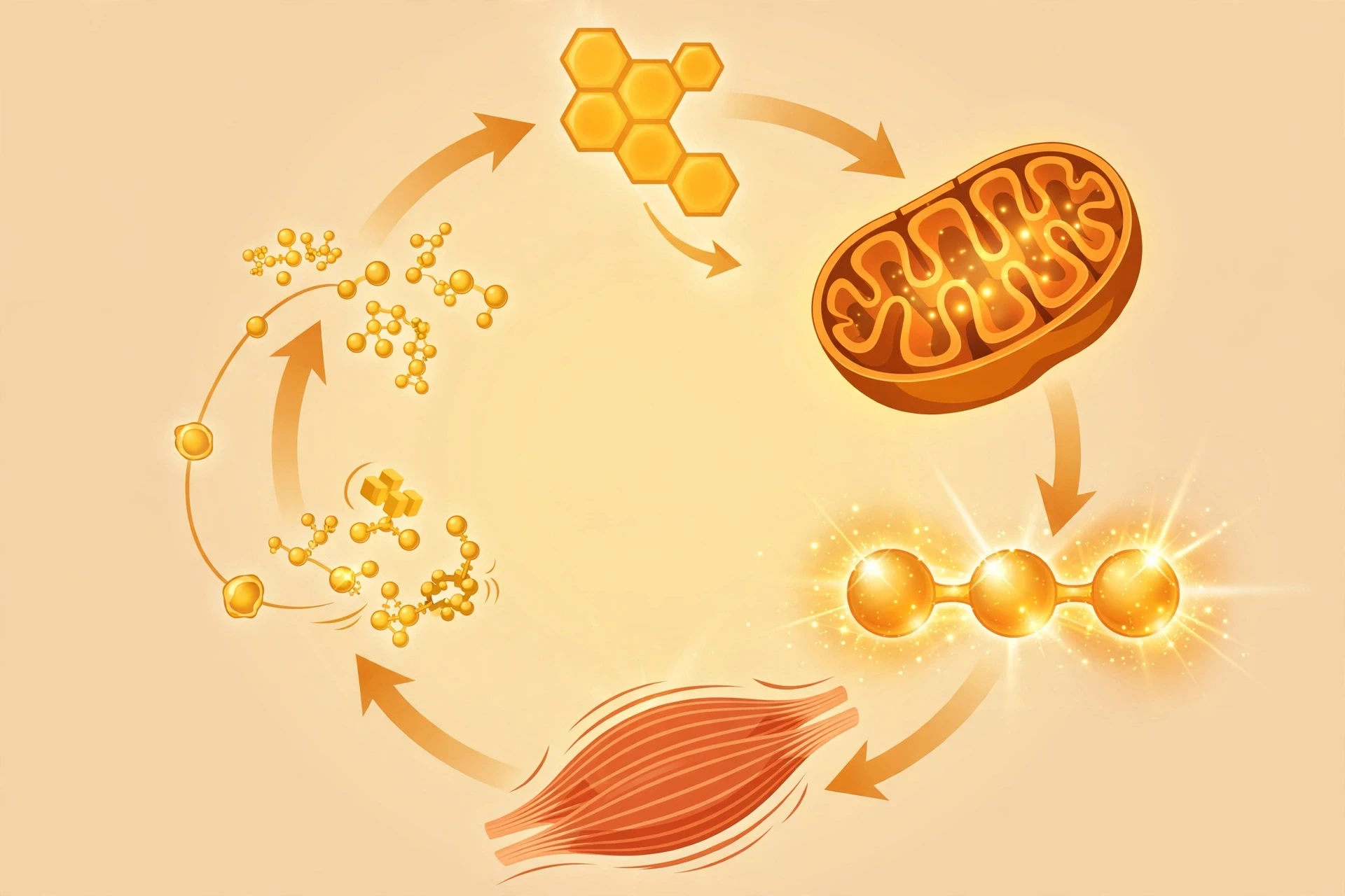
The energy released by breaking down food is not used directly. It is first stored in a molecule called adenosine triphosphate, or ATP — the energy currency of the cell. ATP has three phosphate groups in a row, and the bonds between them hold usable energy. When the cell needs energy, it removes the last phosphate, turning ATP into ADP (adenosine diphosphate) and releasing energy that can drive a reaction, move a muscle, or pump an ion across a membrane. The cell then recharges ADP back into ATP using energy from food. The cycle turns over constantly: a typical cell recycles its own weight in ATP many times a day.
That recycling is why you cannot store ATP the way you store fat. ATP is a short-term currency, made on demand and spent almost immediately. Fat is the savings account; ATP is the cash in your pocket. The mitochondria are the main mint, producing most of the cell's ATP through a process called oxidative phosphorylation, which is described below.
8.3 Glycolysis, the citric acid cycle, and the electron transport chain
The breakdown of glucose follows three main stages. The first is glycolysis, which takes place in the cytoplasm. One glucose molecule is split into two molecules of pyruvate, producing a small net gain of ATP and carrying electrons to a carrier molecule called NADH. Glycolysis needs no oxygen, which is why it can run in cells that are short of oxygen — and why it is probably one of the oldest metabolic pathways on Earth.
The second stage is the citric acid cycle, also called the Krebs cycle, which takes place in the mitochondria. Pyruvate is converted into acetyl-CoA, which enters the cycle and is broken down step by step, releasing carbon dioxide and loading more electrons onto carrier molecules. The cycle also produces a small amount of ATP directly. Its intermediates are shared with other pathways, so the cycle is not only an energy machine but a crossroads: molecules leave it to build amino acids, fatty acids, and other products.
The third stage is the electron transport chain, also in the mitochondria. Electrons from the carriers are passed along a series of protein complexes in the inner mitochondrial membrane, and the energy released is used to pump protons across the membrane, building a gradient. Protons then flow back through ATP synthase, a remarkable molecular turbine, and their flow drives the production of ATP. Oxygen is the final acceptor of the electrons, which is why breathing matters: without oxygen, the chain backs up, the gradient collapses, and ATP production falls sharply. That is the biochemical reason a blocked airway is an emergency within minutes, not hours.
Together, the three stages extract far more energy from glucose than glycolysis alone could. The exact yield depends on conditions and on how it is counted, but the order of magnitude is clear: complete breakdown yields roughly thirty or so ATP molecules per glucose, compared with a net gain of two from glycolysis alone. The cell pays for that efficiency with complexity — dozens of enzymes, membranes, gradients, and control points — and the payoff is a body that can run, think, and heal on the energy in a meal.
8.4 Metabolism beyond glucose
Glucose is not the only fuel. Fatty acids are broken down in the mitochondria through beta-oxidation, producing acetyl-CoA that feeds into the citric acid cycle. Amino acids can also be used for energy when needed, after their nitrogen is removed — a process that produces urea, which the kidneys excrete. The body chooses its fuel by circumstance: glucose for the brain and for quick effort, fatty acids for steady work and for fasting, protein only as a last resort, because burning protein means breaking down the body's own machinery.
Metabolic pathways also interconvert. Excess glucose can be turned into fat. Some amino acids can be turned into glucose. The liver is the great hub of this traffic, taking in nutrients from the gut, storing some, converting others, and releasing what the rest of the body needs. That is why liver disease affects so much more than the liver: when the hub fails, every tissue feels it.
ATP is a short-term currency, made on demand and spent almost immediately. Fat is the savings account; ATP is the cash in your pocket.
9. Water, pH, and the Chemical Environment of Life
9.1 Why water matters
Water is the most abundant molecule in most cells, and its chemistry shapes everything around it. A water molecule is bent, with oxygen at one end and two hydrogens at the other, and oxygen pulls electrons more strongly than hydrogen does. The result is a polar molecule: a slight negative charge near the oxygen, a slight positive charge near the hydrogens. That polarity lets water form hydrogen bonds with itself and with other polar molecules, giving it a high boiling point, a high heat capacity, and a remarkable ability to dissolve salts and sugars.
For biochemistry, water's most important role is as a solvent and a shaper. Ions and polar molecules dissolve in it and move through it. Nonpolar molecules — fats and oils — are pushed together by it, which is what drives membrane formation and protein folding. Water also takes part in reactions directly: hydrolysis reactions split molecules by adding water, and condensation reactions join molecules by removing water. Digestion is largely hydrolysis. Building a protein is largely condensation. Water is not a passive background; it is a participant.
9.2 pH and buffers
The pH scale measures how acidic or alkaline a solution is, based on the concentration of hydrogen ions. A pH of 7 is neutral. Lower values are acidic, higher values are alkaline. The scale is logarithmic, so each whole-number step is a tenfold change in hydrogen ion concentration. Blood is held near pH 7.4, slightly alkaline, and even a small drift can be dangerous, because enzymes and proteins change shape when pH changes.
The body holds pH steady with buffers — mixtures that resist change by absorbing extra hydrogen ions or releasing them as needed. The bicarbonate buffer is the most important in blood: carbon dioxide, bicarbonate, and hydrogen ions sit in equilibrium, and breathing adjusts the balance. If blood becomes too acidic, breathing speeds up to blow off carbon dioxide; if it becomes too alkaline, breathing slows. The kidneys provide slower, longer-term control by excreting or retaining hydrogen ions and bicarbonate. Lungs and kidneys, so different in form, are partners in the same biochemical job.
Stomach acid shows the other side of pH control. The stomach keeps its contents near pH 2, acid enough to activate pepsin and kill many microbes, while the cells lining the stomach protect themselves with a layer of mucus and bicarbonate. The contrast — acid inside the stomach, near-neutral pH just millimetres away in the tissue — is maintained by membranes, pumps, and buffers working together. It is a daily demonstration that biochemistry is local: the same body holds many different chemical environments at once.
9.3 Salts, ions, and gradients
Ions are atoms or molecules with a net electric charge, and they are central to biochemistry. Sodium and potassium gradients across cell membranes drive nerve impulses. Calcium ions act as a signal inside cells, triggering muscle contraction and the release of neurotransmitters. Magnesium ions sit at the heart of chlorophyll and are needed by enzymes that handle ATP. Iron sits at the heart of haemoglobin, binding oxygen. Zinc is needed by hundreds of enzymes, including those that copy DNA.
Cells spend a large share of their ATP maintaining ion gradients — pumping sodium out and potassium in, keeping calcium low inside the cell until a signal is needed. Those gradients are stored energy, like water behind a dam, and the cell releases them in controlled bursts to do work. A nerve impulse is, in biochemical terms, a wave of ion channels opening and closing in sequence, each one letting ions flow down a gradient that ATP built. The electrical language of the nervous system is written in chemistry.
10. Photosynthesis and Respiration: Two Halves of One Cycle
10.1 Capturing sunlight
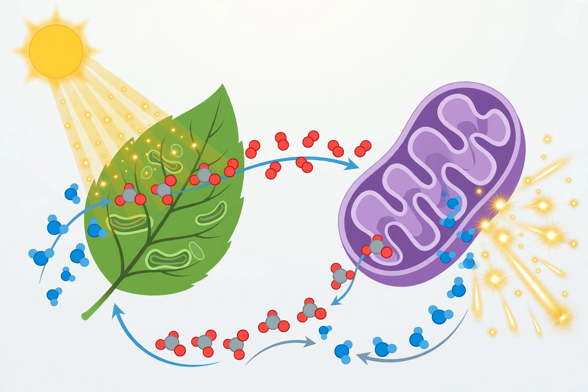
Photosynthesis is the biochemical process by which plants, algae, and some bacteria capture light energy and use it to build sugars from carbon dioxide and water, releasing oxygen as a by-product. The overall equation is simple to write and astonishing to contemplate: light plus carbon dioxide plus water gives glucose plus oxygen. Behind that simplicity lies one of the most complex molecular machines in nature — the photosystems in the chloroplast, which absorb photons, split water, move electrons, and build the ATP and reducing power that the Calvin cycle then uses to fix carbon into sugar.
The details matter because they explain the world we live in. Almost every food chain starts with photosynthesis. The oxygen we breathe is the waste product of water-splitting in photosystem II, released by organisms that have been doing it for billions of years. The fossil fuels we burn are ancient photosynthesis, stored as coal, oil, and gas. When we talk about the carbon cycle, we are talking about photosynthesis and respiration trading carbon back and forth on a planetary scale.
For the full story of how plants capture light and build sugars, see the Sciory article on Photosynthesis.
10.2 Releasing the stored energy
Cellular respiration is the mirror image of photosynthesis: glucose plus oxygen gives carbon dioxide plus water plus usable energy, captured as ATP. The stages described in section 8 — glycolysis, the citric acid cycle, and the electron transport chain — are the machinery of respiration. Together, the two processes form a cycle: photosynthesis stores light energy as chemical energy in sugars; respiration releases that energy to do work. The oxygen released by one is consumed by the other; the carbon dioxide released by one is consumed by the other.
11. Biochemistry in the Body: Signals, Hormones, and the Immune System
11.1 How cells talk to each other
A multicellular body is a community of trillions of cells, and it stays coordinated because cells send signals to one another. The signals are biochemical: hormones that travel in the blood, neurotransmitters that cross the tiny gap between nerve cells, local messengers that act on their neighbours, and contact signals passed directly between touching cells. Each signal has a receptor — a protein that recognises it and starts a response inside the receiving cell.
Hormones illustrate the principle. Insulin, made in the pancreas, tells cells to take up glucose. Adrenaline, made in the adrenal glands, tells the heart to beat faster and the liver to release glucose, preparing the body for sudden effort. Thyroid hormones set the pace of metabolism across the whole body. Cortisol helps the body respond to stress and wakes it in the morning. Each hormone is a chemical message, each receptor is a lock, and each response is a cascade of biochemical steps inside the cell.
11.2 The biochemistry of the immune response
The immune system is biochemistry in defence mode. Antibodies are proteins that recognise specific shapes on invaders — antigens — and mark them for destruction. The complement system is a cascade of proteins in the blood that, once triggered, punches holes in bacterial membranes. Clotting is a cascade of protein-cutting enzymes, each one activating the next, so that a small trigger produces a large, fast response. Inflammation is a biochemical conversation: damaged tissues release signals, blood vessels widen, immune cells arrive, and repair begins.
Vaccines work by teaching this system in advance. A vaccine presents a harmless version of an antigen, the immune system makes antibodies and memory cells against it, and a later encounter with the real pathogen meets a prepared defence. The memory is biochemical too — long-lived cells and circulating antibodies that persist for months or years. That is why a vaccine given in childhood can protect decades later: the chemistry remembers, even when the person has forgotten the injection.
11.3 When signalling goes wrong
When signalling fails, disease follows. In some cancers, a receptor that should switch off stays on, telling cells to divide without pause. In autoimmune disease, the immune system mistakes the body's own molecules for invaders and attacks them. In hormone disorders, too much or too little of a signal throws whole systems out of balance. Biochemists study these failures at the molecular level: which protein is mutated, which pathway is overactive, which step could be blocked by a drug. That is the logic behind targeted therapy — a drug designed for a specific molecular fault, rather than a general poison that harms healthy cells along with diseased ones.
The promise is real, but so is the complexity. A pathway that is overactive in one tumour may be normal in another, and a drug that helps one patient may fail in the next. That is why modern medicine increasingly pairs a treatment with a test: a genetic or biochemical marker that predicts who will benefit. Biochemistry is becoming personal, one molecule at a time.
12. How Biochemists Work: Tools of the Trade
12.1 Seeing the invisible
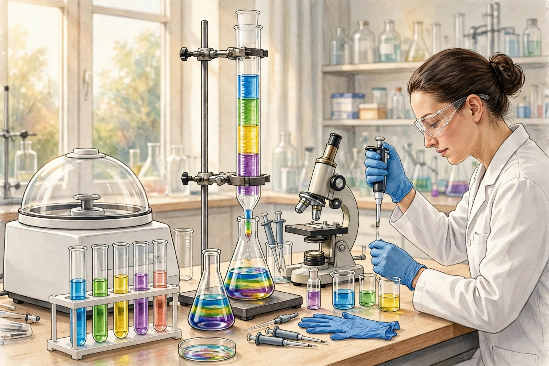
Biochemistry deals with molecules too small to see, so the field has built an arsenal of ways to make them visible, measurable, and manipulable. Chromatography separates mixtures into their components. Electrophoresis separates DNA and proteins by size and charge, producing the banded patterns familiar from crime dramas and paternity tests. Spectroscopy measures how molecules absorb and emit light, revealing their identity and concentration. Mass spectrometry weighs molecules with extraordinary precision, identifying proteins and metabolites from tiny samples. X-ray crystallography and cryo-electron microscopy reveal the three-dimensional shapes of proteins and complexes, atom by atom.
Each tool answers a different question, and a real investigation uses several together. A biochemist studying a new enzyme might purify it by chromatography, check its purity by electrophoresis, measure its speed by spectroscopy, identify its products by mass spectrometry, and solve its structure by crystallography. The answer is not in any one instrument; it is in the agreement between them.
12.2 Reading and rewriting DNA
The ability to read DNA — sequencing — has transformed biochemistry. What once took years and a large team can now be done in hours. Sequencing a genome reveals the genes an organism carries, the variations between individuals, and the mutations behind a disease. It has also revealed how much DNA does not code for protein at all, and how much of the genome's work is regulatory — switching genes on and off in the right place at the right time.
The ability to edit DNA has followed close behind. Techniques that cut and rewrite specific sequences let researchers test what a gene does by removing it, changing it, or replacing it. In medicine, the same ideas are being developed as therapies for inherited disease, aiming to correct the faulty sequence at its source rather than treating its consequences. The science is powerful, and the ethical questions are serious: which changes are treatments, which are enhancements, and who decides? Biochemistry supplies the tools; society must supply the judgement.
13. Biochemistry in Everyday Life, and What Comes Next
13.1 Food, farming, and the kitchen
Biochemistry is at work in every kitchen. Bread rises because yeast enzymes produce carbon dioxide. Cheese forms because an enzyme cuts a milk protein and lets curds form. Meat tenderises because enzymes break down tough connective tissue — which is why pineapple, rich in the enzyme bromelain, can soften meat and can also sting the mouth. Yoghurt forms because bacteria convert lactose into lactic acid, thickening milk and giving it its tang. Cooking itself is biochemistry: heat denatures proteins, gelatinises starch, and drives the browning reactions that give roast food its flavour and colour.
Farming depends on biochemistry too. Fertilisers supply nitrogen, phosphorus, and potassium — the elements plants need in largest amounts. Pesticides target biochemical pathways in pests. Plant breeding and genetic improvement select for biochemical traits: more starch in a grain, more oil in a seed, better resistance to drought. As the climate changes, the biochemistry of crops — how they fix carbon, how they use water, how they cope with heat — will decide what can grow where, and how much food the world can produce.
13.2 Common misunderstandings
A few misunderstandings about biochemistry keep appearing, and they are worth clearing up. First, "chemical" does not mean artificial or harmful. Everything is chemical: water, air, an apple, a vitamin. The question is never whether a substance is a chemical, but what it does, in what amount, and in what context. Second, enzymes are not consumed by the reactions they catalyse, and they are not alive. They are proteins — machines, not organisms — and they stop working when their shape is lost. Third, DNA is not destiny in the simple sense. Genes set possibilities and tendencies, but diet, environment, and chance shape how they are expressed. Two people with the same genetic risk can have very different outcomes, because biochemistry is a conversation between genes and surroundings, not a script read aloud.
13.3 Where biochemistry is heading
The next chapters of biochemistry are already being written. Synthetic biology aims to design new biochemical pathways — microbes that make medicines, fuels, or materials from simple starting materials. Personalised medicine aims to match treatments to a patient's biochemistry, using genetic and metabolic tests to choose the right drug at the right dose. Structural biology is mapping proteins at a speed that was unthinkable a decade ago, opening new targets for drug design. And the study of the microbiome — the trillions of microbes in the gut and on the skin — is revealing that much of human biochemistry is not human at all, but a partnership with other species whose chemistry we are only beginning to understand.
What has not changed is the core idea that opened this article: life is chemistry organised with precision, memory, and purpose. The bread on the breakfast table, the air in your lungs, the thoughts in your head — all of them are biochemistry in action. The more clearly we see that chemistry, the better we can care for the bodies, the food, and the planet that depend on it. That is the promise of biochemistry, and it is a promise that belongs to everyone who eats, breathes, and wonders how it all works.
Frequently Asked Questions
What is biochemistry?
Biochemistry is the study of the chemical processes and molecules inside living things. It explains how carbohydrates, lipids, proteins, and nucleic acids are built, how they react, how they store and spend energy, and how they carry instructions from one generation to the next. It sits where chemistry meets biology, and it covers everything from a single enzyme reaction to the metabolism of a whole organism.
What are the four biomolecules?
The four biomolecules are carbohydrates, lipids, proteins, and nucleic acids. Carbohydrates, built from simple sugars, supply quick energy and structure. Lipids, built from fatty acids and glycerol, store energy long-term and build membranes. Proteins, built from amino acids, do most of the work in the cell, including catalysis, transport, and signalling. Nucleic acids, built from nucleotides, store and transmit genetic information as DNA and RNA.
How does DNA store information?
DNA stores information in the sequence of its four bases — adenine, thymine, guanine, and cytosine. Three bases in a row, a codon, specify one amino acid in a protein, or a start or stop signal. The sequence of bases along a gene therefore spells out the sequence of amino acids in a protein. Because A always pairs with T and G always pairs with C, each strand of the double helix can serve as a template for copying the other, so the information is passed on accurately when cells divide.
What do enzymes do in the body?
Enzymes are protein catalysts that speed up chemical reactions in the body without being used up themselves. They lower the energy barrier of a reaction by binding substrates in an active site and holding them in the right shape to react. In the body, enzymes digest food, copy DNA, build proteins, make ATP, remove toxins, and carry out almost every step of metabolism. Without them, the reactions of life would be far too slow to sustain a living organism.
Why is water so important in biochemistry?
Water is the solvent in which most biochemical reactions take place, and its polarity shapes how molecules behave. It dissolves salts and sugars, forms hydrogen bonds, and drives the folding of proteins and the formation of membranes by pushing water-fearing parts together. Water also takes part in reactions directly, splitting molecules in hydrolysis and being removed when molecules are joined. Cells are mostly water, and life as we know it depends on it.
What is ATP and why do cells need it?
ATP, or adenosine triphosphate, is the energy currency of the cell. Energy from food is used to build ATP from ADP and phosphate, and when the cell needs energy — to move a muscle, pump an ion, or build a molecule — it breaks ATP back to ADP, releasing energy. The cycle turns over constantly. ATP is made mainly in the mitochondria through glycolysis, the citric acid cycle, and the electron transport chain, with oxygen as the final electron acceptor.
Can biochemistry explain disease?
Biochemistry can explain many diseases at the molecular level, though rarely on its own. Diabetes involves insulin and glucose handling; sickle cell disease involves a single amino acid change in haemoglobin; cystic fibrosis involves a faulty transport protein; many cancers involve signalling proteins stuck in the "on" position. Understanding the faulty molecule points to treatments — replacing a missing hormone, blocking an overactive enzyme, or correcting a gene. Biochemistry gives medicine its molecular map, and that map guides diagnosis, treatment, and prevention.